Reading...
![]()
Play button
![]()
Play button
![]()
Use LEFT and RIGHT arrow keys to navigate between flashcards;
Use UP and DOWN arrow keys to flip the card;
H to show hint;
A reads text to speech;
43 Cards in this Set
- Front
- Back
|
carotid sheath consists of
|

vagosympathetic trunk, carotid artery
|
|
|
______ vein (of neck) is not in horse or donkey, but is found dog and cat
|
internal jugular vein is not present horse or donkey (like there is in the dog)
|
|
|
Name boundaries of jugular vein
|
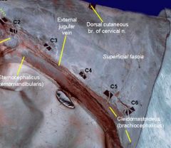
External jugular vein in jugular groove:
- bordered dorsally by cleidomastoideus m. (part of brachiocephalicus) - bordered ventrally by the sternocephalicus - medially by the omohyoideus, which seperates it from carotid sheath - is covered by the cutaneous colli m. which is why venipuncture is done in cranial neck |
|
|
what should superficial cervical lymph node never be called in donkey and horse?
|
Never say "prescapular", only superficial cervical lymph node
|
|
|
_____ muscle is most developed in horse, runs over supraspinatus muscle
|
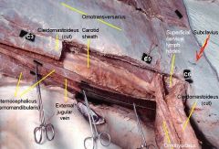
Subclavius muscle is most developed in horse and goat; it is a pectoral muscle, runs over supraspinatus m.
~it is small in cow, absent in carnivores |
|
|
what does sternocephalicus m. in horse extend to and from?
how does structure of sternocephalicus m. vary among species? |
extends from sternum to head
how does structure of sternocephalicus m. vary among species? NOT divided in pig, horse, sheep but divided in carnivore, ox, goat |
|
|
What is sternocephalicus m. called in horse?
specific name(s) What is sternocephalicus m. called in cow and goat? specific name(s) |
sternomandibularus m.
sternomastoid m. sternomandibularis m. |
|
|
What parts of brachiocephalicus m. present in horse?
specific name(s) |
cleiodomastoideus is the upper part of the brachiocephalicus
lower portion known as cleidobrachialis |
|
|
Name 2 extrinsic m. of thoracic. limb that are inseparable in horse?
|
● Brachiocephalicus (outer) overlaps omotransversarius (inner)
|
|
|
Carotid sheath in horse? what is in it?
|

The Carotid sheath
( i). Common carotid (ii). Vagosympathetic trunk NO INTERNAL JUGULAR VEIN IN THE HORSE. |
|
|
what is in visceral space of neck?
what are ventral boundaries of visc.space? |
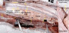
trachea, esophagus,
Carotid sheath (common carotid, vagosympathetic trunk) Recurrent laryngeal nerve Tracheal duct ventral boundaries: Strap muscles (sternohyoideus and sternothyroideus) |
|
|
"Prime mover" (advancing) of thoracic limb in horse?
|
● brachiocephalicus
(omotransversarius m. is adjuvant or assists brachiocephalicus in advancing forelimb) |
|
|
○ Prime muscle of retraction (for equine forelimb)?
|
● Latissimus dorsi m
|
|
|
Name parts of rhomboideus m. in horse:
|
○ cervical and thoracic parts
○ no capitus part like in dog ○ located under (deep to) trapezius m. |
|
|
Nuchal ligament in horse is divided into what two parts?
Describe location relative to each other? Where do they attach? |
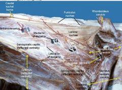
● Nuchal ligament is divided into two parts
FUNICULAR part (is above/dorsal to the laminar part) ○ arises from occipital protuberance, w/o attaching C1-2 ○ paired throughout; fuses with laminar part at C3 LAMINAR part (more ventral) ○ fenestrated from Latin funiculus, dim. of funis “a cord, rope"; and lamina means "thin sheet." |
|
|
What is the vertebral formula of a horse?
|
C7, T18, L6, S5, Ca15-21
|
|
|
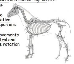
What are the free moving regions of the vertebral column?
Where does the least amount of movement occur? |
* greater flexibility in cervical and caudal
*mov''t in thoracolumbar region is limited b/c of large transverse processes |
|
|
What are the movements of the vertebral column?
|
Dorsal, ventral, and lateral flexion
Some rotation |
|
|
What are the three reasons that the thorax and lumbar region have the least amount of movement?
|
Interspinous ligaments
Intertransverse ligaments Lumbar transverse processes (synovial joints b/t 4th & 5th and 6th & sacrum Also lumbar area is short compared to other species, interverteral disk thin |
|
|
What is the most "mobile" JOINT of the horse vertebral column and why?
|
Lumbosacral joint, for transmission of propulsion from pelvic limbs to cranial part of body
|
|
|
What is the proportion of weight bearing for the forelimbs and hindlimbs in the horse?
Main function of forelimbs? hindlimbs? |

55 : 45
forelimbs bear more weight b/c of large neck and head Forelimbs – for postural function, mainly hindlimbs – for propulsion, mainly. |
|
|
Dorsoscapular ligament:
Arises from where? Inserts where? Identify borders? |
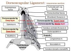
Formed by thoracolumbar fascia! in interscapular region! (withers), extends from *T2-T5*, and arises ventral to ligamentum nuchae and medial to rhomboideus (thoracis) muscle.
Remember T2-T5! |
|
|
What are the three layers of the dorsoscapular ligament and what do they attach to?
|
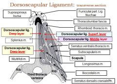
Superficial layer - ribs (laterally)
Middle layer - ribs (proximally) Deep layer - transverse processes |
|
|
What ligament inhibits spread of infection of underlying tissue, but permits craniocaudal spread of an infection?
|
Dorsoscapular ligament
|
|
|
Dorsoscapular ligament is part of the _______mechanism of the forelimb because of its attachment to __________?
|
'concussion absorption mechanism' of the forelimb because of its attachment to TRANSVERSE processes
|
|
|
What membrane must be punctured in order to get to subarachnoid space (to obtain CSF)?
|
atlanto-occipital memrane
(also Dura mater & Arachnoid) |
|
|
What are the muscles of the neck in the horse?
|
Cleidomastoideus (brachiocephalicus)
Sternomandibularis (sternocephalicus) Splenius cervicis Splenius capitis Sternohyoid Sternothyroideus Longus capitis Longus coli |
|
|
What is different in the atlanto-occipital joint of the horse compared to the dog?
|
Presence of longitudinal ligament of the dens and ligament of the apex of the dens
|
|
|
What is continuous (cranially) with the supraspinous ligament?
Where do they meet? Hint: it's origin is the external occipital protuberance, and is significantly widened at the withers? |
FUNICULAR PART of ligamentum nuchae is continuous with the supraspinous ligament at *T3*
p.230 in pasquini's red book FYI: (supraspinous ligament extends along summits of spinous processes of caudal, sacral, lumbar and thoracic vertebrae). |
|
|
What part of the ligamentum nuchae is fenestrated ventrally, b/t C2 and C7, and attaches to the spinous processes of T1 to T4?
|
Laminar part
|
|
|
What are three bursa that are associated with the nuchal ligament?
|
Cranial nuchal bursa
Caudal nuchal bursa Supraspinous bursa |
|
|
What bursa lies between the funicular part of the nuchal ligament and the summit of the spinous process of T3, is congenital, and can cause fistulous withers when inflamed?
|
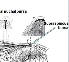
Supraspinous bursa
|
|
|
What muscle covers the external jugular vein caudoventrally?
|
Cutaneous colli muscle
|
|
|
What are the boundaries of the visceral space in the neck dorsally?
ventrally? |
*Longus colli and longus capitis
*strap muscles (sternohyoideus, sternothyroideus) |
|
|
What are the boundaries of the visceral space in the neck laterally?
|
Omohyoideus and sternocephalicus
|
|
|
What are the boundaries of the visceral space in the neck dorsally?
|
Longus colli and longus capitis
|
|
|
How many pairs of ribs does the horse have?
|
18
|
|
|
At standing position, where is the caudal angle of the scapula?
At standing position, where is the point of the elbow? |
7th rib
5th rib |
|
|
Where is the vertex of the diaphragm?
|
lower part of the 6th intercostal space or 7th rib
|
|
|
If the left tracheobonchial ln. is enlarged, what may happen?
|
Pressure on left recurrent laryngeal nerve, roaring
|
|
|
fistulous withers
|
When Supraspinous bursa inflamed, and ruptures
bursa Lies between the funicular part and the summit of the spinous process of T3. Surgical intervention at the level of a vertical line through the tuber spinae of the scapula. |
|
|
What muscle seperates external jugular vein from common carotid in cranial neck?
|
omohyoideus m., only present in horse and ox
|
|
|
How does the nuchal ligament differ between the ruminant and the horse?
|
Horses have:
Cranial bursa Caudal bursa Supraspinous bursa Ligament is paired through entire length |