![]()
![]()
![]()
Use LEFT and RIGHT arrow keys to navigate between flashcards;
Use UP and DOWN arrow keys to flip the card;
H to show hint;
A reads text to speech;
54 Cards in this Set
- Front
- Back
|
Systema urinarium Urinary system comprise? |
Urinary system comprise: ren (2) – the kidney; ureter (2) – the ureter; vesica urinaria (1) – urinary bladder; urethra (1) – the urethra. |
|
|
The urine is eliminated via organs of the urinary tract |
pelvisrenalis, ureter, vesica urinaria, urethra. |
|
|
Kidneys are a ? Function? Measurements? Extremitas superior Extremitas inferior |
The kidneys are paired organs. They produce about 1 – 1,5 l definite urine per day. Thekidneys play a major role in controlling the water and electrolyte balance within the body andmaintaining the acid-base balance of the blood. In an adult thekidney weighs 120 – 200 g, it is 10 – 12 cm long, 5 – 6 cm broad and 4 cm thick. extremitas superior Is thickand round and more near the midline than the lower. It is surrounded by the suprarenal gland,which covers also a small portion of the anterior surface. extremitas inferior is smaller and thinner than the superior pole. |
|
|
2 surfaces 2 margins hilum renale? |
There are two surfaces: facies anterior and facies posterior. The last one is more flat. 2 margins: margo lateralis is convexand directed laterally. The medial margin – margo medialis is concave. In its central partthere is a deep longitudinal fissure – hilum renale: Through hilum renale the blood and lymphvessels, nerves and ureter enter and leave. |
|
|
Kidney is enveloped by? |
Capsula fibrosa Capsula adiposa Fascia renalis |
|
|
Capsula fibrosa |
1. capsula fibrosa – fibrous capsule lies close to cortex renis; |
|
|
Capsula adiposa |
2. capsula adiposa – formed by fat, more thick at the posterior surface and protects kidney from concussion and cooling; |
|
|
Fascia renalis |
3. fascia renalis – fascial envelope consists of two laminas – anterior and posterior lamina. Laterally both laminas are fused together; medially they extend over the kidney`s surfaces. Anterior lamina continues over the renal vessels, aorta abdominalis and v. cava inferior to join the similar plate of the other side. Posterior lamina medially is attached to the vertebral column. Into lower direction they aren’t connected and continue into retroperitoneal tissue. |
|
|
Descensus renis |
Any form of malnutrition by decreasing the deposit of fat in capsula adiposa can cause descending of the kidney |
|
|
Sinus renalis |
Contains: pelvis renalis and calyces renales Is lined by: capsula fibrosa. |
|
|
Cortex |
The cortex – cortex renalis is dark brown in colour, soft and granular in consistence, about 4– 5 mm wide. The parts of it extend into medulla as columnae renales. The stripes radiatingfrom the medulla to the cortex are medullary rays – radii medulares. |
|
|
Medulla |
The medulla is light brown in colour and consists of striated conical formations pyramidesrenales (up to 12 – 15). |
|
|
Lobi renales |
Renal pyramid + renal cortex above it |
|
|
Segmenta renalia |
Lobi renales supplied by 1 a.segmentalis |
|
|
Papilla renalis |
Many apices of lobi renales fuse to form papilla renalis On papilla renalis there are present foramina papillaria through which urin is secreted |
|
|
Calyx renalis minor |
Each papilla is surrounded by this collecting duct |
|
|
Calyx renalis major |
Many calyx renalis minor is surrounded by calyx renalis major(2-3) to form a funnel shaped sac - pelvis renalis (pyelon) |
|
|
Nephronum |
Functional unit of kidney Kidney contains about 1-1,5 million nephrons Each nephron contains a glomerulus and its tubules Glomerulus is a filter through which primary urine is filtered from the blood Secondary urine is produced by reabsorption in tubules |
|
|
Nephronum Parts: |
corpusculum renis; portio nephroni proximalis; ansa nephroni; portio nephroni distalis; pars intercalata. |
|
|
corpusculum renis; |
Corpusculum renis consists of glomerulus and capsula glomeruli. The glomerulus containsabout 30 capillary loops; it has vas afferens and vas efferens. The glomerulus is invaginatedinto the sac – capsula glomeruli. This capsule has pars externa and pars interna and the spacebetween them is the beginning of the tubular system, receiving primary urine. |
|
|
portio nephroni proximalis; |
Portio nephroni proximalis has two parts: curved part – pars convoluta straight part –pars recta. Pars convoluta lies near corpusculum renis and continues into pars recta. |
|
|
ansa nephroni; |
2 parts: pars descendens pars ascendens. |
|
|
portio nephroni distalis; |
which is curved and surrounds corpusculum renis. |
|
|
pars intercalata. |
Pars intercalata is the last part of nephronum and it continues into tubulus renalis colligens. Several tubuli renales colligentes fuse to form ductus papillaris which opens on papillarenalis. |
|
|
Ureter |
A slightly flattened paired tube, which convey the urine from the kidney to theurinary bladder. Its length is about 30 cm. |
|
|
Ureter Path? Male Female |
It commences within pelvis renalis, descends along the posterior abdominal wall anteriorly tom. psoas major. At the level of art. sacroiliaca it crosses linea terminalis, continues into thepelvic cavity passing downward along its lateral wall. Male: Crosses ductus deferens. Female: In female it passes between layers of ligamentum latum ostium ureteris |
|
|
Ureter has 3 parts |
pars abdominalis pars pelvica pars intramuralis |
|
|
pars abdominalis |
lies behind the peritoneum from hilum renale to linea terminalis; |
|
|
pars pelvica |
from linea terminalis till the urinary bladder; |
|
|
pars intramuralis |
passes through the wall of the urinary bladder. |
|
|
Diameter of ureter |
The diameter of the ureter is different in different parts; usually it is about 7 – 8 mm, but thereare three narrowings with the diameter 3 – 4 mm: 1.) upper narrowing is at hilum renale; 2) middle narrowing is at the level of linea terminalis; 3) lower narrowing is in the wall of the urinary bladder. |
|
|
Wall of ureter |
The wall of the ureter is composed of: 1) tunica mucosa, 2) tela submucosa (not welldeveloped), 3) tunica muscularis, 4) tunica adventitia. |
|
|
Vesica urinaria s. Cystis |
The urinary bladder is a sac which acts as a reservoir for the urine. Its capacity is about 250 –500 ml. 3 portions: apex vesicae – projected upward; corpus vesicae; fundus vesicae – directed downward. |
|
|
There are two conditions of the urinary bladder: |
1) Empty 2) Distended |
|
|
Empty |
The empty urinary bladder is situated behind symphysis pubica and has two surfaces – faciesanterior et facies posterior and two lateral margins – margines laterales. |
|
|
Distended |
The distended bladder is directed upward and forward above symphysis pubica and is oval inshape. |
|
|
3 openings at fundus vesicae: |
1) Ostium ureteris dextrum 2) Ostium ureteris sinistrum 3) Ostiumurethrae internum |
|
|
On inner surface at fundus vesicae the openings border a? |
triangle –trigonum vesicae. The mucosa is fused closely with the muscular coat in this part. The twoostia ureteris are joined by the fold plica interureterica. |
|
|
Wall of bladder is composed of 4 layers |
1) tunica mucosa, 2) telasubmucosa, 3) tunica muscularis, 4) tunica adventitia or tunica serosa. |
|
|
The mucosa |
is thin, smooth and covered by transitional epithelium. There are folds in anempty urinary bladder. There are no folds in trigonum vesicae region because of the absenceof the submucosa. The mucosa also contains glands. |
|
|
Tunica muscularis |
The muscular coat is formed by smooth muscular tissue and consists of three layers – externaland internal layers are arranged longitudinally, middle layer – circulary. The fibers of thecircular layer form m. sphincter urethrae internus. |
|
|
Urethra masculina |
Urethra masculina is a tube like organ for eliminating the urine and the reproducting fluid –sperm. The male urethra extends from the urinary bladder (ostium urethrae internum) to ostiumurethrae externum at the glans penis. Its length varies from 15 to 22 cm, diameter about 5 – 7 mm. The male urethra is divided intothree parts: 1) pars prostatica; 2) pars membranacea; 3) pars spongiosa. |
|
|
Pars prostatica |
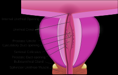
Pars prostatica is the widest and most extended part about of 3 cm long. It passes throughprostata from its base to its apex. On posterior wall of the prostatic part there is longitudinalridge crista urethralis. In its middle part there is an elevation formed by mucous membraneand adjacent tissue – colliculus seminalis, with a slightly depressed fossa on it – utriculusprostaticus (the remnant of ductus paramesonephricus in foetus). On both sides of utriculusprostaticus there are openings of ductus ejaculatorius. In the wall of the prostatic part thereare also orifices of prostatic ducts – ductuli prostatici. |
|
|
Pars membranacea |
is the shortest part about 1,5 – 2 cm long. It extends downward between apex prostatae and bulbus penis, perforating perineum. This part is completely surrounded by m. sphincter urethrae externus formed by striated muscular tissue. |
|
|
Pars spongiosa |
Pars spongiosa is the longest part of the urethra. It is about 15 cm long and extends from parsmembranacea to the external orifice. It is located in corpus spongiosum penis except theupper part directly below perineum surrounded only by the connective tissue. At bulbus penisregion glandula bulbourethralis opens into urethra. |
|
|
Urethra masculina has |
three narrowings about 4 mm in diameter and three enlargements about 10 – 12 mm in diameter: |
|
|
The male urethra has |
two curves |
|
|
The wall of urethra masculina is formed by |
two layers: 1) tunica mucosa, 2) tunica muscularis Lacunae urethrales Corpus spongiosum penis Corpus cavernosum penis fossanavicularis urethrae Ostium urethrae externum |
|
|
Urethra feminina |
It is a narrow canal about 2,5 – 4 cm long and 8 – 12 mm in diameter. |
|
|
Extends from |
It extends from ostiumurethrae internum, passes through diaphragma urogenitalis and terminates by ostiumurethrae externum in vestibulum vaginae 2 cm behind glans clitoridis. Urethra feminina isplaced behind symphysis pubica and imbeded into the anterior wall of the vagina. |
|
|
The wall has three layers: |
1) tunica mucosa 2) tunica muscularis 3) tunica adventitia. |
|
|
Tunica mucosa |
Tunica mucosa lines the urethra and forms longitudinal folds. It has numerous lacunaeurethrales whith glandulae urethrales. |
|
|
Tunica muscularis |
Tunica muscularis consists of external circular and internal longitudinal layers of the smoothmuscular tissue. Circular fibres form m. sphincter urethrae internus. Ostium urethraeexternum is surrounded by sceletal muscular tissue of perineum – m. sphincter urethraeexternus. |
|
|
Differances between urethra masculina and urethra feminina |
urethra masculina – is longer; diameter is smaller; it has curves, narrowings and enlargements; urethra feminina – is shorter; diameter is bigger; it is straight; there are no narrowings and enlargements. Functional differences: urethra masculina – excretes urine and sperm; urethra feminina – excretes urine. |