![]()
![]()
![]()
Use LEFT and RIGHT arrow keys to navigate between flashcards;
Use UP and DOWN arrow keys to flip the card;
H to show hint;
A reads text to speech;
77 Cards in this Set
- Front
- Back
|
What musculomembranous structure separates the thoracic and abdominal cavities? |
Diaphragm |
|
|
What two muscles are located on either side of the lumbar vertebrae and can be fairly well visualized on a quality abdominal image? |
Left and right psoas major |
|
|
What structures constitute the upper GI tract? |
Mouth Pharynx Esophagus Stomach Small intestine |
|
|
What are the parts of the lower GI tract? |
Cecum Ascending colon Hepatic flexure Transverse colon Splenic flexure Descending colon Sigmoid Rectum Anus |
|
|
What is the name of the sphincter muscle located at the junction of the terminal ileum and cecum? |
Ileocecal valve |
|
|
What part of what structure lies within the loop of the duodenum? |
Head of the pancreas |
|
|
What is the largest solid organ of the body that occupies the right upper quadrant? |
Liver |
|
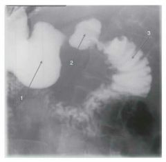
What organ lies in the anatomical loop Illustrated here? Identify the anatomy indicated in the image. |
Pancreas 1. Pylorus 2. Duodenal bulb 3. Descending duodenum |
|
|
What is the double-walled serous membrane associated with the abdomen? |
Peritoneum |
|
|
What is the relationship of the kidneys, ureter, pancreas, duodenum, ascending and descending colon, and aorta to the peritoneum? |
They are all retroperitoneal structures |
|
|
What is the name of the upper, middle region of the abdomen? What is the name of the lower lateral regions? |
Epigastrium Left and right iliac/inguinal |
|
|
What 3 projections are generally included in an acute abdomen survey? |
AP supine abdomen Erect or lateral decubitus abdomen PA chest |
|
|
Images of the abdomen are most generally exposed upon what phase of respiration? |
Expiration |
|
|
What kV range is usually recommended for most abdominal Imaging? |
70 to 80 kV |
|
|
Abnormal accumulation of fluid in the peritoneal cavity is termed __________; abnormal accumulation of air is termed __________. |
Ascites Pneumoperitoneum |
|
|
The condition characterized by telescoping of a portion of bowel into an adjacent portion is termed __________. |
Intussusception |
|
|
What is another name for Crohn's disease? |
Regional enteritis |
|
|
When performing lateral decubitus abdomen to show small amounts of air in the peritoneal cavity, the affected side should be __________. |
Up |
|
|
The CR should be directed to mid-line and __________ for a lateral decubitus projection of the abdomen. |
2" above the iliac crest |
|
|
The CR should be directed to mid-line and __________ for an AP erect projection of the abdomen. |
2" above the iliac crest |
|
|
When evaluating the abdomen for small amounts of air or fluid, both __________ should be visualized. |
Hemidiaphragms |
|
|
Protrusion of a portion of the upper stomach through the esophageal hiatus of the diaphragm describes __________. |
Hiatal hernia |
|
|
List the four layers of GI tissue, from inner to outer. |
Mucosa Submucosa Muscular Serosa |
|
|
Which layer of stomach tissue forms folds called rugae? |
Mucosa |
|
|
Which portion of the small intestine has a feathery appearance when filled with barium? |
Jejunum |
|
|
The large right lobe of the liver is separated from the left by the __________ ligament. |
Falciform |
|
|
One of the principal functions of the liver is to produce __________, which leaves the liver via the right and left __________ ducts. |
Bile Hepatic |
|
|
What two ducts unite to form the common bile duct? |
Cystic duct Common hepatic duct |
|
|
What is the name of the procedure used to examine the biliary and pancreatic ducts by fiber optic means? |
ERCP |
|
|
During ERCP, contrast material is injected into the __________. |
Common bile duct |
|
|
What is the medical term that describes condition of stones in the gallbladder? |
Cholelithiasis |
|
|
Which type of membrane lines body cavities that open to the exterior? |
Mucus |
|
|
The greater curvature forms the __________ aspect of the stomach. What is the name of the distal gastric sphincter? |
Lateral Pyloric |
|
|
What surgical procedure may be done to demonstrate biliary anatomy and rule out residual biliary stones? |
Surgical cholangiography |
|
|
Twisting of the bowel upon itself, causing obstruction, is called __________. |
Volvulus |
|
|
The length of the small bowel is approximately __________ in length. The length of the large intestine is approximately __________. |
10 ft 5 ft |
|
|
What structure is located at the terminus of the small intestine? |
Ileocecal valve |
|
|
List the three parts of the stomach. |
Fundus Body Pylorus |
|
|
List the three parts of the small intestine. |
Duodenum Jejunum Ilium |
|
|
What is the first, most proximal, portion of the large intestine? |
Cecum |
|
|
The muscular ttaeniae coli pulls the large intestine into pouches called __________. |
Haustra |
|
|
Small saccular protrusions of intestinal mucosa through the intestinal wall are called __________. |
Diverticula |
|
|
An abdominal growth of tissue projecting from mucous membrane into a lumen is termed __________. |
Polyp |
|
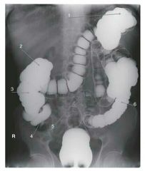
What is the name of the pouches/segmentations seen in this image? Identify the anatomy indicated in the image. |
Haustra 1. Splenic flexure 2. Hepatic flexure 3. Ascending colon 4. Cecum 5. Sigmoid colon 6. Descending colon |
|
|
What type of radiographic examination is required to demonstrate colonic polypoid lesions? |
Double contrast BE |
|
|
What position demonstrates the esophagus projected between the heart and vertebrae? What is usually the optimal of obliquity? |
RAO 35° to 40° |
|
|
A radiographic examination of the esophagus must be performed in the recumbent position in order to demonstrate what two types of pathology? |
Hiatal hernia Esophageal varices |
|
|
What is the best position to demonstrate a barium-filled pylorus and duodenum? To see double contrast of the pylorus and duodenum? |
RAO AP/LPO |
|
|
What position will best demonstrate the retrogastric space? |
Lateral |
|
|
What method of radiologic gastrointestinal examination is used to demonstrate mucosal and other intraluminal lesions? |
Double contrast |
|
|
What method of BE examination will best demonstrate polypoid lesions? |
Double contrast |
|
|
Progressive wave-like movement occurring involuntarily in hollow tubes, especially the alimentary canal, is called __________. |
Peristalsis |
|
|
The term deglutition refers to __________. What does the term aspiration refer to? |
The act of swallowing
The act of inhaling fluid or solid foreign body into the bronchi and lungs |
|
|
Dilation of the esophageal veins, often seen in acute liver disease, is termed __________; the term used to describe difficulty in swallowing is ___________. |
Esophageal varices Dysphagia |
|
|
Modified barium swallow examinations are particularly useful for patients who have suffered what incident? |
Stroke |
|
|
Small bowel series using GI intubation is termed _______. |
Enteroclysis |
|
|
What is the usual patient preparation for an upper gastrointestinal series? |
NPO at least 8 hours |
|
|
Images made during the latter part of a small bowel series require that the CR point of entry is _________ than images made during the first part of the examination. |
Lower |
|
|
What two positions may be used to demonstrate the hepatic flexure without superimposition? |
LPO RAO |
|
|
During double-contrast BE, what part of the large intestine is likely to be filled with barium in the PA recumbent position? |
Transverse |
|
|
During double-contrast BE, what projection may be used to demonstrate the posterior wall of the rectum? |
Ventral decubitus, lateral rectum |
|
|
How much and in what direction should the CR be directed for an AP axial projection of the sigmoid colon? |
30° to 40° cephalad |
|
|
During double-contrast BE, what part of the large intestine is likely to be filled with air in the AP recumbent position? |
Transverse |
|
|
During radiographic examination of the large bowel, what projection is used to open up the sigmoid colon? |
AP or PA axial |
|
|
During double contrast BE, what position will best demonstrate the lateral wall of the ascending colon and medial wall of the descending colon? |
Left lateral decubitus |
|
|
If the surgeon suspects residual biliary stones during cholecystectomy, a catheter can be inserted into the common bile duct with one end extending outside of the body; the patient can later come to the radiology department to rule out biliary stones with what radiologic examination? |
T-tube cholangiogram |
|
|
The __________ kidney is narrower, longer, and in a higher/more superior position in the body than the opposite kidney. |
Left |
|
|
The term nephroptosis means __________. |
Drooping or downward displaced kidney |
|
|
The functional unit of the kidney is termed __________. |
Nephron |
|
|
The term micturition refers to __________. |
Urination |
|
|
The ureters lie __________ to the kidneys and are therefore best demonstrated contrast-filled in the __________ position during intravenous urography. |
Anterior Prone |
|
|
What position will demonstrate the left kidney parallel to the IR, as well as the right ureter free of superimposition? |
RPO |
|
|
What is the correct degree of obliquity used in intravenous urography? |
30° |
|
|
What additional type of image identification is required for intravenous urography? |
Time markers |
|
|
What degree of obliquity is recommended for cystography? |
45° to 60° |
|
|
Radiographic examination of the contrast-filled bladder is termed __________. |
Cystography |
|
|
When examining the contrast build a bladder, how much and what direction is the X-ray tube angled in order to project the pubis inferior to the urinary bladder? |
10° to 15° caudad |