![]()
![]()
![]()
Use LEFT and RIGHT arrow keys to navigate between flashcards;
Use UP and DOWN arrow keys to flip the card;
H to show hint;
A reads text to speech;
38 Cards in this Set
- Front
- Back
- 3rd side (hint)
|
Osteology of the foot Articular surface |
The surface of the bone which is part of a joint |
|
|
|
Osteology of the foot Concave surface |
A rounded depression |
|
|
|
Osteology of the foot Convex surface |
A rounded elevation |
|
|
|
Osteology of the foot Groove |
A elongated depression (trench/gutter) |
|
|
|
Osteology of the foot Tubercle |
small, roughened prominence |
|
|
|
Osteology of the foot Tuberosity |
A larger, roughened prominence |
|
|
|
Osteology of the foot Tibia shape
articulations
landmarks
|
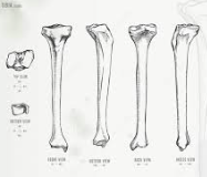
Osteology of the foot Tibia • Triangular in shape • Medial, lateral and posterior surfaces • Anterior, medial and lateral borders
Distal articulations • Fibula • Talus
Distal landmarks • Medial malleolus • Groove for tendon of tibialis posterior |
|
|
|
Osteology of the foot Fibula |
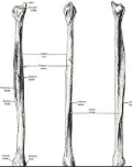
Osteology of the foot Fibula
• Triangular in shape • Medial, lateral and posterior surfaces • Anterior, medial and posterior borders
Distal articulations • Tibia (via fibular notch) • Talus
Distal landmarks • Lateral malleolus • Malleolar fossa • Groove for fibularis longus and brevis |
|
|
|
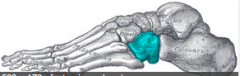
Osteology of the foot Talus Bony landmarks Made up of
|
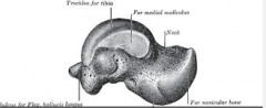
Osteology of the foot Talus • Most superior bone in the foot
Made up of • Head • Neck • Body
Bony landmarks • Talar head • Groove for flexor digitorum longs • Medial tubercle • Lateral tubercle |
|
|
|
Osteology of the foot Superior surface of the talus |
Osteology of the foot Superior surface of the talus
• Also known as the trochlear surface • Articular surface widest anteriorly • Lateral articular surface is larger than the medial articular surface due to the larger lateral malleolus |
Also known as Surface sizes? |
|
|
Osteology of the foot Inferior surface of the talus |
Osteology of the foot Inferior surface of the talus • Anterior facet • Middle facet • Posterior facet • Sulcus tali • Anterior surface (articulation for navicular) |
|
|
|
Osteology of the foot Articular surfaces of the talus
|
Osteology of the foot Articular surfaces of the talus • Superior surface → inferior surface of tibia • Lateral surface → lateral malleolus • Anterior surface → posterior aspect of navicular • Inferior surface → superior aspect of calcaneus |
4 Articulations with other bones |
|
|
Osteology of the foot Articular surfaces of the talus • Medial surface |
• Medial surface → medial malleolus |
|
|
|
Osteology of the foot Articular surfaces of the talus Lateral surface
|
• Lateral surface → lateral malleolus
|
|
|
|
Osteology of the foot Articular surfaces of the talus Anterior surface
|
• Anterior surface → posterior aspect of navicular |
|
|
|
Osteology of the foot Articular surfaces of the talus Inferior surface |
• Inferior surface → superior aspect of calcaneus |
|
|
|
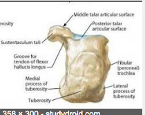
Osteology of the foot Calcaneus |
Osteology of the foot Calcaneus Posterior aspect and land marks
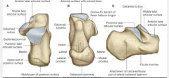
• Heel; largest, strongest bone in the foot
Posterior aspect • Upper part • Middle part (Achilles tendon attachment) • Lower part (plantar aspect) • Calcaneal tuberosity |
Posterior aspect and land marks |
|
|
Osteology of the foot Calcaneus Bony landmarks
Plantar aspect
|
Bony landmarks • Plantar aspect: • Calcanea tuberosity
|
|
|
|
Osteology of the foot Calcaneus Bony landmarks Medial process |
• Medial process (sometimes referred to as medial tubercle) |
|
|
|
Osteology of the foot Calcaneus Bony landmarks Lateral process |

• Lateral process • Tubercle |
|
|
|
Osteology of the foot Calcaneus Bony landmarks Medial aspect
|

Medial aspect
• Sustentaculum tali
• Groove for flexor hallucis longs
|
|
|
|
Osteology of the foot Calcaneus Bony landmarks Lateral aspect
|
Lateral aspect
• Attachment for calcaneofibular ligament • Fibular trochlea |
|
|
|
Osteology of the foot Calcaneus Articulations |
Osteology of the foot Articulations • Anterior, middle and posterior talar surfaces of calcaneus → anterior,middle and posterior facets of the talus • Anterior surface → cuboid |
|
|
|
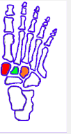
Osteology of the foot Navicular Proximal, concave surface & aspects
|
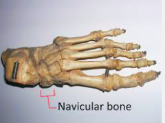
Osteology of the foot Navicular
• Proximal, concave surface → talus • Dorsal/medial aspects wider than plantar/lateral aspects
|
|
|
|
Osteology of the foot Navicular Bony landmarks
|
Bony landmarks
• Navicular Tuberosity; medial plantar aspect |
|
|
|
Osteology of the foot Navicular Articulations
|
• Articulations: • Posterior aspect → talus • Anterior aspect → posterior surfaces of the cuneiforms *May articulate with the cuboid |
|
|
|
Osteology of the foot Cuneiforms |
Three (3) cuneiforms Shaped like a wedge (wider superiorly than inferiorly) |
|
|
|
Cuneiforms Medial cuneiform Articulations
|
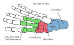
Medial cuneiform Articulations
Posteriorly with the navicular Anteriorly with bases of 1st and 2nd metatarsals Laterally with intermediate cuneiform |
|
|
|
Intermediate cuneiform Articulations
|
Intermediate cuneiform -Posteriorly with the navicular
-Anteriorly with the base of the 2nd metatarsi
-Medially with the medial cuneiform
- Laterally with the lateral cuneiform |
|
|
|
Lateral cuneiform Articulations
|
Lateral cuneiform Posteriorly with the navicular
Anteriorly with the base of the2nd, 3rd, 4th metatarsals
Medially with the intermediate cuneiform
Laterally with the cuboid
|
|
|
|
Cuboid Most lateral tarsal bone Articulations
|
Osteology of the foot Cuboid • Most lateral tarsal bone • Articulations: • Posterior aspect of cuboid → calcaneus • Medial aspect of cuboid → lateral cuneiform •Anterior aspect of cuboid → bases of the 4th and 5th metatarsals *May articulate with the navicular |
|
|
|
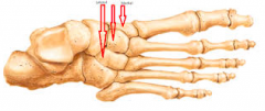
Osteology of the foot Cuboid Bony landmarks
|
Bony landmarks • Tuberosity (lateral aspect) • Groove for fibularis longus (anterior to tuberosity on the inferior surface) |
|
|
|
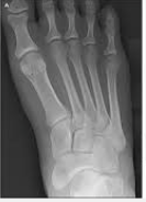
Osteology of the foot Metatarsals Comprises of |
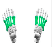
Osteology of the foot Metatarsals • Numbered 1 to 5 (medial → lateral) 1st metatarsal is the thickest, shortest 2nd metatarsal is the longest 5th has a prominent tuberosity (lateral aspect of base – Styloid process) • Comprises of: • Base (proximal), shaft, head (distal)
|
|
|
|
Osteology of the foot Metatarsals Proximal articulations |
• Proximal articulations: Bases of metatarsals 2-5 articulate with each other Cuneiforms, cuboid
|
|
|
|
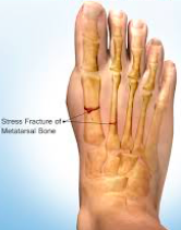
Osteology of the foot Metatarsals Distal articulations |
• Distal articulations: Head of 1st metatarsal → base of 1st proximal phalanx, sesamoid bones Head of 2nd metatarsal → base of 2nd proximal phalanx |
|
|
|
Phalanges (toes) comprises of
|
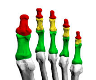
Phalanges (toes)
• Each toe comprises of three bones • Proximal phalanx, middle phalanx, distal phalanx • The big toe (hallux) comprises of proximal phalanx and distal phalanx • Each phalanx comprises of: • Base(proximal)• Shaft• Head (distal)
|
|
|
|
Osteology of the foot Phalanges (toes) Articulations |
• Articulations Base of the proximal phalanx → head of the corresponding metatarsal Head of the proximal phalanx → base of the middle/distal phalanx Non-articular, crescent shaped head of distal phalanx (end of your toe) |
|
|
|
Sesamoid bones Where and what do they do |
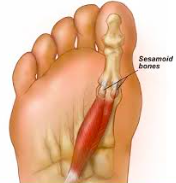
Osteology of the foot Sesamoid bones • Located under the 1st metatarsal head • Medial (tibial) sesamoid • Lateral (fibular) sesamoid • Lie within the tendon of flexor hallucis brevis • Assist in joint function by: • Altering the ‘pull’ of muscles • Reducing friction • Resisting compressive forces
|
|