Reading...
![]()
Play button
![]()
Play button
![]()
Use LEFT and RIGHT arrow keys to navigate between flashcards;
Use UP and DOWN arrow keys to flip the card;
H to show hint;
A reads text to speech;
83 Cards in this Set
- Front
- Back
|
Why may cytology NOT lead to a diagnosis?
|
Not a good representative sample
Multiple interpretations of cell types |
|
|
T or F:
Only intact nucleated cells are evaluated in cytology. |
True!
|
|
|
When should cytology samples be heat fixed?
|
Never! Always air-dried!
|
|
|
What is the approach (ie: 4 questions asked) to a cytological evaluation?
|
Are cells normal?
Is process inflammatory or not? If non-inflammatory, are criteria of malignancy present? If malignant, what type? |
|
|
What is the normal cell morphology for corneal cells?
|
Pseudostratefied columnar epithelium
|
|
|
Which cell type should be present whenever a body cavity fluid is collected?
|
Mesothelial cells
|
|
|
What is the normal morphology of most respiratory cells?
|
Tall columnar with microvilli
|
|
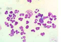
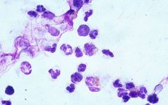
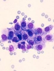
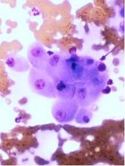
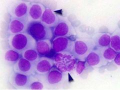
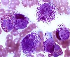
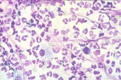
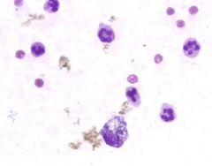
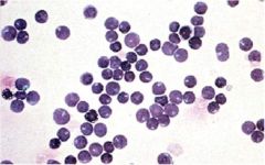
What type of cells are predominant here? If this is an inflammatory process, describe the type of inflammation!!!
|

Neutrophils (degenerative);
indicative of suppurative inflammation (degenerative neutrophils have encountered bacteria) |
|

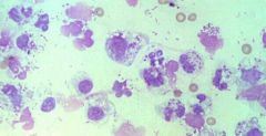
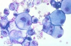
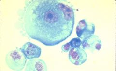
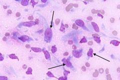
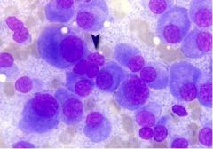
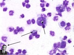
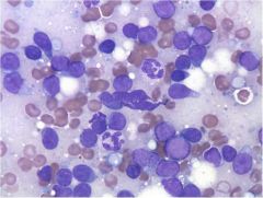
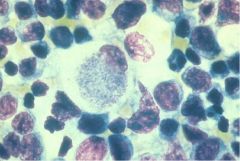
What cells are indicated by the arrows? What type of inflammation is present here?
|
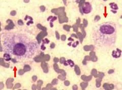
Big ol' Macrophages
Macs + Neutrophils = GRANULOMATOUS INFLAMMATION |
|
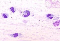
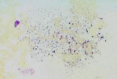
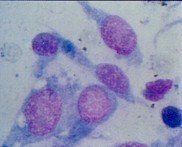
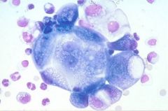
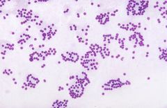
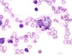
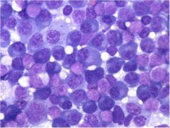
What type of inflammation is present here?
|

Septic, suppurative inflammation (septic = bacteria present, suppurative = neutrophils present)
|
|
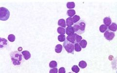
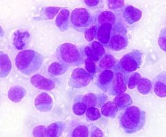
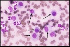
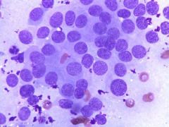
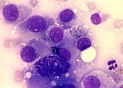
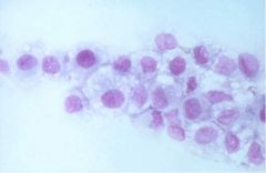
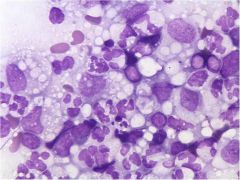
What type of inflammation is depicted here?
|

Lymphocytic
|
|
What type of inflammation is depicted here?
|

Eosinophilic inflammation
|
|
What type of inflammation is depicted here?
|

Septic, suppurative inflammation
|
|
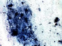
What type of inflammation is present in this image?
|

NONE! Just stain precipitate!
|
|
|
Which of the following are morphologic criteria of malignancy?
a) pleiomorphism (cellular or nuclear) b) abnormalities of the nucleolus c) increased cytoplasmic eosinophilia d) decreased nucleus to cytoplasm ratio e) multiple nuclei |
A, B, and E
|
|
|
What are the three types of malignant cells?
|
Epithelial
Mesenchymal Round cells |
|
|
What are some characteristics of epithelial cells?
|
occur in clusters with cell-to-cell adhesion;
large; distinct cell borders; generally round nucleus; moderate to abundant cytoplasm |
|
|
The condenser lens should be _______ when evaluating a cytology slide.
|
UP
|
|
Evaluate this bone sample in terms of:
Is there inflammation present? If so, what type of inflammation? If not, is it neoplastic? |

Not inflammatory.
Neoplastic (osteosarcoma); note anisocytosis, high nucleus:cytoplasm, basophilic cytoplasm, and mitotic figure! |
|
Evaluate this FNA of a prostate in terms of:
inflammation (yes/no/what type)? neoplasia (yes/no) |

No inflammation;
Cells are HYPERPLASTIC; no anisocytosis or anisokaryosis |
|
|
What are the three cell types that can become malignant? What are the cancerous names of each (type of 'oma)?
|
Epithelial (carcinoma)
Mesenchymal (sarcoma) Round cells (round cell tumor) |
|
|
What are cytologic characteristics of epithelial cells?
|
Large
distinct cell borders cell-to-cell adhesion round nucleus (generally) moderate to abundant cytoplasm |
|

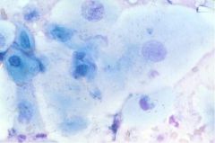
Which cell line is depicted here (epithelial, mesothelial, or round cell)?
|
Epithelial; note cell-to-cell adhesion!
|
|

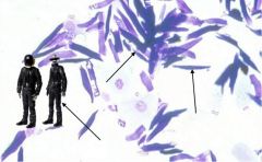
Which cell line is depicted here (epithelial, mesenchymal, or round cells)?
|
Mesenchymal; note lack of cell-to-cell adhesion and spindle shape
|
|

What cell type (epithelial, mesenchymal, or round cell) is depicted here?
|
Round cell; note lack of cell-to-cell adhesion and, um, they're ROUND!
|
|
Characterize this slide in terms of:
inflammation cell lineage malignancy |

not inflammatory
epithelial cells malignant carcinoma!!! (note cell-to-cell adhesion, marked anisocytosis, binuclate cell) |
|

Characterize this anal sac sample in terms of:
cell lineage inflammation malignancy |
epithelial lineage
not inflammatory (neuts are normal from anal region) this is an adenoma but probably not too malignant (not a lot of pleiomorphism) |
|

Characterize this anal sac sample in terms of:
cell lineage, inflammation, and malignancy |
Epithelial, non-inflammatory, malignant!
Perianal adenocarcinoma (lots of anisokaryosis, mitotic figures, some binucleate cells) |
|
Characterize this sample in terms of:
Cell lineage, inflammation, and malignancy. |

Epithelial
Granulomatous inflammation Malignant! Metastatic mammary adenocarcinoma |
|

What cell lineage is this from? What tissue?
|
Epithelial (thyroid)
|
|
|
What are characteristics of mesenchymal cells?
|
Usually singular (may be in small clumps);
Indistinct borders; Spindle-shaped to polygonal; Oval to round nuclei moderate amt of cytoplasm |
|
|
What are characteristics of round cells?
|
single, small to medium sized cells;
distinct cell borders, and they are triangular shaped....NO DUMBASS!!! THEY'RE ROUND!!! |
|
|
What are examples of round cells?
|
Mast cell
Lymphocytes Histiocytes Melanoma TVT |
|
What lineage are these cells from?
|

Mesenchymal
|
|
|
T or F:
Lipomas are a tumor of mesenchymal cells. |
True!!! Even though they're not spindle shaped and tend to stick together!!
|
|
|
T or F:
Histiocytomas are pretty much unique to dogs. |
True!
|
|

What cell type is depicted here? Is this malignant?
|
These are histiocytes (but a bad example of a histiocyte clump...they are usually more single)!
This is a histiocytoma; not malignant. |
|

What lineage is this cell from? What is it indicative of?
|
Round cell;
This is a BUTT CELL indicative of histiocytoma |
|

What type of tumor would this be?
|
Mast cell tumor!!!
|
|

What type of tumor is depicted here? What lineage?
|
Melanoma! A round cell tumor type.
|
|

What type of tumor would this be?
|
Melanoma!
|
|

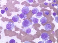
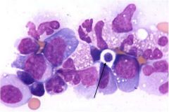
What type of tumor is depicted here (ignore the arrow for now)?
|
Plasmacytoma (tons of pleiomorphic plasma cells)
|
|

What type of tumor is shown here? Are both indicated cells normal?
|
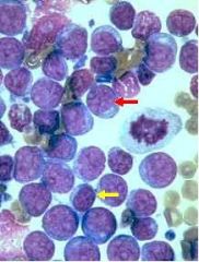
Lymphoma
Red arrow is neoplastic; yellow arrow is normal |
|

What type of tumor is depicted here?
|
Transmissible Venereal Tumor (looks like histiocytoma but with VACUOLES)
|
|
|
An accumulation of fluid in the body is known as...
|
...an EFFUSION
|
|
|
What are the three types of effusions?
|
Transudate;
Modified transudate; Exudate |
|
|
Which fluid has low cellularity and low protein?
|
Transudate
|
|
|
What is the most common cause of a transudate?
|
hypoalbuminemia
|
|
|
What is a major cause of a modified transudate?
|
cardiovascular disease
|
|
|
Which fluid has a high protein content but low cellularity?
|
modified transudate
|
|
|
What type of effusion does FIP cause?
|
modified transudate
|
|
|
Which effusion has high protein and high cellularity?
|
Exudate
|
|
|
T or F:
Exudates are always inflammatory. |
False. They can be inflammatory or non-inflammatory (due to neoplasia).
|
|
|
How can an equine gut rupture be determined via cytology of an abdominocentesis sample?
|
Absence or presence of feed material.
|
|

What type of effusion would this canine thoracocentesis be?
|
septic suppurative exudate
|
|

Characterize this sample of thoracic fluid from a cat?
|
Neoplastic lymphoma
|
|
Characterize this sample of canine abdominal fluid!
|

Bile peritonitis (non-septic)
|
|

What are these weird things from the abdominal fluid of a foal? What is it indicative of?
|
CaCO3 crystals and Sammy Davis Jr.;
Indicates uroperitoneum |
|

What kind of effusion is shown here? What is the crap in the macrophage?
|
Hemorrhagic effusion;
Mac contains hemosiderin |
|
|
How can iatrogenic hemorrage from performing a sampling of body cavity fluid be distinguished from a chronic process?
|
Look for platelets; if there are platelets then it was likely iatrogenic
|
|
|
What cell lines body cavities?
|
Mesothelial cells
|
|
|
T or F:
Mesothelial cells are ciliated. |
False! They have microvilli!
|
|
|
What are characteristics of reactive mesothelial cells?
|
Larger
Vacuolated Binucleate Basophilic |
|

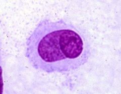
What kind of cells are these?
|
Probably reactive mesothelial cells but safer to call them large mononuclear cells since they look so much like macrophages
|
|
|
T or F:
Horses usually have more abdominal fluid than other species. |
True!
|
|

What are these things?
|
Amniotic fluid + Daft Punk
|
|

What does Lemmy seem to be admiring here?
|
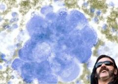
Mesothelioma
|
|
If this is seen in abdominal fluid, what do you think?
|

Bad thoughts....its metastatic squamous cell carcinoma
|
|
|
What causes cells to line-up in joint fluid? What is this called?
|
hyaluronic acid;
called windrowing |
|
|
What types of cells predominate in joint fluid?
|
Large mononuclear cells
|
|
|
Characterize joint fluid grossly.
|
Transparent to slightly yellow;
Viscous |
|
|
Characterize normal CSF grossly and cytologically.
|
Clear colorless,
low cellularity normally mononuclear cells |
|

What is this weird thing in some dog CSF?
|
Cryptococcus neoformans
|
|
posterior staphylmoas or oblique insertion of optic nerve/ Fuch's spots, lacquer cracks
|

high myopia (degenerative)
|
|
|
What are three major reasons for lymph node enlargement?
|
Hyperplasia
Neoplasia Inflammation |
|
|
T or F:
Most lymphocytes in a normal lymph node are small and mature. |
Tru dat!
Once they get stimulated they get bigger....heh heh |
|

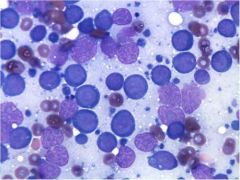
What is going on with this lymph node?
|
Nothing! It is normal! Note that most lymphocytes are small and mature. A few segs are normal.
|
|

What is going on with this lymph node?
|
Hyperplastic and reactive (note all the plasma cells but no anisokaryosis or anisocytosis)
|
|

What gives with this lymph node?
|
Lymphadenitis (macrophages and neutrophils)
|
|
What gives with this lymph node?
|

Allergic reaction (note eosinophils)
|
|

What has this dog been eating?
|
Salmon! Neorickettsia helminthoeca is here!!
|
|

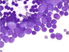
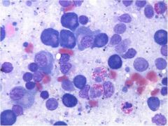
Is this lymphoma or hyperplasia?
|
Lymphoma!
Note all the large lymphocytes!!! |
|
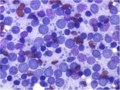
Is this lymphoma or hyperplasia?
|

Could be really reactive or early lymphoma
|