Reading...
![]()
Play button
![]()
Play button
![]()
Use LEFT and RIGHT arrow keys to navigate between flashcards;
Use UP and DOWN arrow keys to flip the card;
H to show hint;
A reads text to speech;
37 Cards in this Set
- Front
- Back
|
From the Nernst equation membrane potential at equilibrium = with K+ alone
|
90 mV
|
|
|
Positive ions entering a cell cause an inward current and
|
DEPOLARIZATION
|
|
|
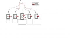
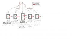
In both cardiac and smooth muscle cells, depolarization results from opening of channels that allow influx of
|
Na+ and Ca++ ion
|
|
|
Pacemaker cells
|
Non contractile cardiac muscle cells specialized for initiating and conducting action potentials responsible for contraction of cardiac muscle cells
|
|
|
Action potentials in pacemaker and contractile cardiac cells is different because of difference in selective movement over time
|
of ions (particularly Na and Ca) into the cell
|
|
|
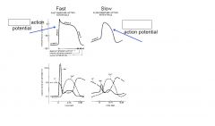
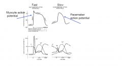
Fast response action potentials
|
Describe the action potentials in contracting myocytes and in conducting (purkinje) fibers
|
|
|
Slow action potentials
|
Occur in the sino-atrial node ( the natural pacemaker activity of the heart) and the atrio ventricular node ( the specialized tissue that conduct cardiac impulses from the atria to the ventricle
|
|
|
|
|
|
|
|
|
The action potential of cardiac myocytes is initiated by opening of ...and a sudden increase of intracellular ...and ...
|
voltage gated sodium channels
sodium depolarization |
|
|
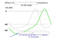
The resting membrane potential is unstable in pacemaker cells due to
|
Results from a slightly greater permeability to sodium and calcium in pacemaker cells
As a result membrane potential tens toward threshold for opening of voltage gated channels |
|
|
Rate of depolarization is slower in pacemaker cells due to
|
to the fact that here are no functional voltage gated Na+ channel in pacemaker membranes
Depolarization is caused by Ca++ enter via slowly activating voltage gated Ca channels |
|
|
Amplitude of the ....is lower in pace maker cells
|
action potential
|
|
|
Plateau phase is .... in pacemaker cells
|
shorter
|
|
|
Threshold is more positive in pacemaker cells because
|
Because voltage gated Ca channels have more positive threshold than voltage gated Na+ channels
|
|
|
SA node >... > ... > ...
|
AV node
Bundle of His Purkinje fiber |
|
|
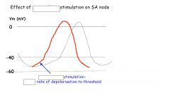
THE RATE OF DECAY OF THE RESTING POTENTIAL OF THE SA NODE DETERMINES
|
THE RATE AT WHICH ACTION POTNETIALS ARE GENERATED AND HENCE HEART BEAT
|
|
|
Cells are in an absolute refractory period during....This is because... are rapidly inactiving in phase...and do not reactivate until the membrane potential becomes more negative than...
|
most of the actions potential
Na+ channels 0 -65mN |
|
|
The refractory period and the length of the AP compared to the contraction means that unlike skeletal muscle, cardiac muscle cannot be...Because it needs to
|
tetanized.
relax to fill. |
|
|
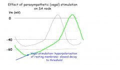
parasympathetic fibers ( of the vagus nerve) release ... on SA node
this increases...causing ... of the membrane potential and ... rate of spontaneous depolarization leading to ... heart rate |
acetylcholine (ACh)
resting membrane permeability to K+, hyperpolarisation slowed slower |
|
|
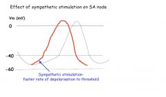
Sympathetic nerves release ..., that promotes...permeability to Na ... rate of depolarization and hence... HR
|
noradernaline
increased increased increased |
|
|
CONDUCTION VELOCITY
|
IS THE SPEED AT WHICH AN ACTION POTNETIAL PROPGATES THROUGH A REGION
|
|
|
The... of the conducting fiber is one determinant of conduction speed
... .... such as the AV node conduct more.... than... such a purkinje fibers |
diameter
Smaller diameters slowly larger diameters |
|
|
... initiates depolarization, contraction begins in the ...
|
SA node
atrial muscle |
|
|
The wave of depolarization spreads across the atrial muscle and is directed to the ... due to the insulating property of the cardiac skeleton
|
AV node
|
|
|
The slower rate of conduction in the...ensures that atrial contraction and therefore emptying of the blood in the ventricle is completed before ventricular contraction begins
|
AV node
|
|
|
From the ... conduction continues through the... pathway,to the... and ...the ... and finally ... of the ventricles.
|
AV node
ventricular conduction bundle of His right and left bundle branches purkinje fibers cardiac myocytes |
|
|
SA node
|
Small mass of nodal myocytes
Lateral wall of right atrium at junction of cranial vena cava |
|
|
AV node
|
Club shaped mass of nodal myocytes
At the junction of interatrial septum, floor of the right atrium near opening of coronary sinus |
|
|
AV bundle or bundle of His
|
Runs from AV node to the ventricles
Pierces the fibrous skeleton where right atrium joins the IA septum Emerges dorsal to the IV septum Immediately divides right and left branches (cura) |
|
|
Right crus or right branch bundle
|
Runs to apex, subendocardium of IV septum
Major branches- right ventricular papillary muscles arising from septum, branches to IV septum, to right septomarginal trabecula to supply papillary muscle and right ventricle |
|
|
Left crus or left bundle branch
Major branches: |
subendocardial ramifying branches over surface of left ventricle, Left septomarginal trabecula, two papillary muscles on the outer wall
Apex curves back to reach all parts of left ventricle |
|
|
Purkinje fibers
|
Final extension of right and left cura
Network of subendocardial conducting fibers Individual fiber bundles loosely enclosed in connective tissue Fibers pass into cardiac muscle cells |
|
|
The cardiac skeleton is a
|
fibrous plate that is reinforced in some species with cartilage or bone
|
|
|
Functions of the cardiac skeleton
|
Electrical discontinuity between atria and the ventricles so that all electrical activity is conducted though the AV node
Provides attachment for the ventricle and atrial muscle Support for the valves |
|
|
|
|
|
|