Reading...
![]()
Play button
![]()
Play button
![]()
Use LEFT and RIGHT arrow keys to navigate between flashcards;
Use UP and DOWN arrow keys to flip the card;
H to show hint;
A reads text to speech;
232 Cards in this Set
- Front
- Back
|
Collagen
|
ropelike protein of the extracellular matrix
|
|
|
Proteoglycans
|
large molecules consisting of polysaccharides attached to core proteins. attract and retain large amounts of water.
|
|
|
tendons
|
tough connective tissue band connecting a muscle to a bone.
|
|
|
ligaments
|
tough connective tissue band usually connecting bone to bone
|
|
|
cartilage
|
firm, smooth, resilient, nonvascular connective tissue
|
|
|
long bones
|
longer than wide. upper and lower limbs
|
|
|
short bones
|
broad as they are long; in wrist and ankles
|
|
|
Flat bones
|
thin, flattened shape; skull, ribs, scapulae, sternum
|
|
|
Irregular bones
|
vertebrae and facial bones
|
|
|
diaphysis
|
shaft of a long bone
|
|
|
epiphysis
|
end of a bone; separated from the remainder of the bone by the epiphyseal plate or epiphyseal line.
|
|
|
articular cartilage
|
covers the ends of the epiphyses where the bone articulates with other bones. acts as a cushion.
|
|
|
epiphyseal plate
|
Site at which bone growth in length occurs; located between the epiphysis and diaphysis of a long bone; area of cartilage growth is followed by ossification; also called the growth plate
|
|
|
epiphyseal line
|
Dense plate of bone in a bone that is no longer growing, indicating the former site of the epiphyseal plate.
|
|
|
medullary cavity
|
large, marrow-filled cavity in the diaphysis of a long bone
|
|
|
marrow
|
soft tissue in center of bone
|
|
|
Yellow marrow
|
consists mostly of adipose tissue
|
|
|
Red marrow
|
blood-forming cells; children have more
|
|
|
periosteum
|
thick, double layered connective tissue sheath covering the entire surface of a bone, except the articular surface, which is covered by cartilage
|
|
|
endosteum
|
membranous lining of the medullary cavity and the cavities of spongy bone.
|
|
|
osteoblasts
|
cell that makes bone
|
|
|
osteocytes
|
mature bone cell surrounded by bone matrix
|
|
|
lamellae
|
thin sheet or layer of bone
|
|
|
lacunae
|
small space, cavity, or depression; a space in cartilage in which a chondrocyte is located; a space in bone matrix in which an osteocyte is located;
|
|
|
canaliculi
|
tiny canal in bone between osteocytes containing osteocyte cell processes; a cleftlike lumen between the cells of each hepatic cord, connects medial corner of the eye to the lacrimal sac.
|
|
|
compact bone
|
Bone that is denser and has fewer spaces than spongy bone
|
|
|
spongy bone
|
Bone with a latticelike appearance.
|
|
|
central canal
|
small canal containing blood vessels, nerves, and loose connective tissue and running parallel to the long axis of a bone; also called a haversian canal.
|
|
|
osteon
|
single central canal, with its contents, and the associated lamellae and osteocytes surrounding it. Also called a haversian system.
|
|
|
trabeculae
|
spongy bone consists of delicate interconnecting rods or plates of bone; resemble the beams or scaffolding of a building; add strength without adding weight
|
|
|
ossification
|
bone formation
|
|
|
intramembranous ossification
|
bone formation within connective tissue membranes.
|
|
|
ossification centers
|
osteoblasts line up on the surface of connective tissue fibers and begin depositing bone matrix to form trabeculae; process begins in these areas
|
|
|
endochondral ossification
|
bone formation within cartilage
|
|
|
chondrocytes
|
cartilage cells
|
|
|
primary ossification center
|
center part of the diaphysis, where bone first begins to appear
|
|
|
osteoclasts
|
cell that digests and removes bone
|
|
|
secondary ossification centers
|
form in the epiphyses
|
|
|
bone remodeling
|
involves the removal of existing bone by osteoclasts and the deposition of new bone by osteoblasts; responsible for changes in bone shape, the adjustment of bone to stress, bone repair, and calcium ion regulation in body fluids.
|
|
|
parathyroid hormone
|
hormone produced by the parathyroid gland; increases bone breakdown and blood calcium levels.
|
|
|
calcitonin
|
hormone released from cells of the thyroid gland, that acts on tissues, especially bone to cause a decrease in blood levels of calcium ions
|
|
|
foramen
|
hole; referring to a hole or opening in a bone.
|
|
|
hydroxyapatite
|
most of the mineral in bone; form of calcium phosphate crystals
|
|
|
canal/meatus
|
elongated hole, a tunnel-like passage through the bone
|
|
|
fossa
|
Depression below the level of the surface of a bone; usually longitudinal in shape.
|
|
|
tubercle
|
lump or knob on a bone
|
|
|
tuberosity
|
lump on a bone, usually larger than a tubercle
|
|
|
process
|
projection on a bone
|
|
|
condyle
|
rounded, articulating surface of a joint
|
|
|
braincase
|
encloses the cranial cavity; consists of 8 bones that immediately surround and protect the brain
|
|
|
facial bones
|
form the structure of the face
|
|
|
parietal bones
|
form the sides and roof of the cranium. Each bone is roughly quadrilateral in form, and has two surfaces, four borders, and four angles.
|
|
|
temporal bones
|
situated at the sides and base of the skull, and lateral to the temporal lobes of the cerebrum.
|
|
|
squamous suture
|
arches backward from the pterion and connects the temporal squama with the lower border of the parietal bone: this suture is continuous behind with the short, nearly horizontal parietomastoid suture, which unites the mastoid process of the temporal with the region of the mastoid angle of the parietal bone.
|
|
|
frontal bone
|
a bone in the human skull. The name comes from the Latin word frons (meaning "forehead"). The bone resembles a cockleshell in form, and consists of three portions:[1]
a large vertical portion, the squama frontalis, corresponding with the region of the forehead. an orbital or horizontal portion, the pars orbitalis, which enters into the formation of the roofs of the orbital and nasal cavities. a nasal portion, it articulates with the nasal bones and the frontal process of the maxilla to form the root of the nose. |
|
|
coronal suture
|
a dense, fibrous connective tissue joint that separates the frontal and parietal bones of the skull. At birth, the bones of the skull do not meet.
|
|
|
occipital bone
|
a saucer-shaped membrane bone situated at the back and lower part of the cranium, is trapezoidal in shape and curved on itself. It is pierced by a large oval aperture, the foramen magnum, through which the cranial cavity communicates with the vertebral canal.
The curved, expanded plate behind the foramen magnum is named the squama occipitalis. The thick, somewhat quadrilateral piece in front of the foramen is called the basilar part of occipital bone. On either side of the foramen are the lateral parts of occipital bone. |
|
|
lambdoid suture
|
a dense, fibrous connective tissue joint on the posterior aspect of the skull that connects the parietal and temporal bones with the occipital bone.
Its name comes from its lambda-like shape. |
|
|
external auditory canal
|
a tube running from the outer ear to the middle ear. The adult human ear canal extends from the pinna to the eardrum and is about 2.5 centimetres (1 in) in length and 0.7 centimetres (0.3 in) in diameter.
|
|
|
mastoid process
|
a conical prominence projecting from the undersurface of the mastoid portion of the temporal bone. It is located just behind the external acoustic meatus, and lateral to the styloid process. Its size and form vary somewhat; it is larger in the male than in the female. This part of the skull projects from the temporal bone and is roughly pyramidal or conical in shape. One important role for this bone is as a point of attachment for several muscles - the splenius capitis, longissimus capitis, digastric posterior belly, and sternocleidomastoid. These muscles are one reason the mastoid process tends to be larger in men, because men have bigger muscles as a general rule and thus require larger points of attachment.
|
|
|
sphenoid bone
|
an unpaired bone situated at the front middle of the skull in front of the temporal bone and basilar part of the occipital bone. The sphenoid bone is one of the seven bones that articulate to form the orbit. Its shape somewhat resembles that of a butterfly or bat with its wings extended. It is divided into the following parts:
a median portion, known as the body of sphenoid bone, containing the sella turcica which houses the pituitary gland two greater wings and two lesser wings Pterygoid processes of the sphenoides which project from it posteriorly (below) |
|
|
zygomatic bone
|
a paired bone which articulates with the maxilla, the temporal bone, the sphenoid bone and the frontal bone. The zygomatic is homologous to the jugal bone of other tetrapods. It is situated at the upper and lateral part of the face and forms the prominence of the cheek, part of the lateral wall and floor of the orbit, and parts of the temporal and infratemporal fossa. It presents a malar and a temporal surface; four processes, the frontosphenoidal, orbital, maxillary, and temporal; and four borders.
|
|
|
zygomatic arch
|
formed by the zygomatic process of temporal bone (a bone extending forward from the side of the skull, over the opening of the ear) and the temporal process of the zygomatic bone (the side of the cheekbone), the two being united by an oblique suture;[1] the tendon of the Temporalis passes medial to the arch to gain insertion into the coronoid process of the mandible.
|
|
|
maxilla
|
a fusion of two bones along the palatal fissure that form the upper jaw. The maxilla assists in forming the boundaries of three cavities:
the roof of the mouth the floor and lateral wall of the nasal antrum the wall of the orbit |
|
|
mandible
|
a bone forming the skull with the cranium. The mandible consists of:
a curved, horizontal portion, the body. (See body of mandible). two perpendicular portions, the rami, which unite with the ends of the body nearly at right angles. (See ramus mandibulae). The angle formed at this junction is called gonial angle. Alveolar process, the tooth bearing area of the mandible (upper part of the body of the mandible) Condyle, superior (upper) and posterior projection from the ramus, which makes the temporomandibular joint with the temporal bone Coronoid process, superior and anterior projection from the ramus. This provides attachment to the temporalis muscle. |
|
|
orbits
|
he cavity or socket of the skull in which the eye and its appendages are situated. "Orbit" can refer to the bony socket, or it can also be used to imply the contents. In the adult human, the volume of the orbit is 30 ml, of which the eye occupies 6.5 ml. The base, which opens in the face, has four borders. The following bones take part in their formation:
Superior margin: frontal bone Inferior margin: maxilla and zygomatic Medial margin: frontal, lacrimal and maxilla Lateral margin: zygomatic and frontal |
|
|
nasal cavity
|
cavity divided by the nasal septum and extending from the external nares anteriorly to the nasopharynx posteriorly; bounded inferiorly by the hard palate.
|
|
|
superior and inferior orbital fissures
|
foramen in the skull, although strictly it is more of a cleft, lying between the lesser and greater wings of the sphenoid bone. transmits the maxillary nerve and its zygomatic branch, and the ascending branches from the pterygopalatine ganglion. The infraorbital vessels are found in the inferior orbital fissure, and travel down the infraorbital groove into the infraorbital canal and exit through the infraorbital foramen.
It is formed by the sphenoid bone and maxilla. |
|
|
optic foramen
|
The superior surface of the sphenoid bone is bounded behind by a ridge, which forms the anterior border of a narrow, transverse groove, the chiasmatic groove (optic groove), above and behind which lies the optic chiasma; the groove ends on either side in the optic foramen, which transmits the optic nerve and ophthalmic artery (with accompanying sympathetic nerve fibres) into the orbital cavity.
|
|
|
nasolacrimal canal
|
The canal containing the nasolacrimal duct. It is formed by indentations in the inferior nasal conchae, maxilla and lacrimal bone. It drains to into the nasal cavity through the anterior portion of the inferior meatus which is between the inferior concha and the floor of the nasal cavity.
|
|
|
lacrimal bone
|
the smallest and most fragile bone of the face, is situated at the front part of the medial wall of the orbit. It has two surfaces and four borders.
|
|
|
nasal septum
|
separates the left and right airways in the nose, dividing the two nostrils. It is depressed by the Depressor septi nasi muscle. composed of five structures:
perpendicular plate of ethmoid bone vomer bone cartilage of the septum crest of the maxillary bone crest of the palatine bone |
|
|
vomer
|
one of the unpaired facial bones of the skull. It is located in the midsagittal line, and articulates with the sphenoid, the ethmoid, the left and right palatine bones, and the left and right maxillary bones.
|
|
|
perpendicular plate
|
a thin, flattened lamina, polygonal in form, which descends from the under surface of the cribriform plate, and assists in forming the septum of the nose; it is generally deflected a little to one or other side. The anterior border articulates with the spine of the frontal bone and the crest of the nasal bones.
|
|
|
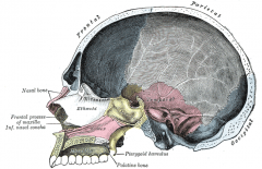
ethmoid bone
|
a bone in the skull that separates the nasal cavity from the brain. It is located at the roof of the nose, between the two orbits. The cubical bone is lightweight due to a spongy construction. The ethmoid bone is one of the bones that makes up the orbit of the eye. The ethmoid has three parts: the cribriform plate, the ethmoidal labyrinth, and the perpendicular plate.
|
|
|
nasal bones
|
two small oblong bones, varying in size and form in different individuals; they are placed side by side at the middle and upper part of the face, and form, by their junction, "the bridge" of the nose.
Each has two surfaces and four borders. |
|
|
nasal conchae
|
a long, narrow and curled bone shelf (shaped like an elongated sea-shell) that protrudes into the breathing passage of the nose. the turbinates divide the nasal airway into four groove-like air passages, and are responsible for forcing inhaled air to flow in a steady, regular pattern around the largest possible surface of cilia and climate-controlling tissue. A rapidly dilating arteriolar circulation to these bones may lead to a sharp increase in the pressure within in response to acute cooling of the body core - the pain from this pressure is often referred to as "brain freeze", and is frequently associated with the rapid consumption of ice cream.
|
|
|
Paranasal sinuses
|
a group of four paired air-filled spaces that surround the nasal cavity (maxillary sinuses), above the eyes (frontal sinuses), between the eyes (ethmoid sinuses), and behind the ethmoids (sphenoid sinuses). The sinuses are named for the facial bones in which they are located.
|
|
|
mastoid air cell
|
A section of the mastoid process of the temporal bone of the cranium shows it to be hollowed out into a number of spaces. exhibit great variety in their size and number.
|
|
|
foramen magnum
|
a large opening in the occipital bone of the cranium. It is one of the several oval or circular apertures in the base of the skull (the foramina), through which the medulla oblongata (an extension of the spinal cord) enters and exits the skull vault.
Apart from the transmission of the medulla oblongata and its membranes, the foramen magnum transmits the spinal accessory nerve, vertebral arteries, the anterior and posterior spinal arteries, the membrana tectoria and alar ligaments. |
|
|
sella turcica
|
a saddle-shaped depression in the sphenoid bone of the human skull and of the skulls of other Hominidae including chimpanzees, orangutans, and gorillas. The seat of the saddle is known as the hypophyseal fossa, which holds the pituitary gland. The hypophyseal fossa is located in a depression in the body of the sphenoid bone. Located anteriorly to the hypophyseal fossa is the tuberculum sellae.
Completing the formation of the saddle posteriorly is the dorsum sellae which is continuous with the clivus, inferoposteriorly. The dorsum sellae is terminated laterally by the posterior clinoid processes. |
|
|
occipital condyles
|
undersurface facets of the occipital bone in vertebrates, which function in articulation with the superior facets of the atlas vertebra.
|
|
|
styloid processes
|
usually serving as points of attachment for muscles, refers to the slender, pointed process (protrusion) of :
temporal bone of the skull - Temporal styloid process radius bone of the lower arm - Radial styloid process ulna bone of the lower arm - Ulnar styloid process Third metacarpal - Third metacarpal styloid process Tibia and Fibula - Tibial process, fibular process. |
|
|
mandibular fossa
|
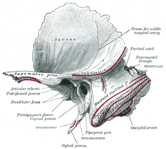
the depression in the temporal bone that articulates with the mandibular condyle. In the temporal bone, the mandibular fossa is bounded, in front, by the articular tubercle; behind, by the tympanic part of the bone, which separates it from the external acoustic meatus; it is divided into two parts by a narrow slit, the petrotympanic fissure (Glaserian fissure). The mandibular fossa is also referred to as the glenoid fossa, especially in dental literature.
|
|
|
hard palate
|
a thin horizontal bony plate of the skull, located in the roof of the mouth. It spans the arch formed by the upper teeth.
It is formed by the palatine process of the maxilla and horizontal plate of palatine bone. It forms a partition between the nasal passages and the mouth. Also on the anterior portion of the roof of the hard palate is the Rugae which are the irregular ridges in the mucous membrane that help facilitate the movement of food backwards towards the pharynx. This partition is continued deeper into the mouth by a fleshy extension called the soft palate. |
|
|
palatine bones
|
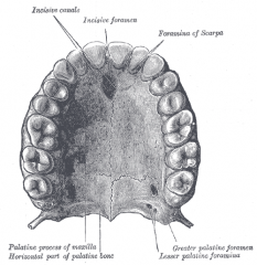
It is situated at the back part of the nasal cavity between the maxilla and the pterygoid process of the sphenoid. The human palatine articulates with six bones: the sphenoid, ethmoid, maxilla, inferior nasal concha, vomer and opposite palatine.
|
|
|
soft palate
|
the soft tissue constituting the back of the roof of the mouth. distinguished from the hard palate at the front of the mouth in that it does not contain bone.
|
|
|
hyoid bone
|
a horseshoe-shaped bone situated in the anterior midline of the neck between the chin and the thyroid cartilage. At rest, it lies at the level of the base of the mandible in the front and the third cervical vertebra (C3) behind.
Unlike other bones, the hyoid is only distantly articulated to other bones by muscles or ligaments. The hyoid is anchored by muscles from the anterior, posterior and inferior directions, and aids in tongue movement and swallowing. |
|
|
vertebral column
|
the vertebral column usually consists of 24 articulating vertebrae,[1] and 9 fused vertebrae in the sacrum and the coccyx. It is situated in the dorsal aspect of the torso, separated by intervertebral discs. It houses and protects the spinal cord in its spinal canal, and hence is commonly called the spine, or simply backbone.
There are normally thirty-three (33) vertebrae in humans, including the five that are fused to form the sacrum (the others are separated by intervertebral discs) and the four coccygeal bones that form the tailbone. |
|
|
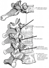
cervical vertebrae
|
vertebrae immediately inferior to the skull. first seven.
|
|
|
thoracic vertebrae
|
the middle segment of the vertebral column, between the cervical vertebrae and the lumbar vertebrae.[1] In humans, they are intermediate in size between those of the cervical and lumbar regions; they increase in size as one proceeds down the spine, the upper vertebrae being much smaller than those in the lower part of the region. the first one (T1) located closest to the skull and higher numbered vertebrae (T2-T12) proceeding away from the skull and down the spine.
|
|
|
lumbar vertebrae
|
the five vertebrae between the rib cage and the pelvis. They are the largest segments of the vertebral column and are characterized by the absence of the foramen transversarium within the transverse process, and by the absence of facets on the sides of the body. They are designated L1 to L5, starting at the top. The lumbar vertebrae help support the weight of the body, and permit movement.
|
|
|
sacral bone
|
a large, triangular bone at the base of the spine and at the upper and back part of the pelvic cavity, where it is inserted like a wedge between the two hip bones. Its upper part connects with the last lumbar vertebra, and bottom part with the coccyx (tailbone). It consists of usually five initially unfused vertebrae which begin to fuse between ages 16–18 and are usually completely fused into a single bone by age 34.
|
|
|
coccygeal bone
|
the final segment of the vertebral column in tailless primates. Comprising three to five separate or fused vertebrae (the coccygeal vertebrae) below the sacrum, it is attached to the sacrum by a fibrocartilaginous joint, the sacrococcygeal symphysis, which permits limited movement between the sacrum and the coccyx.
|
|
|
Kyphosis
|
condition of over-curvature of the thoracic vertebrae (upper back). It can be either the result of degenerative diseases (such as arthritis), developmental problems (the most common example being Scheuermann's disease), osteoporosis with compression fractures of the vertebrae, or trauma.
In the sense of a deformity, it is the pathological curving of the spine, where parts of the spinal column lose some or all of their lordotic profile. This causes a bowing of the back, seen as a slouching posture. |
|
|
Lordosis
|
the inward curvature of a portion of the lumbar and cervical vertebral column.[1] Two segments of the vertebral column, namely cervical and lumbar, are normally lordotic, that is, they are set in a curve that has its convexity anteriorly (the front) and concavity posteriorly (behind), in the context of human anatomy.
|
|
|
Scoliosis
|
a medical condition in which a person's spine is curved from side to side. Although it is a complex three-dimensional deformity, on an X-ray, viewed from the rear, the spine of an individual with scoliosis may look more like an "S" or a "C", rather than a straight line.
|
|
|
intervertebral disks body
|
lie between adjacent vertebrae in the spine. Each disc forms a cartilaginous joint to allow slight movement of the vertebrae, and acts as a ligament to hold the vertebrae together. Discs consist of an outer annulus fibrosus, which surrounds the inner nucleus pulposus. The annulus fibrosus consists of several layers of fibrocartilage. The strong annular fibers contain the nucleus pulposus and distribute pressure evenly across the disc. The nucleus pulposus contains loose fibers suspended in a mucoprotein gel with the consistency of jelly. The nucleus of the disc acts as a shock absorber, absorbing the impact of the body's daily activities and keeping the two vertebrae separated. The disc can be likened to a jelly doughnut
|
|
|
vertebral arch
|
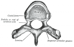
the posterior part of a vertebra.
It consists of a pair of pedicles and a pair of laminae, and supports seven processes: four articular processes two transverse processes one spinous process |
|
|
vertebral foramen
|
the foramen (opening) formed by the anterior segment (the body), and the posterior part, the vertebral arch.
The vertebral foramen begins at cervical vertebra #1 (atlas) and continues inferior to lumbar vertebra #5. Within this foramen the spinal cord and associated meninges are housed. |
|
|
vertebral canal
|
the space in vertebrae through which the spinal cord passes. It is a process of the dorsal human body cavity. This canal is enclosed within the vertebral foramen of the vertebrae. In the intervertebral spaces, the canal is protected by the ligamentum flavum posteriorly and the posterior longitudinal ligament anteriorly.
|
|
|
pedicles
|

two short, thick processes, which project dorsally, one on either side, from the superior part of the vertebral body at the junction of its posterior and lateral surfaces. They connect the body of the spinal vertebra to the arch. It is often used as a radiographic marker and entry point in vertebroplasty, kyphoplasty, and spinal fusion procedures.
|
|
|
laminae
|

two broad plates, extending dorsally and medially from the pedicles, fusing to complete the roof of the vertebral arch.
|
|
|
transverse process
|
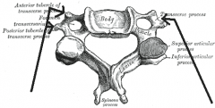
project one at either side from the point where the lamina joins the pedicle, between the superior and inferior articular processes. They serve for the attachment of muscles and ligaments.
|
|
|
spinous process
|
directed backward and downward from the junction of the laminae (in humans), and serves for the attachment of muscles and ligaments
|
|
|
intervertebral foramina
|
articulated with each other the bodies form a strong pillar for the support of the head and trunk, and the vertebral foramina constitute a canal for the protection of the medulla spinalis (spinal cord). Between every pair of vertebrae are two apertures (openings),
|
|
|
articular process
|
![projections of the vertebra that serve the purpose of fitting with an adjacent vertebra. The actual region of contact is called the articular facet.[1]
Articular processes spring from the junctions of the pedicles and laminæ, and there are two right and](https://images.cram.com/images/upload-flashcards/909691/571629_m.png)
projections of the vertebra that serve the purpose of fitting with an adjacent vertebra. The actual region of contact is called the articular facet.[1]
Articular processes spring from the junctions of the pedicles and laminæ, and there are two right and left, and two superior and inferior. These stick out of an end of a vertebra to lock with a zygapophysis on the next vertebra, to make the backbone more stable. The superior processes or prezygapophysis project upward from a lower vertebra, and their articular surfaces are directed more or less backward. The inferior processes or postzygapophysis project downward from a higher vertebra, and their articular surfaces are directed more or less forward and outward. The articular surfaces are coated with hyaline cartilage. |
|
|
articular facet
|
a surface where two anatomical structures (usually bones) meet.
|
|
|
atlas
|
the most superior (first) cervical vertebra of the spine.
|
|
|
axis
|
It forms the pivot upon which the first cervical vertebra (the atlas), which carries the head, rotates. the second cervical vertebra.
|
|
|
dens
|
a protuberance (process or projection) of the axis (second cervical vertebra). It exhibits a slight constriction or neck, where it joins the main body of the vertebra. The condition, where the dens is separated from the body of the axis, is called os odontoideum, and may cause nerve and circulation compression syndrome.
|
|
|
median sacral crest
|
surmounted by three or four tubercles, the rudimentary spinous processes of the upper three or four sacral vertebrae.
|
|
|
sacral hiatus
|
The laminae of the fifth sacral vertebra, and sometimes those of the fourth, fail to meet behind, and thus a sacral hiatus occurs in the posterior wall of the sacral canal.
|
|
|
sacral promontory
|
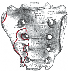
the anatomical term for the superiormost portion of the sacrum. It marks part of the border of the pelvic inlet. The rectosigmoid junction is at the level of the sacral promontory.
|
|
|
thoracic cage
|
a bony and cartilaginous structure which surrounds the thoracic cavity and supports the pectoral girdle, forming a core portion of the human skeleton. A typical human rib cage consists of 24 ribs, the sternum (with Xiphoid process), costal cartilages, and the 12 thoracic vertebrae. It, along with the skin and associated fascia and muscles, makes up the thoracic wall and provides attachments for the muscles of the neck, thorax, upper abdomen, and back.
|
|
|
true ribs
|
The first seven ribs are connected posteriorly with the vertebral column, and anteriorly, through the intervention of the costal cartilages, with the sternum
|
|
|
false ribs
|
interconnected membranous structures that are connected with the sternum process.
|
|
|
floating ribs
|
four atypical ribs (two lowermost pairs, XI-XII) in the human ribcage. They are called so because they are attached to the vertebrae only, and not to the sternum or cartilage coming off the sternum.
|
|
|
sternum
|
breastbone
|
|
|
manubrium
|
the broad, upper part of the sternum. Located ventrally with a quadrangular shape, wider superiorly and narrower inferiorly, it articulates with the clavicles and the cartilages of the first pair of ribs.
|
|
|
xiphoid process
|
a small cartilaginous process (extension) of the lower part of the sternum which is usually ossified in the adult human. By age 15 to 29, the xiphoid usually fuses to the body of the sternum with a fibrous joint.
|
|
|
jugular notch
|
found at the superior border of the manubrium of the sternum, between the clavicular notches
|
|
|
sternal angle
|
the anterior angle formed by the junction of the manubrium and the body of the sternum[1] (the manubriosternal junction) in the form of a secondary cartilaginous joint (symphysis). This is also called the manubriosternal joint or Angle of Louis. The sternal angle is a palpable clinical landmark.
|
|
|
appendicular
|
relating to an appendage, such as the limbs and their associated girdles
|
|
|
pectoral girdle
|
the set of bones which connects the upper limb to the axial skeleton on each side. It consists of the clavicle and scapula in humans and, in those species with three bones in the pectoral girdle, the coracoid
|
|
|
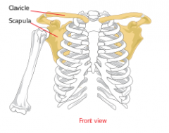
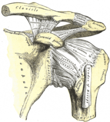
scapula
|
shoulder blade
|
|
|
glenoid cavity
|
part of the shoulder. It is a shallow pyriform, articular surface, which is located on the lateral angle of the scapula. It is directed laterally and forward and articulates with the head of the humerus; it is broader below than above and its vertical diameter is the longest.
|
|
|
spine
|
a ridge that runs across the posterior surface of the scapula.
|
|
|
acromion process
|
extends from the scapular spine to form the point of the shoulder
|
|
|
clavicle
|
articulates with the scapula at the acromion process. the proximal end is attached to the sternum providing the only bony attachment of the scapula to the reminder of the skeleton.
|
|
|
coracoid process
|
curves below the clavicle and provides for the attachment of arm and chest muscles
|
|
|
arm
|
the region between the shoulder and the elbow
|
|
|
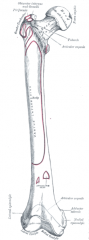
humerus
|
a long bone in the arm or forelimb that runs from the shoulder to the elbow.
onnects the scapula and the lower arm (consisting of the radius and ulna), and consists of three sections. The upper extremity consists of a rounded head, a narrow neck, and two short processes (tubercles, sometimes called tuberosities.) Its body is cylindrical in its upper portion, and more prismatic below. The lower extremity consists of 2 epicondyles, 2 processes (trochlea & capitulum), and 3 fossae (radial fossa, coronoid fossa, and olecranon fossa). As well as its true anatomical neck, the constriction below the greater and lesser tubercles of the humerus is referred to as its surgical neck due to its tendency to commonly get fractured, thus often becoming the focus of surgeons. |
|
|
head
|
proximal end of the humerus. smooth, rounded. attaches the humerus to the scapula at the glenoid cavity
|
|
|
greater tubercle
|
situated lateral to the head of the humerus and posterolateral to the lesser tubercle.
|
|
|
lesser tubercle
|

more prominent than the greater tubercle: it is situated in front, and is directed medially and anteriorly.
|
|
|
deltoid tuberosity
|
a rough, triangular area on the anterolateral (exterior-front) surface of the middle of the humerus to which the deltoid muscle attaches.
|
|
|
epicondyles
|
on the distal end of the humerus, just lateral to the condyles, provide attachment sites for forearm muscles
|
|
|
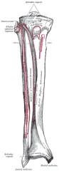
forearm
|
has two bones; ulna (medial) and radius (lateral)
|
|
|
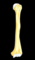
ulna
|

one of the two long bones in the forearm, the other being the radius. It is prismatic in form and runs parallel to the radius, which is shorter and smaller. In anatomical position (i.e. when the arms are down at the sides of the body and the palms of the hands face forward) the ulna is located at the side of the forearm closest to the body (the medial side), the side of the little finger.
|
|
|
radius
|
one of the two large bones of the forearm, the other being the ulna. It extends from the lateral side of the elbow to the thumb side of the wrist and runs parallel to the ulna, which exceeds it in length and size. It is a long bone, prism-shaped and slightly curved longitudinally. The radius articulates with the capitulum of the humerus, the radial notch and the head of the ulna. The corresponding bone in the lower leg is the tibia.
|
|
|
trochlear notch
|
proximal end of the ulna. fits tightly over the end of the humerus, forming most of the elbow joint.
|
|
|
olecranon process
|
proximal to the trochlear notch, an extension of the ulna. felt as the point of the elbow.
|
|
|
coronoid process
|
distal to the trochlear notch. completes the grip of the ulna on the distal end of the humerus..
|
|
|
styloid process
|
the distal end of the ulna. located on medial side
|
|
|
radial tuberosity
|
distal to the radial head. biceps brachii attaches. articulates with the wrist bones.
|
|
|
wrist
|
eight carpal bones. two rows of four bones each.
|
|
|
carpal bones
|
eight bones of the wrist
|
|
|
metacarpal bones
|
attached to the carpal bones and form the bony framework of the hand. Align with five digits.
|
|
|
digits
|
thumb and fingers
|
|
|
phalanges
|
three small bones in each digit
|
|
|
pelvic girdle
|
place where lower limbs attach to the body.
|
|
|
coxal bones
|
a large, flattened, irregularly shaped bone, constricted in the center and expanded above and below. In some vertebrates (including humans) it is composed of three bones; ilium, ischium, and pubis.
|
|
|
ilium
|
the uppermost and largest bone of the pelvis,
|
|
|
ischium
|
the lower and back part of the hip bone
|
|
|
pubis
|
the ventral and anterior of the three principal bones composing either half of the pelvis.
|
|
|
iliac crest
|
the superior border of the wing of ilium and the superolateral margin of the greater pelvis
|
|
|
anterior superior iliac spine
|

refers to the anterior extremity of the iliac crest of the pelvis, which provides attachment for the inguinal ligament, and the sartorius muscle. The Tensor Fasciae Latae muscle attaches about 5cm away at the iliac tubercle.
|
|
|
pubic symphysis
|
he midline cartilaginous joint (secondary cartilaginous) uniting the superior rami of the left and right pubic bones. It is located anterior to the urinary bladder and superior to the external genitalia; for females it is above the vulva and for males it is above the penis.
|
|
|
sacroiliac joints
|
in the bony pelvis between the sacrum and the ilium of the pelvis, which are joined by strong ligaments. In humans, the sacrum supports the spine and is supported in turn by an ilium on each side. The joint is a strong, weight bearing synovial joint with irregular elevations and depressions that produce interlocking of the two bones. The human body has two sacroiliac joints, one on the left and one on the right, that often match each other but are highly variable from person to person.
|
|
|
acetabulum
|
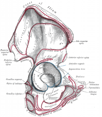
a concave surface of the pelvis. The head of the femur meets with the pelvis at the acetabulum, forming the hip joint.
|
|
|
obturator foramen
|
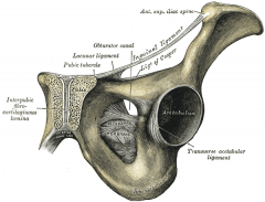
he hole created by the ischium and pubis bones of the pelvis through which nerves and blood vessels pass.
|
|
|
pelvic inlet
|
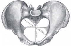
a planar surface which defines the boundary between the pelvic cavity and the abdominal cavity (or, according to some authors, between two parts of the pelvic cavity, called lesser pelvis and greater pelvis).
Its position and orientation relative to the skeleton of the pelvis is anatomically defined by its edge, the pelvic brim. The pelvic brim is an approximately apple-shaped line passing through the prominence of the sacrum, the arcuate and pectineal lines, and the upper margin of the pubic symphysis. |
|
|
pelvic outlet
|
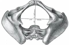
The lower circumference of the lesser pelvis is very irregular; the space enclosed by it is named the inferior aperture or pelvic outlet.
|
|
|
thigh
|
the area between the pelvis and the knee.
|
|
|
femur
|
the most proximal (closest to the center of the body) bone of the leg. articulates with the acetabulum in the pelvic bone forming the hip joint, while the distal part of the femur articulates with the tibia and patella forming the knee joint.
|
|
|
head
|
the highest part of the thigh bone (femur). It is supported by the neck of the femur.
|
|
|
condyles
|
the round prominence at the end of a bone, most often part of a joint - an articulation with another bone
|
|
|
epicondyles
|
|
|
|
trochanters
|
large, irregular, quadrilateral eminence and a part of the skeletal system.
It is directed a little lateralward and backward, and, in the adult, is about 1 cm lower than the head. Because the pelvic outlet in the female is larger than in the male, there is a greater distance between the greater trochanters in the female. |
|
|
patella
|
a thick, circular-triangular bone which articulates with the femur and covers and protects the anterior articular surface of the knee joint. It is the largest sesamoid bone in the human body.
|
|
|
leg
|
knee to ankle
|
|
|
tibia
|
the larger and stronger of the two bones in the leg below the knee in vertebrates (the other being the fibula), and connects the knee with the ankle bones. The tibia is named for the Greek aulos flute, also known as a tibia. Medial of the two leg bones
|
|
|
fibula
|
a leg bone located on the lateral side of the tibia, with which it is connected above and below. It is the smaller of the two bones, and, in proportion to its length, the most slender of all the long bones. Its upper extremity is small, placed toward the back of the head of the tibia, below the level of the knee joint, and excluded from the formation of this joint. Its lower extremity inclines a little forward, so as to be on a plane anterior to that of the upper end; it projects below the tibia, and forms the lateral part of the ankle joint.
|
|
|
tibial tuberosity
|
a large oblong elevation on the proximal, anterior aspect of the tibia, just below where the anterior surfaces of the lateral and medial tibial condyles end.
|
|
|
medial malleolus
|
the bony prominence on each side of the ankle. Formed by the tibia.
|
|
|
lateral malleolus
|
the prominence on the outer side of the ankle, formed by the lower end of the fibula.
|
|
|
ankles
|
consists of seven tarsal bones
|
|
|
tarsal bones
|
a cluster of seven articulating bones in each foot situated between the lower end of tibia and fibula of the lower leg and the metatarsus. In the foot the tarsus articulates with the bones of the metatarsus, which in turn articulate with the bones of the individual toes. The joint between the tibia and fibula above and the tarsus below is referred to as the ankle joint.
In humans the largest bone in the tarsus is the calcaneus, which is the weight-bearing bone within the heel of the foot. |
|
|
talus
|
a bone in the collection of bones in the foot called the tarsus. The tarsus forms the lower part of the ankle joint through its articulations with the lateral and medial malleoli of the two bones of the lower leg, the tibia and fibula. Within the tarsus, it articulates with the calcaneus below and navicular in front within the talocalcaneonavicular joint. Through these articulations, it transmits the entire weight of the body to the foot.[2]
The second largest of the tarsal bones, it is also one of the bones in the human body with the highest percentage of its surface area covered by articular cartilage. Additionally, it is also unusual in that it has a retrograde blood supply, i.e. arterial blood enters the bone at the distal end.[citation needed] |
|
|
calcaneus
|
a bone of the tarsus of the foot which constitute the heel.
|
|
|
cuboid
|
The dorsal surface, directed upward and lateralward, is rough, for the attachment of ligaments.
The plantar surface presents in front a deep groove, the peroneal sulcus, which runs obliquely forward and medialward; it lodges the tendon of the peroneus longus, and is bounded behind by a prominent ridge, to which the long plantar ligament is attached. The ridge ends laterally in an eminence, the tuberosity, the surface of which presents an oval facet; on this facet glides the sesamoid bone or cartilage frequently found in the tendon of the peroneus longus. The surface of bone behind the groove is rough, for the attachment of the plantar calcaneocuboid ligament, a few fibers of the flexor hallucis brevis, and a fasciculus from the tendon of the tibialis posterior. The lateral surface presents a deep notch formed by the commencement of the peroneal sulcus. The posterior surface is smooth, triangular, and concavo-convex, for articulation with the anterior surface of the calcaneus (the calcaneocuboid joint); its infero-medial angle projects backward as a process which underlies and supports the anterior end of the calcaneus. The anterior surface, of smaller size, but also irregularly triangular, is divided by a vertical ridge into two facets, forming the fourth and fifth tarsometatarsal joints: the medial facet, quadrilateral in form, articulates with the fourth metatarsal; the lateral, larger and more triangular, articulates with the fifth. The medial surface is broad, irregularly quadrilateral, and presents at its middle and upper part a smooth oval facet, for articulation with the third cuneiform; and behind this (occasionally) a smaller facet, for articulation with the navicular bone; it is rough in the rest of its extent, for the attachment of strong interosseous ligaments. |
|
|
navicular
|
located on the medial side of the foot, and articulates proximally with the talus, distally with the three cuneiform bones, and laterally with the cuboid.
|
|
|
cuneiforms
|
located between the navicular bone and the first, second and third metatarsal bones and are medial to the cuboid bone.
|
|
|
metatarsal bones
|
a group of five long bones in the foot located between the tarsal bones of the hind- and mid-foot and the phalanges of the toes. Lacking individual names, the metatarsal bones are numbered from the medial side (side of great toe): the first, second, third, fourth, and fifth metatarsal. The metatarsals are analogous to the metacarpal bones of the hand.
|
|
|
arches
|
formed by the positions of the tarsal bones and metatarsal bones and held in place by ligaments. two longitudinal arches extend from the heel to the ball of the foot and a transverse arch extends across the fott.
|
|
|
joint
|
a place where two bones come together. usually moveable.
|
|
|
synarthrosis
|
non moveable joint
|
|
|
amphiarthrosis
|
slightly moveable joint
|
|
|
diathrosis
|
freely moveable joint
|
|
|
fibrous joints
|
consist of two bones that are united by fibrous tissue and that exhibit little or no movement. 3 divisions: sutures, syndesmoses or gomphoses
|
|
|
sutures
|
fibrous joints between the bones of the skull
|
|
|
fontanels
|
one of several membranes gaps between bones of the skull
|
|
|
syndesmoses
|
fibrous joints in which the bones are separated by some distance and held together by ligaments.
|
|
|
gomphoses
|
consist of pegs fitted into sockets and held in place by ligaments. Ex tooth and socket.
|
|
|
cartilaginous joints
|
unite two bones by means of cartilage. only slight movement can occur at these joints. Ex. epiphyseal plates
|
|
|
synovial joints
|
freely movable joint
|
|
|
articular cartilage
|
efers to the hyaline cartilage on the articular surfaces of bones.
Though it is often found in close contact with menisci and articular disks, articular cartilage is not considered a part of either of these structures, which are made entirely of fibrocartilage. |
|
|
joint cavity
|
filled with fluid. space between articular cartilage in joints.
|
|
|
joint capsule
|
an envelope surrounding a synovial joint. Each capsule consists of two layers:
an outer layer (stratum fibrosum) composed of avascular white fibrous tissue an inner layer (stratum synoviale) which is a secreting layer, and is usually described separately as the synovial membrane. On the inside of the capsule, articular cartilage covers the end surfaces of the bones that articulate within that joint. The outer layer is highly innervated by the same nerves which perforate through the adjacent muscles associated with the joint. |
|
|
synovial membrane
|
membrane that lines the inside of a joint cavity; produces synovial fluid.
|
|
|
synovial fluid
|
somewhat viscous substance serving as a lubricant in movable joints, tendon sheaths and bursae.
|
|
|
bursa
|
closed sac or pocket containing synovial fluid; usually found in areas where friction occurs.
|
|
|
bursitis
|
inflammation of a bursa
|
|
|
tendon sheath
|
Synovial membrane that is extended along some tendons associated with joints.
|
|
|
plane joints
|
consist of two opposed flat surfaces that glide over each other. Ex. vertebrae
|
|
|
saddle joints
|
consist of two saddle-shaped articulating surfaces oriented at right angles to each other. Ex. thumb
|
|
|
hinge joints
|
movement in one plane only. Convex cylinder of one bone applied to a corresponding concavity of the other bone. Ex. knee and elbow
|
|
|
menisci
|
shock absorbing fibrocartilage pads
|
|
|
pivot joints
|
restrict movement to rotation around a single axis. Ex. shaking head no.
|
|
|
ball and socket joints
|
consist of a ball at the end of oen bone and a socket in an adjacent bone into which a portion of the ball fits. Ex. shoulder and hip joints
|
|
|
ellipsoid joints
|
elongated ball and socket joints. Ex atlas and occipital bone
|
|
|
flexion
|
moves a part of the body in the anterior direction from the frontal plane.
|
|
|
extension
|
moves a part in the posterior direction from the frontal plane.
|
|
|
plantar flexion
|
movement of the foot toward the plantar surface
|
|
|
dorsiflexion
|
movement of the foot toward the shin
|
|
|
abduction
|
movement away from the median or midsagittal plane.
|
|
|
adduction
|
movement toward the median plane
|
|
|
pronation
|
a rotational movement of the forearm at the radioulnar joint, or of the foot at the subtalar and talocalcaneonavicular joints.[1][2] For the forearm, when standing in the anatomical position, pronation will move the palm of the hand from an anterior-facing position to a posterior-facing position without an associated movement at the shoulder (glenohumeral joint). This corresponds to a counterclockwise twist for the right forearm and a clockwise twist for the left (when viewed superiorly). For the foot, pronation will cause the sole of the foot to face more laterally than when standing in the anatomical position. Pronation is the opposite of supination.
|
|
|
supination
|
a position of either the forearm or foot. When the arms are unbent and at the sides, the forearm is in supination when the palm faces to the front (anteriorly), or faces up. Supination in the foot occurs when a person appears "bow-legged" with their weight supported primarily on the lateral side of their feet (5th Metatarsal)
|
|
|
eversion
|
turning the foot so that the plantar surface
|
|
|
inversion
|
turning the foot so that the plantar surface faces medially.
|
|
|
rotation
|
the turning of a structure around its long axis, as shaking the head no.
|
|
|
circumduction
|
occurs at freely movable joints, such as the shoulder. ex the arm moves so that it describes a cone with the shoulder joint at the apex.
|
|
|
protraction
|
a movement in which a structure, such as the mandible, glides anteriorly.
|
|
|
retraction
|
the structure glides posteriorly
|
|
|
elevation
|
movement of a structure in a superior direction.
|
|
|
depression
|
movement of a structure in an inferior direction. Opening the mouth involves depression of the mandible.
|
|
|
excursion
|
movement of a structure to one side, as in moving the mandible from side to side
|
|
|
opposition
|
a movement unique to the thumb and little finger. It occurs when the tips of the thumb and little finger are brought toward each other across the palm of the hand.
|
|
|
reposition
|
return digits to anatomical position.
|
|
|
hyperextension
|
If a part of the body such as a joint is overstretched or "bent backwards" because of exaggerated extension motion
|