Reading...
![]()
Play button
![]()
Play button
![]()
Use LEFT and RIGHT arrow keys to navigate between flashcards;
Use UP and DOWN arrow keys to flip the card;
H to show hint;
A reads text to speech;
43 Cards in this Set
- Front
- Back
|
Which type of adult brain tumor occurs in the convexities of hemispheres (near the surfaces of the brain) and parasagittal region?
|

Meningioma
|
|
|
What kind of cells are involved in a Meningioma? Significance for location of tumor?
|
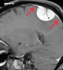
- Arises from arachnoid cells
- Extra-axial (external to brain parenchyma) - May have dural attachment ("tail") |
|
|
What are the symptoms and prognosis for a Meningioma?
|
- Typically benign
- Often asymptomatic - May present with seizures or focal neurologic signs |
|
|
How do you treat Meningioma?
|
Resection and/or radiosurgery
|
|
|
What is the histologic appearance of a Meningioma?
|
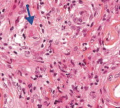
- Spindle cells, concentrically arranged in a whorled pattern
- Psammoma bodies (laminated calcifications) |
|
|
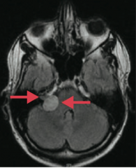
What type of adult brain tumor is often cerebellar and is associated with von Hippel-Lindau syndrome when found with retinal angiomas?
|
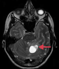
Hemangioblastoma
|
|
|
What brain tumor can lead to polycythemia? How?
|
Hemangioblastoma - can produce erythropoietin → 2° polycythemia
|
|
|
What is the histologic appearance of a Hemangioblastoma?
|
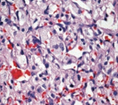
Closely arranged, thin-walled capillaries with minimal interleaving parenchyma
|
|
|
Which type of adult brain tumor is often found at the cerebellopontine angle and can be localized to CN VIII? Origin of cells?
|
Schwannoma (if localized to CN VIII it is an acoustic schwannoma / acoustic neuroma)
- Schwann cell origin |
|
|
Which adult brain tumor is S-100 (+)?
|
Schwannoma
|
|
|
How do you treat a Schwannoma?
|
Resected or treated with stereotactic radiosurgery
|
|
|
What should you think of if you see bilateral acoustic Schwannomas?
|
NF-2
|
|
|
Which adult brain tumor is often found in the frontal lobes?
|
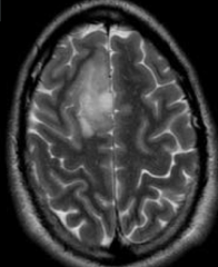
Oligodendroglioma
|
|
|
What is the histologic appearance of an Oligodendroglioma?
|
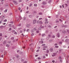
- Chicken-wire capillary pattern
- Oligodendrocytes = "fried egg" cells with round nuclei and clear cytoplasm - Often calcified |
|
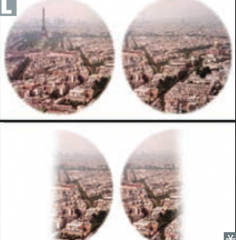
What type of adult brain tumor can put pressure on the optic chiasm causing bitemporal hemianopia?
|

Pituitary Adenoma (most commonly a prolactinoma)
|
|
|
What are the possible sequelae of a pituitary adenoma?
|
Hyper or hypo-pituitarism
|
|
|
What is the location of the adult brain tumors?
|
- Glioblastoma Multiforme: cerebral hemispheres and corpus callosum
- Meningioma: external to brain parenchyma - Hemangioblastoma: cerebellar - Schwannoma: cerebellopontine angle, may localize to CN VIII - Oligodendroglia: frontal lobes - Pituitary adenoma: pituitary / optic chiasm |
|
|
What are the types of childhood primary brain tumors?
|
- Pilocytic (low-grade) astrocytoma
- Medulloblastoma - Ependymoma - Craniopharyngioma |
|
|
Which type of childhood brain tumor is GFAP (+)?
|
Pilocytic (low-grade) Astrocytoma
|
|
|
Where are Pilocytic (low-grade) Astrocytoma usually found? Prognosis?
|
- Most often in posterior fossa (eg, cerebellum), but can be supratentorial
- Benign with good prognosis |
|
|
Which type of childhood brain tumor is associated with Rosenthal fibers (eosinophilic, corkscrew fibers)?
|

Pilocytic (low-grade) Astrocytoma
|
|
|
What is the gross and histologic appearance of Pilocytic (low-grade) Astrocytoma?
|
- Usually well circumscribed
- Rosenthal fibers: eosinophilic, corkscrew fibers - Cystic + solid |
|
|
Which type of childhood brain tumor is a form of primitive neuroectodermal tumor?
|
Medulloblastoma
|
|
|
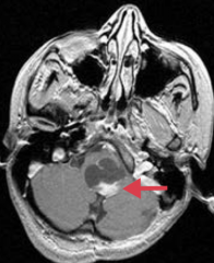
What can a Medulloblastoma cause?
|
- Can compress the 4th ventricle → hydrocephalus
- Can send "drop metastases" to spinal cord |
|
|
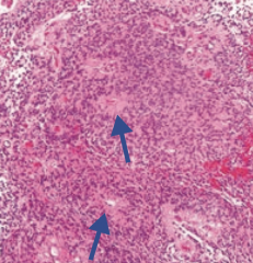
What type of childhood brain tumor is associated with Homer-Wright rosettes? Prognosis?
|
Medulloblastoma - highly malignant
|
|
|
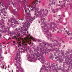
What is the gross and histologic appearance of Medulloblastoma?
|
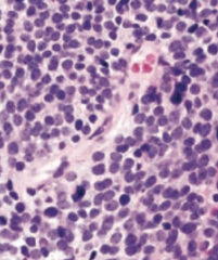
- Solid cerebellar tumor
- Homer-Wright rosettes - Small blue cells |
|
|
What type of childhood brain tumor is derived from ependymal cells? Prognosis?
|
Ependymoma - poor prognosis
|
|
|
What can an Ependymoma cause?
|
Most commonly found in 4th ventricle so it can cause hydrocephalus
|
|
|
What type of childhood brain tumor is associated with perivascular rosettes
|
Ependymoma
|
|
|
What is the gross and histologic appearance of an Ependymoma?
|

- Commonly in 4th ventricle
- Perivascular rosettes - Rod-shaped blepharoblasts (basal ciliary bodies) found near nucleus |
|
|
What type of childhood brain tumor may be confused with a pituitary adenoma? Source?
|
Craniopharyngioma - derived from remnants of Rathke pouch
|
|
|
What is the prognosis and clinical syndrome caused by Craniopharyngioma?
|
- Benign tumor
- May be confused with pituitary adenoma because they both cause bitemporal hemianopia |
|
|
What is the most common childhood supratentorial brain tumor?
|
Craniopharyngioma
|
|
|
What is the histologic appearance of a Craniopharyngioma?
|
Calcification is common (tooth-enamel like)
|
|
|
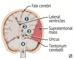
What are the types of herniation syndromes?
|

1. Cingulate (subfalcine) herniation under falx cerebri
2. Downward transtentorial (central) herniation 3. Uncal herniation 4. Cerebellar tonsillar herniation into foramen magnum |
|
|
What type of herniation can compress the anterior cerebral artery?
|
Cingulate (subfalcine) herniation under falx cerebri (#1)
|
|
|
What type of herniation can compress the ipsilateral CN III causing a blown pupil and down and out gaze?
|

Uncal Herniation (#3) - medial temporal lobe
|
|
|
What type of herniation can compress the posterior cerebral artery causing contralateral homonymous hemianopsia)?
|

Uncal Herniation (#3) - medial temporal lobe
|
|
|
What type of herniation can compress the contralateral crus cerebri causing ipsilateral paralysis / false localization sign?
|

Uncal Herniation (#3) - medial temporal lobe
|
|
|
What type of herniation can compress the brainstem, inhibiting respiration, and possibly causing coma and death?
|

Cerebellar tonsillar herniation into the foramen magnum (#4)
|
|
|
What are the potential consequences of a cingulate (subfalcine) herniation under the falx cerebri?
|
Can compress anterior cerebral artery
|
|
|
What are the potential consequences of an uncal herniation?
|
Compresses:
- Ipsilateral CN III → blown pupil and down and out gaze - Ipsilateral PCA → contralateral homonymous hemianopsia - Contralateral crus cerebri → ipsilateral paralysis, "false localization sign" |
|
|
What are the potential consequences of a cerebellar tonsillar herniation into the foramen magnum?
|
Coma and death result when these herniations compress the brain stem (and inhibit respiration)
|