![]()
![]()
![]()
Use LEFT and RIGHT arrow keys to navigate between flashcards;
Use UP and DOWN arrow keys to flip the card;
H to show hint;
A reads text to speech;
38 Cards in this Set
- Front
- Back
|
Abdomen |
--Inferior trunk b/t thorax and pelvis CONSISTS OF: 1) Abdominal Wall - musculotendinous; thick muscles located on either side of lumbar vertebrae. The only bones are posterior vertebrae 2) Abdominal Cavity - digestive organs, spleen, kidneys
*when supine, the abdominal cavity extends superiorly to about the 5th anterior intercostal space |
|
|
Regions of the Abdomen |
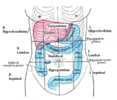
1- Right hypochondrium 2- Epigastrium 3- Left hypochondrium 4- Right lumbar region 5- Umbilical region 6- Left lumbar region 7- Right inguinal region 8- Hypogastrium 9- Left inguinal region |
|
|
Layers of Abdominal Wall |
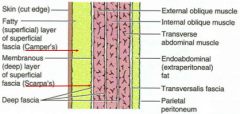
-Skin -Fatty (superficial) layer of superficial fascia (Camper's) -Membranous (deep) layer of superficial fascia (Scarpa's) -Deep fascia -External oblique muscle -Internal oblique -Transverse abdominal -Endoabdominal fat -Transversalis fascia -Parietal peritoneum |
|
|
Abdominal Muscles |
NEW: -Quadratus lumburom
|
|
|
Muscles of Abdominal Wall |
-Forms support and protection to the abdominal viscera -Formed by 3 flat muscles (EAO, IAO, TA) and 1 strap muscle (RA) -Segmentally innervated by T6-T12 and L1 -Flat muscles end anteriorly in an aponeurosis which interlaces to form linea alba in midline -The aponeurosis forms a rectus sheath which encases the rectus abdominus muscle |
|
|
External Oblique |
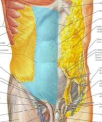
yellow=muscle blue=aponeurosis
-the lower edge of aponeurosis of EO forms the inguinal ligament |
|
|
Inguinal Ligament |
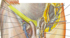
-Inferior margin of EO apo -Spans from the anterior superior iliac spine to pubic tubercle -Triangular opening at the medial is superficial inguinal ring, the exit of the spermatic cord in males |
|
|
Abdomen Movements |
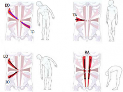
|
|
|
Rectus Sheath |
-Rectus abdominus is surrounded by the aponeuroses of the EO, IO and TA -EOA is in front of RA -IOA splits and passes in front and behind RA -TAA passes behind RA
|
|
|
Arcuate line |
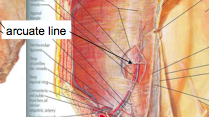
-At a point halfway b/t umbilicus and pubis, the apo layers that were behind RA change so that they're all in front RA |
|
|
Functions of Abdominal Wall muscles |
-Support anterolateral abdominal wall -Protect abdominal viscera -Compresses abdominal contents/increases abdominal pressure: -Oppose/assist diaphragm during inspiration -Helps to bring out gastric contents during vomiting -Bearing down to empty the rectum or bladder, or during childbirth -Move the trunk and maintain posture -Guards inguinal canal
|
|
|
Inguinal Canal |
Contains: -Ilioinguinal nerve -Round ligament of the uterus (females) -Spermatic cord (males)
|
|
|
Contents of Spermatic Cord |
-Ductus deferens -carries sperm from testis to urethra -Artery of ductus deferens -Testicular artery -Pampiniform plexus of veins -Cremasteric artery -Sympathetic nerve fibers -Genital branch of genitofemoral n. -Lymphatic vessels
|
|
|
3 Coverings of Spermatic Cord |
1) Internal spermatic fascia -derived from transversalis fascia 2) Cremasteric fascia -from IO 3) External spermatic fascia -from EOA |
|
|
Inguinal hernia |
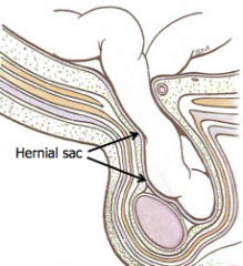
*happens if abdominal muscles are weak -Intestines or peritoneal fat can push into inguinal canal, forming a hernia -During standing, coughing or vigorous straining, contraction of IO & TA cause the roof of the canal to become lower & taut
|
|
|
Homologous female anatomy |
-Embryological remnants of primordial ovaries descend into labia majora and become round ligament of uterus
-Femoral hernias: weak ant. abdom wall associated with passage of large femoral vessels |
|
|
Abdominal Cavity |
-Limited superiorly by diaphragm and continous inferiorly w/ pelvic cavity completely filled with abdominal viscera -Lined by peritoneum |
|
|
Peritoneum |
-Serous membrane consisting of: -Parietal peritoneum - lines abdominal wall, pelvic wall & inferior surface of diaphragm -Visceral peritoneum - covers the viscera, spleen & stomach -Mesentary - general term for a double layer of peritoneum that suspends an organ |
|
|
Intraperitoneal organs |
-Liver -Gallbladder -Stomach -Small intestine (except duodenum) -Transverse colon -Sigmoid colon -Cecum and appendix -Spleen -Superior part of uterus |
|
|
Retro/Subperitoneal organs |
-Duodenum -Descending colon -Abdominal aorta -Rectum -Inferior vena cava -Pancreas -Inferior part of uterus -Kidneys -Ureters -Bladder -Ascending colon
|
|
|
Peritoneal Cavity |
-potential space b/t parietal & visceral peritoneum -contains peritoneal fluid (lubricates surface and facilitates free movement of viscera) -completely enclosed in males -communicates w/ exterior thru openings in uterine tubes (fallopian tubes) in females |
|
|
Omentum |
-a double layer of peritoneum that extends from the stomach to the adjacent organs -lesser - lesser curvature of stomach and proximal part of duodenum to liver -greater - greater curvature of stomach to transverse colon |
|
|
Greater and Lesser sacs in peritoneal cavity |
-greater - main part of cavity
-lesser (omental bursa) - lies posterior to lesser omentum and stomach, allows free movement of stomach
-omental foramen (epiploic foramen) - opening b/t 2 sacs
|
|
|
Mesentary |
-connects peritoneal organs w/ abdominal wall -encloses blood vessels & nerves to organs it surrounds: -small intestine (except duodenum) -liver -appendix -stomach -spleen -transverse colon -sigmoid colon
|
|
|
Peritoneal ligaments |
-connects an organ with another organ -lacks connective tissue like mesentary -may contain blood vessels |
|
|
4 parts of GI tract |
-esophagus -stomach -small intestine -large intestine |
|
|
Esophagus |
-passes food from pharynx to stomach -begins at C6 -passes thru diaphragm at esophageal hiatus and ends here (T10) -enters stomach at cardial orifice (T11 and 7th left costal cartilage) -right & anterior to descending thoracic aorta *esophageal nerve plexus (vagus n) |
|
|
Stomach |
location: superior, left & center function: enzymatic digestion -muscular walls of stomach -break down particles, increase surface area of food, convert food to chyme -low pH of stomach activates enzymes -gastric enzymes break down proteins into amino acids
|
|
|
Parts of stomach |
-cardia -adjacent to esophagogastric junction -fundus -body -pyloric part -plyoric antrum and canal -plyroic sphincter -pyloric orifice
|
|
|
Small Intestine |
-extends from pylorus of stomach to ileocecal junction with large intestine
-location of complete digestion -fats broken down -most products of digestion are absorbed -water, electrolytes, minerals also absorbed *(duodenum, jejunum, ileum) |
|
|
Duodenum |
-shortest, widest part -from pyloric opening to duodenojejunal junction -c shaped -surrounds head of pancreas -4 regions: superior, descending, horizontal, ascending |
|
|
Jejunum and ileum |
6-7 meters long 2/5 jejunum 3/5 ileum
attached to posterior abdominal wall by the mesentery |
|
|
Large Intestine |
-ileocecal junction to anus -~1.5 meters long -functions to convert liquid contents of ileum into semisolid feces by absorbing fluid and electrolytes -consists of: cecum, appendix, colon, rectum, anal canal |
|
|
Cecum |
-blind pouch of large intestine -lies in the right iliac fossa -usually surrounded by peritoneum (intraperitoneal) |
|
|
Appendix |
-"vermiform appendix" -narrow, hollow, muscular tube -suspended from terminal ileum by mesoappendix -most often it is behind cecum |
|
|
4 parts of colon |
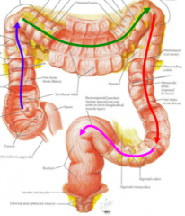
ascending transverse descending sigmoid
teniae coli pull colon to form haustra (pouches) |
|
|
Rectum |
-rectosigmoid junction at S3
-s-shaped
-supports and retains fecal mass before it is expelled during defecation |
|
|
Anal canal |
-terminal part of large intestine, inferior to pelvic diaphragm
-internal anal sphincter muscles (involuntary & smooth muscle)
-external anal sphincter muscles (voluntary & skeletal muscle) |