Reading...
![]()
Play button
![]()
Play button
![]()
Use LEFT and RIGHT arrow keys to navigate between flashcards;
Use UP and DOWN arrow keys to flip the card;
H to show hint;
A reads text to speech;
60 Cards in this Set
- Front
- Back
|
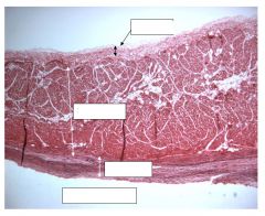
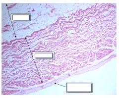
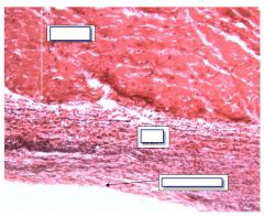
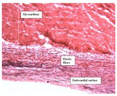
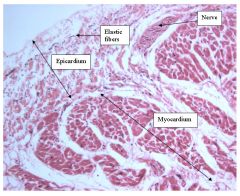
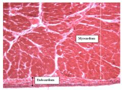
Wall of the left atrium (has thickest endocardium)
*endocardium: connective tissue, abundant elastic fibers (stained black here) *myocardium: middle and thickest layer, cardiac muscle *epicardium: thin layer but thick near AV sulcus |
|
|
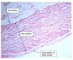
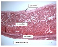
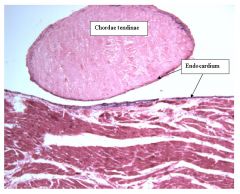
Right Atrium
*endocardium is thinner than left atrium *myocardium is more loosely organized in the right than the left atrium *epicardium has considerable adipose tissue |
|
|
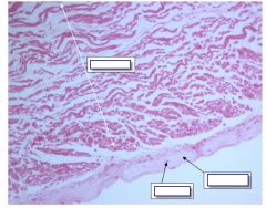
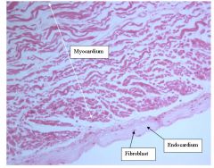
left atrium endocardial surface
*dense irregular FECT (w/ fibroblast *ELASTIC FIBERS - stained black (increase in concent closer to AVsulcus) *dense myocardium->left atrium |
|
|
right atirum
*smooth walled portion is shown (as opposed to the rough portion) *loose myocardium->right atrium |
|
|
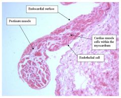
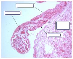
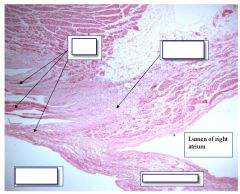
rough walled portion of right atrium
*pectinate muscle which includes myocardium and endocardium *can make out endothelial cells *myocardium is so thin some areas appear missing->right atrium |
|
|
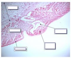
trabecular muscle of right atrium
*pectinate muscle is a simple projection, here the trabecular muscle reestablishes continuity with myocardium |
|
|
Outer portion of left atrial wall
*epicardium is more loosely arranged than the endocardium also elastic fibers are less densely concentrated * nerves and blood vessels are found in the epicardium (are rare in the endocardium) |
|
|
image near AV sulcus
*endocardium becomes quite large as you approace the AV sulcus |
|
|
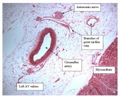
left AV sulcus
*wide epicardium *epicardium because vessels and nerve |
|
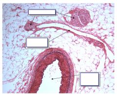
|
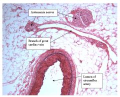
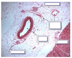
left AV sulcus
*arteries and veins are distinguished by the structure of their walls *thicker wall in arteries *nerve has no neurons so not ganglion |
|
|
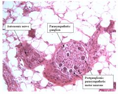
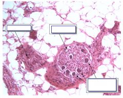
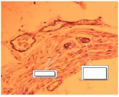
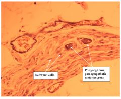
left AV sulcus
*here is ganglion because neurons are found *nuclei are eccentric nuclei not surrounded by satellite cells which is postganlionic sympathetic or parasympathetic motor *in the wall of an organ so parasympathetic motor *autonomic nerve- because cant distinguish if PS motor (pre or post gangi), S motor |
|
|
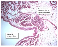
left AV sulcus
*coronary sinus is the only blood vessel that has cardiac and not smooth m in its wall *large epicardium->left AV sulcus |
|
|
endocardial surface of left ventricle
*ventricle because--thinner endocardium, fewer elastic fibers *left v because--left v is relatively smoother than right v |
|
|
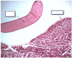
left ventricle
*seperate dense regular FECT -->cordae tendinae->must be vventricle *very thin endocardium->closer to heart apex |
|
|
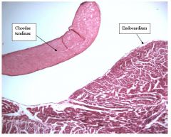
ventricle
*cordae tendinae-> dense regular FECT surrounded by thin endocardium |
|
|
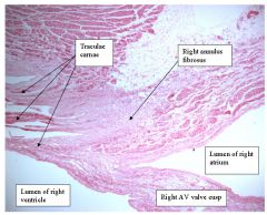
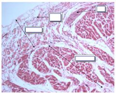
junction between right atrium/right ventricle
*spaces-->trabeculae carnae (like trabecular muscles of atria but in ventricle) *trabeculae carnae are more common in the right ventricle |
|
|
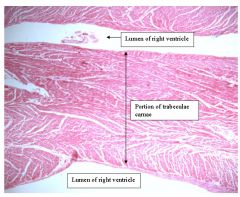
wall of right ventricle closer to apex
*crabeculae carnaee->right ventricle |
|
|
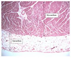
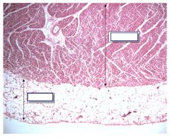
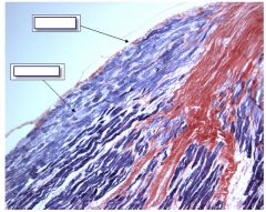
outer portion of wall of left ventricle
*myocardium densely packed (relative to atrium) *epicardium similar to that seen in atria->NOT distinguishing feature *closer to AV sulcus thicker epicardium is |
|
|
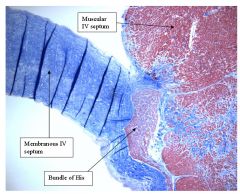
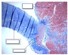
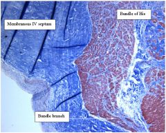
interventicular septum
*connective tissue stained blue-> membranous IV septum *cardiac muscle stained red->muscular IV septum *bundle of his at base of membranous IV *myocardium of IV is similar to myocardiium of left atrium (thickness, density, lack of trabeculation) |
|

|
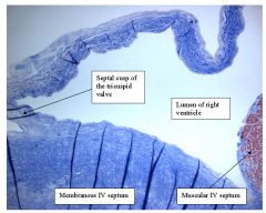
IV septum
*septal cusp arises frm membranous IV septum->divides right atrium from right ventricle |
|
|
membranous IV septum
*root of aorta consists of only connective tissue (stained blue here) *connective fibers above cusp to become wall of aorta |
|
|
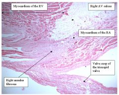
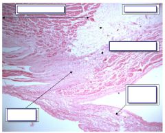
right AV valves (tricuspid)
*valve cusps arise from annulus fibrosus (right of connective tissue) *tissue is difference of myocardiumo of RV or RA |
|
|
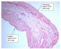
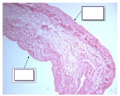
image of valve cusp of tricuspid
*holding surface: collagen fibers are relatively large *deforming surface: collagen fibers are less noticeable (small diameter) enriched in elastic fibers |
|
|
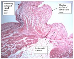
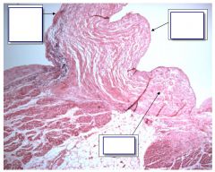
left AV valve cusp (mitral valve)
*annulus fibrosus connective tissue which intervenes between A and V myocardia *can distingiush holding and deforming surface by collagen fibers and elastic fibers on deforming side |
|
|

Valve cusp of mitral valve
*elastic fibers are black which are concentrated in the deforming surface and less apparent in the holding surface *deforming surface faces the atrium, holding surface faces the ventricle |
|
|
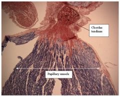
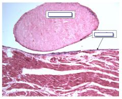
Papillary muscles
*arise out of the floor of ventricles *dense regular FECT stains red *cardiac muscle stains blue *collagen fibers of the chordae tendinae project into cardiac papillary muscle |
|
|

Higher mag of papillary muscle
*lined by a layer of endocardium *layer of purkinje cells just deep to endocardium |
|
|
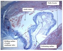
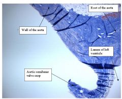
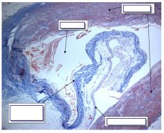
Orgin of Aorta
*arises out of upper portion of IV septum *valve cusp of aortic seminulnar valve is seen *holding/deforming surfaces distinguished by differences of collegen content *aortic sinus is the portion of lumen that lies just above the aortic valve *wall of aorta has distinct structure |
|
|
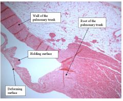
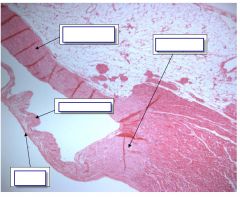
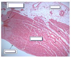
pulmonary trunk
*arises from tubular extension of roof of right ventricle (the conus arteriosus) *pulmonary trunk arises from ring-shaped loop *differences in collagen distinguish the surfaces of the semilunar pulmonary valve |
|
|
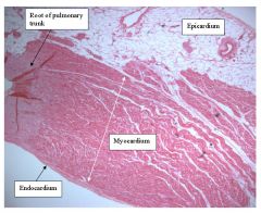
just proximal to root of pulmonary trunk
*all three hart layers prior to pulmonic root (unlike aortic origin) --- has myocardium and epicardium |
|
|
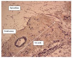
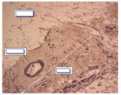
SA node
*mass of connective tissue located at the approximate junction of epicardium and myocardium of right atrium *nodal artery runs length of this tissue *conducting cells scattered within (lack staining relative to working cardiac muscle) |
|
|
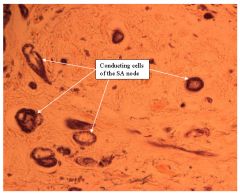
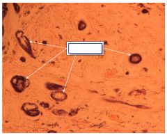
SA node at higher magnification
*conducting cells scattered *lack of staining because of reduced number of myofibrils *conducting cells within connective tissue --> node *junction of epicardium/myocardium ->SA node |
|
|
parasympathetic ganglion
*wall of organ + neurons -> postganglionic parasympathetic -> whole structure is ganglion |
|
|
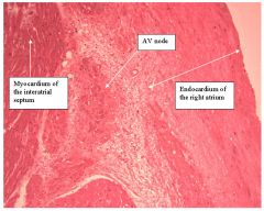
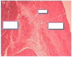
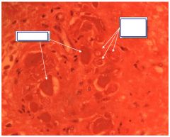
AV node
*AV node at junction of myocardium/epdocardium of right atrium w/ connective cells *NO epicardium in vicinity (unlike SA node) |
|
|
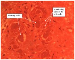
AV node (at higher magnification)
*conducting cells visible (lack of staining compared to working cells) *conducting cells embedded in connective tissue->node *between endocardium/myocardium -> AV node |
|

|
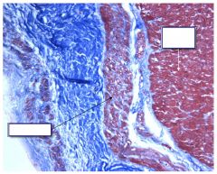
IV septum
*bundle of His located at the base of membranous IV septum *conducting cells of bundle of His are NOT surrounded by connective tissue *entire note is surrounded by connective tissue of membranous IV septum *branch of bundle of his runs down into muscular IV septum will break up into Purkinje |
|
|
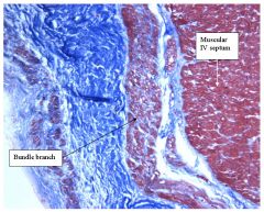
Bundle branch of bundle of His
*branch runs at the interface between endocardium and myocardium *stain lighter displaying "hollow centers" as compared to surrounding working cells of muscular IV septum |
|
|
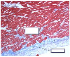
Bundle of His where bundle branch has become Purkinje fibers
*stain lightly displaying "hollow centers" *form superficial layer upon the myocardium just below endocardium *not specifically encased by connective tissue *thick layer of Purkinje cells -> IV septum or papillary muscle |
|

|
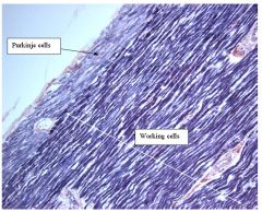
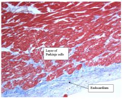
papillary muscle
*Purkinje cells lie deep to endocardium *Purkinje cells stain lighter than the deeper working myocardial cells |
|
|
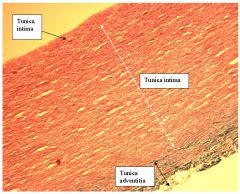
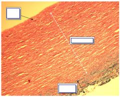
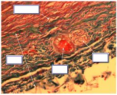
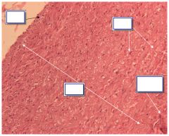
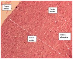
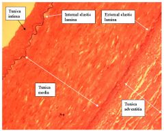
Elastic artery
*three basic layers - thinest bordering lumen is tunica intima (endothelium and connective tissue) -middle layer is tunica media and is the thickest (vascular smooth muscle/elastic fibers) -outermost layer is tunica adventitia (variable in thickness, corarser collagen (stained green here) ) |
|
|
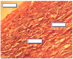
Elastic artery at higher magnification
*numerous elastic fibers within tunica media which comprise the elastic lamina |
|
|

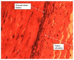
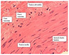
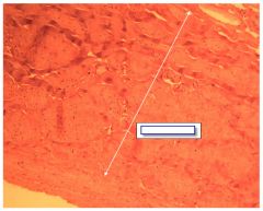
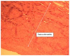
outer portion of elastic artery (tunica adventitia)
*more collagen than elastic fibers *blood vessel and nerve embedded within tunica adventitia called vasa vasorum and nervi vasorum |
|
|
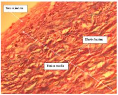
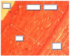
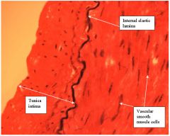
elastic artery (stained with H&E)
*distinct elastic lamina (hallmark of this type of vessel) *tunica adventitia lacks elastic lamina same for tunica intima |
|
|
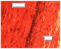
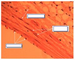
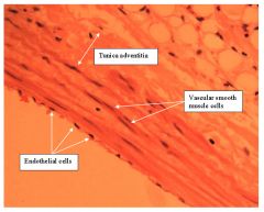
large muscular artery
*tunica media does not contain abundance of elastic lamina *elastic fibers are contentrated at internal elastic lamina and external elastic lamina which serve as boundaries between tunica intima tunica intima/media and tunica media/adventitia resp. |
|
|
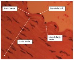
inner layers of muscular artery
*presence of internal elastic lamina *few elastic fibers within tunica media instead can see vascular smooth muscle cells without obscured by elastic lamina |
|
|
outer layers of muscular artery
*external lamina more substantial collection of elastic fibers compared to internal (so is wider) *tunica adventitia ends indistinctly |
|
|
(smaller) muscular artery
*lack of elastic lamina *has internal elastic lamina *external elastic lamina moderate in content *blood vessel (vasa vasorum) and nerve (nervi vasorum) in tunica adventitia |
|

|
inner two layers of muscular artery
* tunica intima is thin -- as muscular arteries become aterioles the tuica intima can become a single layer of endothealial cells (with minor amounts of connective tissue) *internal elastic lamina visible * |
|
|
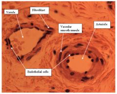
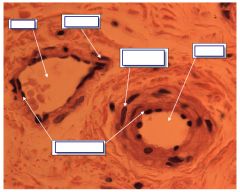
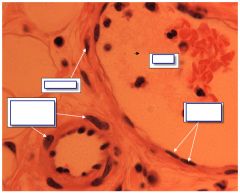
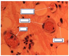
ateriole and venule
*lined by endothelium which constitutes tunica intima *primary layer of the venule is tunica adventitia (fibroblasts) *primary layer of arteriole is tunica media (vascular smooth muscle cells) *this difference -> wall thickness/lumen ratio is much higher for the ateriole than venule |
|
|
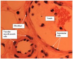
ateriole and venule
*venule has wall which is tunica intima (endothelium only) and olnly a layer or two of fibroblasts within tunica adventitia |
|

|

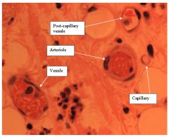
example of microcirculation: arteriole
*smallest ateriole possible with only one smooth muscle cell layer *capillary is only a single layer of endothelium (lumen only big enough for a single blood cell) *same structure but bigger-> post capillary venule |
|
|
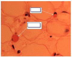
Addipose tissue
*capillary and post capillary venule have same structure but differ in size |
|
|
|
|
|
heart |
heart |
|
|
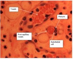
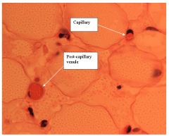
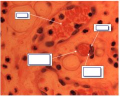
venule and post capillary venule
*post-capillary venule is too large to be a capillary (ie more than one RBC can fit in lumen) *post-capillary venule also has a second cell outside endothelial cell (the pericyte) |
|
|
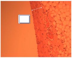
wall thickness of medium size vein
*unlike venules, has tunica media consisting of a few layers of vascular smooth muscle *like venules, primary layer is the tunica adventitia *tunica intima consits of a single layer of endothelial cell w/ little connective tissue. |
|
|
wall of medium sized vein (higher magnification)
*tunica intima is only a single layer *smooth muscle cells present (tunica media) which are loosely arranged compared to arteriole *relative difference in smooth muscle is more apparent in vein/artery case vis.arteriole/venuole *like all veins tunica adventitia blends with surrounding tissues |
|
|
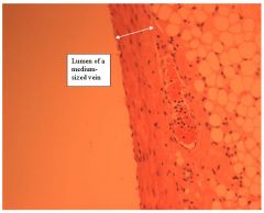
low magnification of vena cava
*shows three layers, tunica adventitia is biggest |
|
|
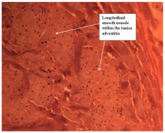
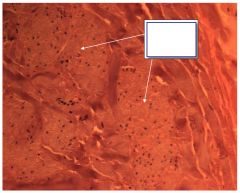
outer portion of vessel wall of vena cava
*longitudinal smooth muscle within tunica adventitia *dense connective tissue between these longitudinal pockets *no other blood vessel has this arrangment of smooth muscle in the tunica adventitia |
|
|
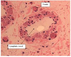
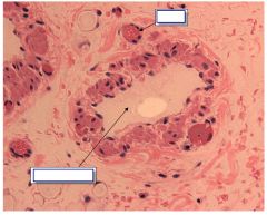
lymphatic vessel
*key distinguishing feature: asymmetry of smooth muscle within tunica media--smooth muscle has variable thickness *smooth muscle is not circular because smooth muscle is arranged in spiral fashion around the long axis *lined by endothelium (tunica intima), variably thick layer of connective tissue comprising the tunica adventitia, primary layer |