![]()
![]()
![]()
Use LEFT and RIGHT arrow keys to navigate between flashcards;
Use UP and DOWN arrow keys to flip the card;
H to show hint;
A reads text to speech;
28 Cards in this Set
- Front
- Back
|
Review: Pleural tap taken from which recess? |
Costodiaphragmatic recess 7th- mid-clavicular intercostal space 9th- mid-axillary intercostal space 10th-paraveterbral intercostal space |
|
|
Review: Which pleura changes names? |
parietal pleura- cervical, costal, mediastinal, and diaphragmatic |
|
|
what separates both superior and inferior lobes on the lungs? |
oblique fissure |
|
|
what separates the superior and medial lobe? on which lung? |
transverse fissure- right lung only |
|
|
pulmonary ligament |
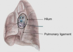
visceral and parietal pleura meet during inspiration
2 layers come apart slightly so structures can expand inferiorly |
|
|
Left hilium |
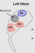
pulmonary artery on top |
|
|
R hilium |
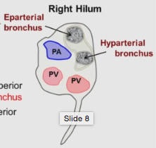
2 bronchi; epiarterial bronchus on top PA in between |
|
|
medial lobe - ribs? superior lobe- ribs? |
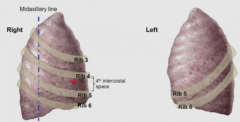
4th intercostal space ; ribs 4-6 superior lobe- rib 3 |
|
|
percussion solid? fluid filled? |
solid- flat sound fluid filled- dull sound |
|
|
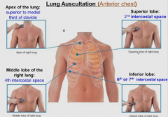
auscultation of lung |
Are Surgeons More Intelligent?
Apex- 1/3 medial supraclavicular Superior- 2nd intercostal Middle lobe- 4th intercostal Inferior- 6th-7th intercostal. posteriorly- triangle of auscultation |
|
|
Open pneumothorax |
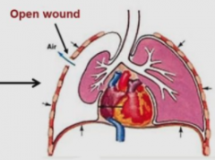
affected lung- tracheal shift opening- fistula lung collapses air is sucked in and out |
|
|
Tension pneumothorax |
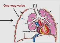
non affected lung - tracheal shift wound/ruptured bleb- valve like opening for entry of air into pleural cavity widening of intercostal spaces affected lung diaphragm placed inferiorly |
|
|
Spontaneous pneumothorax |
small, no tracheal shift |
|

Tracheal deviation |
moves towards affected side: 1. lung agenesis, 2. open pneumothorax, 3. pneumonectomy
move towards unaffected side: 1. tension pneumothorax and 2. Pleural effusion
|
|
|
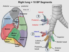
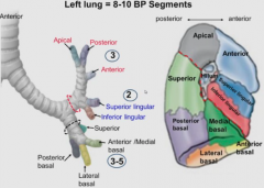
Bronchopulmonary segments |
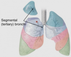
18- 20 segmental tertiary bronchi surgical: segmentectomy one single segment- pulmonary artery adjacent segments- pulmonary vein lung cancer in early stages- one bronchopulmonary segment from a lung will have cancer |
|
Aspiration of foreign bodies |
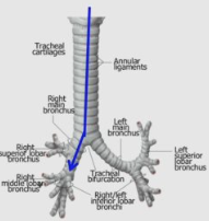
most likely to lodge into right bronchus R bronchus is wide, short and more vertical |
|
|
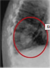
Pneumonia |
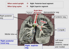
Aspirated fluid/vomitus aspiration locations/ ausculation or percussion: supine/laying down: superior segment on R sitting upright- posterior basal segment on R |
|
|
Pancoast tumor (syndrome) |
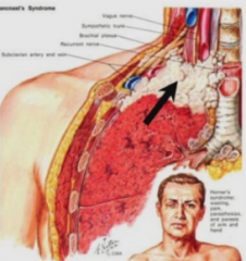
lung cancer in the apex of the lungs associated compression syndromes: 1. compression of sympathetic trunk 2. compression of brachial plexus (claw hand; klumpke palsy - C8 T1) 3. compression of subclavian a & v (pallor, pulselessness, pain) |
|
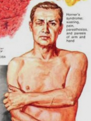
Horner's syndrome |
Compression of sympathetic trunk Miosis (a constricted pupil), ptosis (a weak, droopy eyelid), apparent anhidrosis (decreased sweating) Horner syndrome may reflect serious disease in the neck or chest (such as a Pancoast tumor (tumor in the apex of the lung) |
|
|
Carinal tumors |
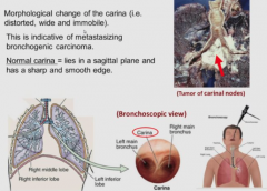
if carina changes shape indicative of bronchogenic carcinoma (lung cancer) |
|
|
Pulmonary embolism (PE) |
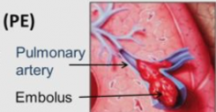
pulmonary artery obstruction with a blood clot |
|
|
Common sources of PE |
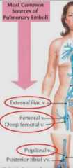
1. External iliac v 2. Femoral v 3. Deep femoral v 4. Popliteal v Common sources are from lower limb deep veins |
|
|
R lung |
|
|
|
L lung |
|
|
|
What are the 2 plexus of lung lymphatic system? |
Superficial and Deep plexus |
|
|
Superficial plexus drains? First node? |
1. parenchyma (packing) 2. visceral pleura *First node: Hilar/broncho-pulmonary |
|
|
Deep plexus drains? First node? |
1. submucosa (beneath epithilea) 2. CT bronchi *First node: pulmonary |
|
|
Lymphatic drainage of lung (deep pathway) |
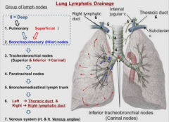
1. pulmonary nodes 2. hilar/bronchopulmonary* (superficial plexus starts) 3. tracheobronchal (superior* & inferior-carinal) 4. parabronchal (on the side) 5. bronchomediastinal lymph trunk 6. L and R thoracic duct 7. venous angle of subclavian veins (internal jugular veins as well) *involved in arch of azygos vein compression |