Reading...
![]()
Play button
![]()
Play button
![]()
Use LEFT and RIGHT arrow keys to navigate between flashcards;
Use UP and DOWN arrow keys to flip the card;
H to show hint;
A reads text to speech;
70 Cards in this Set
- Front
- Back
|
Identify the 9 functions of the integumentary system.
|
Protection
Barrier Temperature regulation Wound repair Absorption/excretion Production of vitamin D Perception/sensation Identification Communication |
|
|
epidermis layer
|
outer most layer, thin but tough, replaced every 4 weeks
|
|
|
subcutaneous layer
|
adipose (fat) tissue
|
|
|
dermis layer
|
inner supportive layer, consist of connective tissue (collagen), nerves, sensory receptors, blood vessels, lymphatics, hair follicles, sebaceous glands, and sweat glands
|
|
|
If patient is reporting of pain in lesion on skin, what does this tell you about the skin layer it is in?
|
It must be in the dermis because that is where nerve endings are.
|
|
|
Identify the 4 appendages to the skin in the integumentary system.
|
Hair
Sebaceous glands - produce sebum Sweat glands Nails - hard, keratin plates |
|
|
Identify the 2 types of hair.
|
Vellum - fine hair all over the body
Terminal - scalp, eyebrows |
|
|
Identify the 2 types of sweat glands.
|
Eccrine glands - produce sweat (diluted saline solution) active at 2 months of age
Apocrine glands - produce thick milky secretions, activated in puberty |
|
|
primary lesion
|
first lesion that appears
|
|
|
secondary lesion
|
lesion that occurs due to changes over time or changes because of a factor such as scratching or infection
|
|
|
macule
|
color change, flat, <1cm (freckle)
|
|
|
patch
|
flat, macule that is >1cm (Mongolian spot, café au lait, birthmark)
Note: Same as macule, just large |
|
|
papule
|
elevated, solid lesion <1cm, lesion you can feel (mole, wart)
|
|
|
plaque
|
papule >1cm in width (psoriasis)
Tip: Basically a large papule |
|
|
nodule
|
solid, elevated, hard or soft >1cm, extends deeper into dermis
|
|
|
tumor
|
>2cm, firm or soft mass (lipoma, hemangioma)
|
|
|
wheal
|
superficial, raised lesion (PPD, insect bite)
|
|
|
urticaria
|

multiple wheal like lesions, very itchy (hives)
|
|
|
vesicle
|
fluid filled, elevated lesion, <1cm (herpes, chicken pox, small blister)
|
|
|
bulla
|
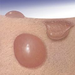
>1cm vesicle, (burn, large blister, bullous impetigo)
|
|
|
cyst
|
fluid filled cavity extending to dermis or subcutaneous layer (sebaceous cyst)
|
|
|
pustule
|
pus filled lesion (acne, pimple); any size
|
|
|
crust
|
thickened, dried out exudate left when vesicles/pustules burst or dry up (impetigo)
|
|
|
scale
|
flakes of skin, silvery or white (psoriasis, eczema, seborrhea, dermatitis)
|
|
|
fissure
|
linear crack with abrupt edges (athletes foot, cracks in
corners of mouth) |
|
|
erosion
|
shallow depression, usually no scar (superficial abrasion)
|
|
|
ulcer
|
deep depression, leaves scar usually (decubitus ulcer, bed sore)
|
|
|
excoriation
|
self inflicted abrasion, superficial crusting secondary
|
|
|
scar
|
healed lesion, replaced with collagen/connective tissue
|
|
|
atrophic scar
|
skin level depressed with loss of tissue, thinning (striae/stretch marks)
|
|
|
lichenification
|
prolonged intense scratching eventually thickens the skin; common in people with eczema; found in elbows, behind knees
|
|
|
keloid
|
elevated scar, feels rubbery; common in darker pigmented people
|
|
|
vascular lesion
|
lesions vascular in nature
Tip: Since vascular, there will mostly be a color change |
|
|
spider angioma
|
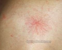
red, star shaped with solid circular center
|
|
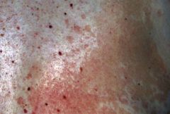
|
purpura
|
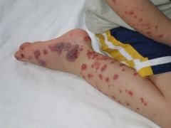
red/purple patch, flat macular hemorrhage >1cm
|
|
|
petechiae
|
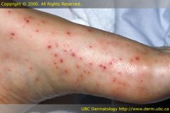
tiny, pinpoint, hemorrhage <1-2mm, “little flat blood spots ranging in color - red, purple, or brown
|
|
|
ecchymosis
|
flat macular lesion of various colors depending on stage of bruise
|
|
|
hemiangioma
|
reddish/blue, solid, spongy collection of benign blood vessels; typically benign
|
|
|
Identify the 4 color variations of the skin.
|
Pallor - pale, white
Erythema - redness Cyanosis - bluish Jaundice - yellow color |
|
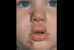
Identify and define the pictured condition.
|
Tinea corporis - "ringworm of the body"; skin infection due to fungi
|
|
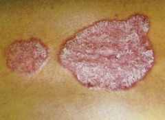
Identify and define the pictured condition.
|
psoriasis - scaly, erythematous patch with silvery scales on top
|
|
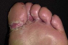
Identify and define the pictured condition.
|
Tinea pedis - "athlete's foot', fungal infection that first appears as small vesicle and then grows scaly and hard
|
|
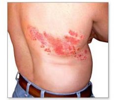
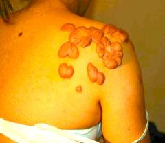
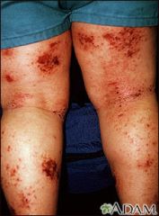
Identify and define the pictured condition.
|
Herpes zoster (shingles) - small, grouped vesicle that emerge along route of cutaneous sensory nerve; pustules then crusts
|
|
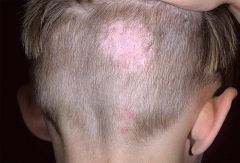
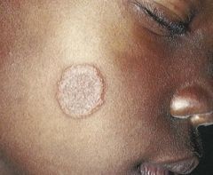
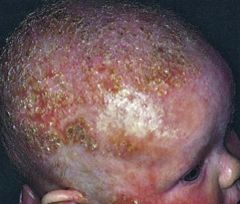
Identify and define the pictured condition.
|
Tinea capitis - fungal infection of the scalp, "ringworm of the scalp"
|
|
Identify and define the pictured condition.
|
impetigo - bacterial infection of the skin; moist, thin-roofed vesicle with red base; forms honey-colored crusts; common in infants/kids; caused by streptococcus (strep) or staphylococcus (staph) bacteria
|
|

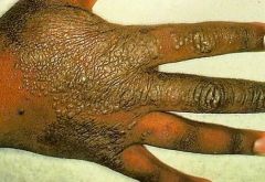
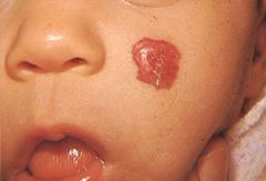
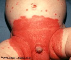
Identify and define the pictured condition.
|
diaper/contact dermatitis - inflammatory disease caused by skin irritation from heat, moisture, and diapers
red, moist, maculopapular patch with no defined borders along inguinal and gluteal folds |
|
Identify and define the pictured condition.
|
candidiasis - Candida skin infection affecting superficial skin layers
Scalding red, moist patches with marked borders with some loose scales |
|
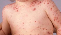
Identify and define the pictured condition.
|
varicella (chicken pox) - infectious disease, esp. of children, causing a mild fever and a rash of itchy inflamed blisters
|
|
Identify and define the pictured condition.
|
eczema - long-term (chronic) skin disorder that involves scaly and itchy rashes; red papules and vesicle with weeping, oozing, crusting
|
|
Identify and define the pictured condition.
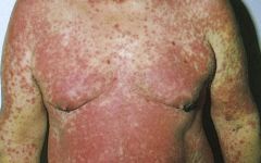
|
allergic drug reaction - caused by an allergic reaction to a drug; erythematous and symmetric rash, typically generalized
|
|
Identify and define the pictured condition.
|
tinea versicolor - long-term (chronic) fungal infection of the skin caused by a fungus found normally on skin; patches of pink/tan/white with scaling
|
|
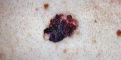
Identify and define the pictured condition.
|
melanoma - most dangerous type of skin cancer caused by changes in cells called melanocytes; common in individuals with excess UV exposure
|
|
Identify and define the pictured condition.
|
Kaposi's sarcoma - (patch stage) vascular tumor; most common in HIV-infected persons; AIDS-defining illness; easily mistaken for bruises or nevi and ignored
|
|
Identify and define the pictured condition.
|
seborrheic dermatitis - "cradle cap"; common, inflammatory skin condition that causes flaky, white to yellowish scales to form on oily areas such as the scalp or inside the ear; occurs with or without reddened skin
|
|
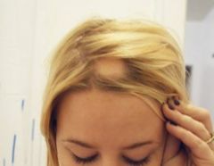
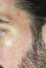
Identify and define the pictured condition.
|
alopecia areata - sudden apperance of sharply, circumscribed round/oval hairless patch; unknown cause
|
|
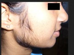
Identify and define the pictured condition.
|
hirsutism - excessive body hair in females forming a sexual pattern (upper lip, chest, arms, legs); caused by endocrine/metabolic dysfunction
|
|
|
Identify the medical term for a bruise.
|
ecchymosis
|
|
|
Identify the medical classification for a freckle.
|
macule
|
|
|
Identify the medical classification for acne.
|
pustule
|
|
|
Identify the medical classification for a mole or wart.
|
papule
|
|
|
Identify the medical classification for a Mongolian spot.
|
patch
|
|
|
Identify the medical classification for a stretch marks.
|
striae (atrophic scarring)
|
|
|
Identify the medical classification for a birthmark.
|
patch
|
|
|
Identify what needs to documented about each lesion found during the physical assessment. (8)
|
Location
Color Size Symmetry Pattern Elevation Odor Drainage or discharge |
|
|
Identify the 10 aspects of the objective data/physical exam portion a skin assessment.
|
1. Color/General Pigmentation
2. Lesions 3. Temperature 4. Moisture/Dryness 5. Texture 6. Edema 7. Skin Mobility/Skin Turgor 8. Vascular Lesions/Bruising 9. Inspect and Palpate Hair 10. Inspect and Palpate Nails |
|
|
Identify what needs to documented about hair found during the physical assessment. (4)
|
Color
Texture Distribution Scalp lesions |
|
|
Identify what needs to documented about nails found during the physical assessment. (5)
|
Shape
Contour (angle 160') Consistency/texture Color Check capillary refill |
|
|
___________ have mild body odor compared to Caucasians and African Americans. (2)
|
Asians and Native Americans
|
|
|
In regards to hair, African American tend to be more _______; Asians have _______.
|
In regards to hair, African American tend to be more _______; Asians have _______.
course and dry; silky and straight |
|
|
Identify the ABC's of melanoma,
|
• Asymmetry – cut in half, should be same
• Border – well-defined, regular borders • Color – uniform color throughout • Diameter - <5mm is normal; >5mm is abnormal • Elevation/Changes – raised, growing, |