![]()
![]()
![]()
Use LEFT and RIGHT arrow keys to navigate between flashcards;
Use UP and DOWN arrow keys to flip the card;
H to show hint;
A reads text to speech;
211 Cards in this Set
- Front
- Back
|
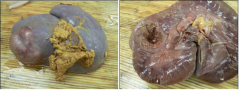
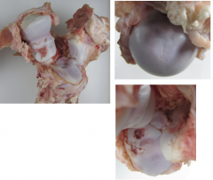
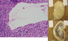
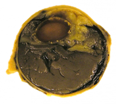
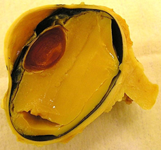
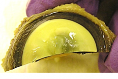
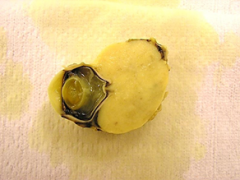
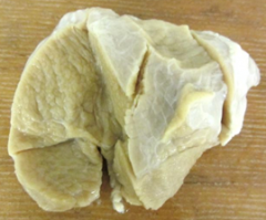
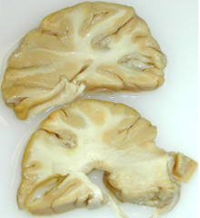
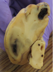
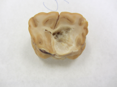
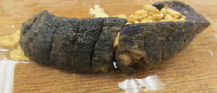
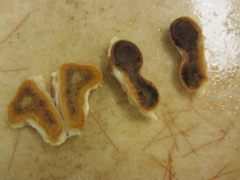
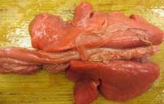
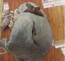
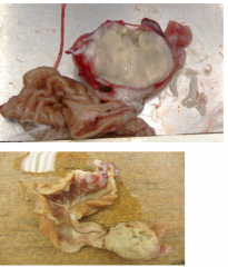
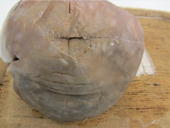
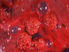
MDx = left kidney: chronic focal renal nodule DDx = neoplastic (renal carcinoma, fibroma, fibrosarcoma, hemangiosarcoma), metastatic site (not likely with focal distribution), inflammation (abscess, granuloma) |
|
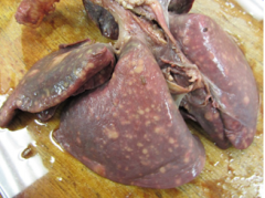
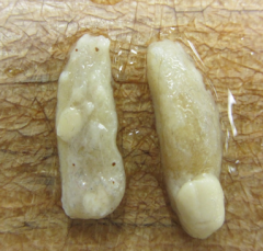
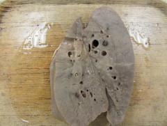
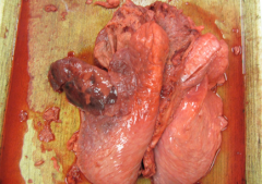
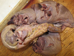
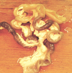
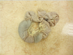
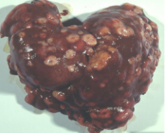
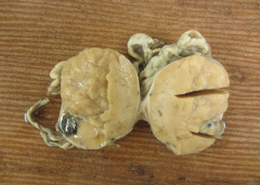
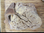
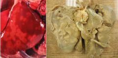
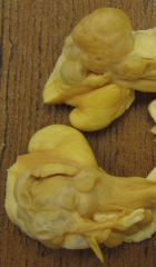
Lung with multifocal to coalescing white foci. nodules with firm, friable, cream colored caseous material. Mdx? Ddx? |
mdx = multifocal pulmonary nodules
ddx= granuloma/pyogranuloma (neosporosis, toxo., blasto., coccidiomycosis, tuberculosis), neoplasia, abscesses |
|
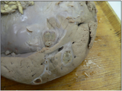
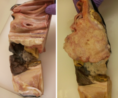
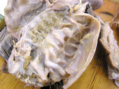
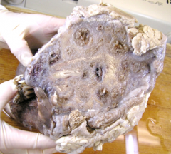
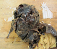
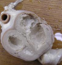
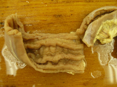
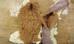
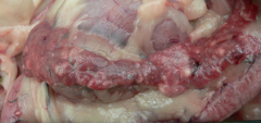
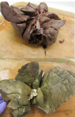
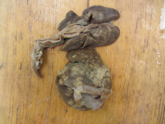
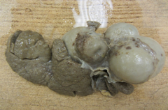
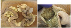
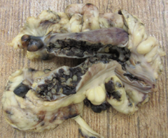
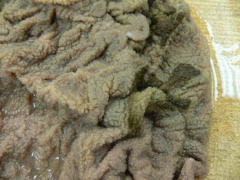
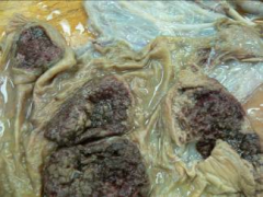
on the cut surface of the liver, there are multiple cavitations lined by tan friable material lined by prominent band of fibrous tissue. Mdx? |
Mdx = liver: chronic multifocal hepatic abscesses
|
|
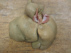
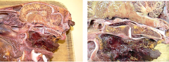
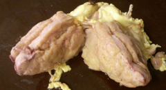
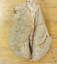
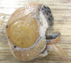
Liver is swollen, pale, yellow, greasy, friable, and has accentuated lobar pattern. Mdx? |
Mdx= liver: chronic diffuse hepatic lipidosis |
|
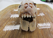
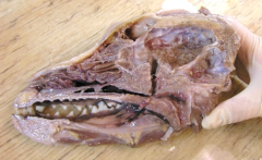
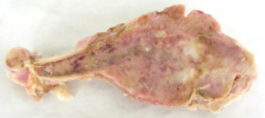
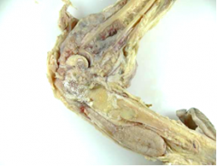
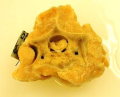
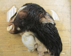
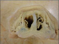
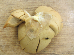
large firm pale mass, no capsule, not well demarcated. extends from gingival mucosa through maxilla, & fills sinuses/turbinates. Mdx? Ddx? |
Mdx= severe, chronic, locally extensive maxillary mass
Ddx= SCC, fibrosarc, osteosarc, malignant melanoma |
|
|
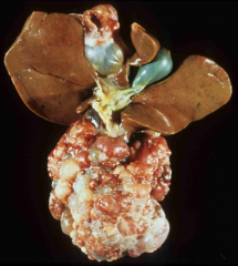
Provide a possible pathogenesis for epistaxis related to chronic multifocal hepatic abscesses in a feedlot steer. |
rumenitis in feedlot steer --> fusibacterium necrophorum invades rumen wall --> enters portal circulation --> forms liver abscesses --> erode through caudal vena cava --> pus and bacteria embolized to lungs --> vascular thrombi/hypertension/and pulmonary aneurysm --> acute epistaxis. |
|
|
provide a possible mechanisms for hepatic lipidosis in an anorectic cat. |
1. increased uptake/synthesis of lipids (eg increased mobilization of FA from peripheral fat droplets during fasting) 2. decreased hepatic output of lipids (impaired vldl formation and release) 3. decreased fatty acid oxidation |
|
|
What are ddx for maxillary neoplasia? |
fibrosarcoma, osteosarcoma, malignant melanoma, ameloblastoma, odontoma, oral lymphoma. |
|
|
What are some risk factors for osteochondrosis? |
tends to affect young, male, rapidly growing, large breeds of animals, especially dogs, cattle, horses, pigs, chickens and turkeys. |
|
|
What is the pathogenesis of fibrous osteodystrophy? |
decreased vitamin D --> decreased intestinal calcium absorption --> decreased serum calcium --> increased PTH release --> increased osteoclastic resorption from bone to increase serum Ca --> decreased bone density --> replaced by fibrous CT |
|
|
what is the common name for cervical vertebral stenosis and instability? |
wobblers syndrome |
|
|
what is the common name for mandibular osteomyelitis? |
lumpy jaw |
|
|
what is the pathogenesis of suppurative mandibular osteomyelitis? |
trauma to oral cavity --> Actinomyces bovis (oral commensal) enters--> persistent, progressive infection and inflammation of mandible --> necrosis --> proliferation of new bone and formation of granulation tissue |
|
|
what are potential ddx for canine bone neoplasm? |
neoplastic: osteosarc, chondrosarc, fibrosarc, histiocytic sarcoma, hemangiosarc, plasma cell myeloma, metastatic carcinoma
non-neoplastic: chronic ostemyelitis, fx bone callus. |
|
|
Provide a pathogenesis for necrotizing osteomyelitis as a sequelae of chronic laminaits |
downward rotation of p3 penetrates sole --> solar abscess forms --> inflammation and infection extends upwards into P3 --> osteomyelitis |
|
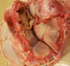
articular cartilage with areas of separation and focal cavitation with reddened subchondral bone. thick fibrotic jt capsule. Mdx? |
Mdx = humerus, multifocal osteochondrosis dissecans |
|
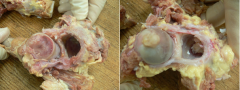
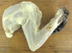
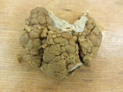
bilateral enlargement of left cd mandible. proliferation of mandibular cortical bone. MDx? DDx? |
Mdx: mandibular hyperostosis, bilateral locally extensive, chronic severe
Ddx: fibrous osteodystrophy, osteosarcoma, chronic osteomyelitis |
|
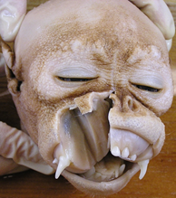
maxilla thick, firm, but not hard. bulging gum-line on upper palate, teeth protrude at 45 degree angle, jaw unable to close. Mdx? |
Diffuse fibrous osteodystrophy |
|
ribs are unevenly thickened, string of beads along costochondral joints. soft and pliable. widened tibial physes and metaphysis. Mdx? |
fibrous osteodystrophy and physeal chondrodystophy |
|
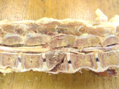
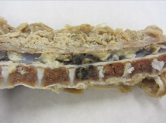
focal cavitation in sternebrae, filled with bone and CT capsule. spicules of bone radiate outward forming a firm mass. tract from fragment to larger cavity. mdx? |
sternum: chronic focal purulent osteomyelits with sequestrum and hyperostosis |
|
cervical vertebrae narrowed at c3-4. can decrease canal width by 60% by flexing joint. Mdx? |
mdx= vertebrae, C3-4: cervical vertebral stenosis and cervical vertebral instability |
|
ribs: multifocal coalescing firm raised white masses surrounded by congestion & hemorrhage. spine: complete fx at L5 surrounded by hemorrhage & fibrin. Mdx? |
ribs: t2-t5 fracture callus vertebrae: complete fx through L5 |
|
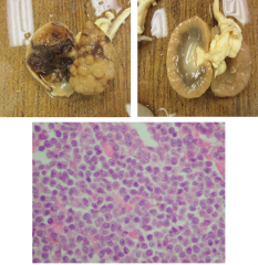
tissue from 2 yo mix breed MN dog, swollen joints, anorexia, lethargy, blindness. Mdx? |
mdx calvarium: chronci locally extensive pyogranulomatous osteomyelitis |
|
histology from 2 yo k9 with extensive exudate covering the ventrolateral calvarium surface, the bones of which are cavitated and exudate filled. Mdx? |
mdx: intralesional yest bodies. (Cocidiomycosis) |
|
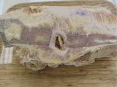
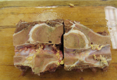
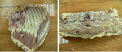
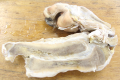
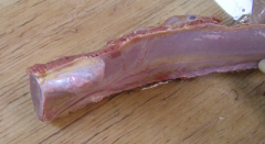
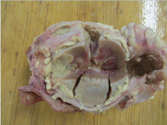
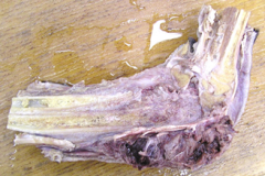
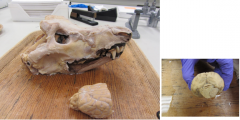
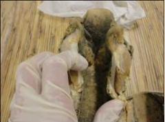
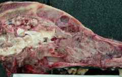
mandible from holstein cow sent to slaughter. Mdx? |
Mdx: chronic multifocal suppurative mandibular osteomyelitis |
|
Tissue from 5 mo great dane puppy with forelimb lameness, fever, bilateral painful, swollen distal antebrachia. mdx? |
mdx radius and ulna: chronic necrosuppurative metaphyseal osteomyelitis with cortical hyperostosis and periosteal fibrosis |
|
Tissue from 10 yo mn k9 with 4 month hx of right forelimb lameness and swelling. mdx? ddx? |
mdx- right humerus: chronic locally extensive osteosarcoma
ddx- osteosarcoma, chronic osteomyelitis, fracture callus, fibrosarcoma, hemangiosarcoma, plasma cell myeloma, metastatic carcinoma. |
|
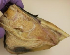
Tissue from horse with acute, weight bearing lameness. mdx? |
mdx: acute, severe, comminuted fx of the right radial carpal bone with acute multifocal ulceration of articular cartilage. |
|
tissue from horse wtih long hx of lameness. mdx |
chronic laminitis with ventral rotation of P3 |
|

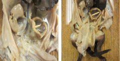
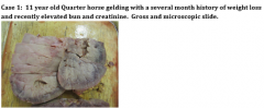
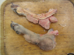
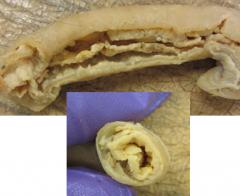
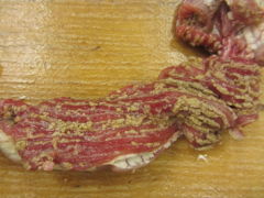
tissue from 9 yo QH gelding with laceration on plantar surface of distal LH foot. MDx? |
left hind foot: chronic tenosynovitis of the superficial digital flexor, deep digital flexor, and suspensory ligament. Chronic proliferative arthritis and bursitis of the distal interphalangeal joint and navicular bursa |
|
tissue from 12 yo k9 euthanized for unrelated neoplasm. mdx? |
coxofemoral joint: bilateral osteoarthritis, severe, diffuse chronic. |
|
tissue from 9 yo mastiff with hx of right rear leg lameness. mdx? |
right tarsus: locally extensive neoplasia |
|
tissue from 2 yo tb filly presented for necropsy. |
rib: multifocal osteochondromatosis (multiple osteochondromas) |
|
tissue from steer from group of 50 feedlot steers that had gone through an outbreak of respiratory dz a few weeks before. mdx? |
stifle: severe subacute locally extensive fibrinous arthritis. |
|
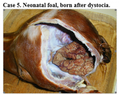
tissue from 7 mo foal. foal was anorectic. Mdx? DDx? |
Mdx= diffuse hepatic and renal lipidosis
ddx = mobilization of lipid secondary to anorexia; hypoxia' protein deficiency |
|
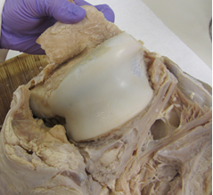
1 yo persian cat with recent weight loss, lethargy, anorexia. moderate-severe dehydration. mdx? congential vs acquired? |
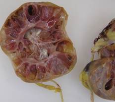
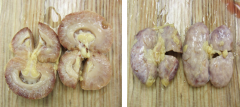
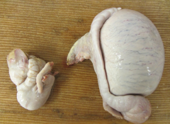
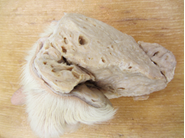
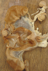
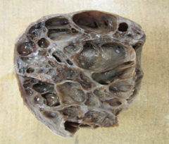
mdx= bilateral multifocal renal cysts (polycystic kidney)
Congenital- they are many and large, with normal intervening renal parenchyma. acquired cysts are fewer and smaller and associated with fibrosis. |
|
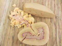
from 6 mo foal with heart murmur. mdx? causes? |
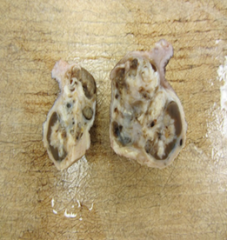
mdx = kidney: renal infarcs, multiple, acute
cause = sepsis will cause endothelial cell damage --> increased coagulation/thrombi/infarcts. endocarditis of left AV can cause emboli --> renal artery infarction |
|
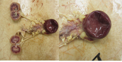
4 yo ewe with hx of wt loss and enlarged prescap. LN. MDx? |
mdx = kidney: multiple chronic renal abscesses |
|
tissue from 11 yo MN DSH. 2 day hx of constipation and 5 day hx of anorexia. bx reveals densely cellular infiltrative sheet of uniform round cells expanding and disrupting kidney parenchyma. mdx? ddx? |
mdx= multifocal renal cortical nodules (multifocal nodular renomegaly)
ddx= renal lymphoma, FIP, metastatic neoplasm (eg carcinoma, mct) |
|
tissue from 2 yo cat. microscopically the cortex is multifocally expanded by macrophage and pmn infiltrates centered on blood vessels. mdx? ddx? |
mdx = kidney - vasculocentric nephritis, diffuse chronic pyogranulomatous severe.
ddx - FIP, lymphosarc., toxoplasmosis. |
|
tissue from 7 yo lab. anorexic, lethargic, and had multifocal oral lesions. mdx? |
mdx- chronic diffuse bilateral renal fibrosis |
|
|
what is the pathogenesis of oral ulcers secondary to azotemia? |
azotemia --> damage small vessels (fibrinoid necrosis) --> multifocal mucosal ischemia and necrosis.
azotemia --> urea accumulate in saliva --> urea-splitting bacteria metabolize urea to ammonia --> mucosal damage |
|
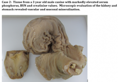
mdx? |
stomach: chronic multifocal gastritis with mineralization.
tongue: multifocal glossal ulcers (multifocal ulcerative glossitis) |
|
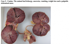
mdx? ddx? |
mdx = bilateral diffuse renal mass/renomegaly
ddx= lymphosarc., renal cell carcinoma, tcc, granulomatous or pyogranulomatous nephritis |
|
tissue from neonatal puppy. mdx? cause? |
mdx = urinary bladder distention, bilateral hydronephrosis and hydroureter.
cause; congenital obstruction in lower urinary tract |
|
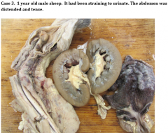
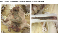
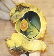
tissues from a 4.5 yo MI DSH that had been straining to urinate unsuccessfully. mdx? cause? |
mdx= acute diffuse serosal hemorrhagic cystitis and urinary bladder distension
cause= urethral obstruction secondary to urolithiasis |
|
mdx?
|
chronic multifocal to diffuse granulomatous interstitial nephritis with nematodes (halicephalobus sp.) |
|
most likely dx? |
urinary bladder: TCC |
|
mdx? |
-diffuse hydronephrosis and focal hemorrhagic pyelonephritis -acute diffuse necrohemorrhagic cystitis - acute focal necrotizing urethritis - acute locally extensive fibrinohemorrhagic peripenile cellulitis |
|
mdx? ddx? |
acute, multifocal petechial and ecchymotic hemorrhages
ddx: porcine circavirus 2, salmonellosis, acute erysipelas, african swine fever, classical swine fever. |
|
|
- Bladder: fibrinohemorrhagic cystitis, acute, locally extensive - utrethra: fibrionhemorrhagic urethritis, acute locally extensive - kidney: fibrinopurulent pyelonephritis, acute, diffuse, hydronephrosis - ureter: bilateral hydroureter |
|
tissue from 5 yo clyde. gelding w/ hx of right eye discharge. mdx? |
squamous cell carcinoma of the palpebral conjunctiva and 3rd eyelid |
|
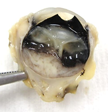
5 yo MN lab after unilateral enucleation. mdx? |
intraocular (uveal) melanoma with retinal detachment, intraocular hemorrhage and uveitis with hypopyon. |
|
tissue from 12 y.o. mare with hx of recurrent uveitis. tx with dexsp implant. recently, horse had been squinting both eyes and had purulent d/c from one eye. mdx? |
chronic diffuse fibrinous endophthalmitis with posterior syechiae and corneal neovascularization |
|
tissue from 4 yo jersey bull euthanized for unrelated resons. mdx? |
acute focal corneal ulcer; acute diffuse hyphema; acute multifocal subretinal hemorrhage with retinal detachment. |
|
tissue from 8 yo SF lab that underwent unilateral enucleation of the eye for exopthalmos. mdx? |
retrobulbar meningioma |
|
tissue from newborn crossbreed male calf with arthrogryposis of all four limbs. mdx? |
micropthalmia |
|
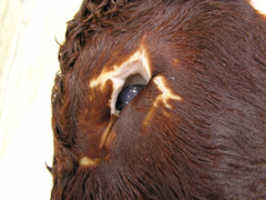
tissue from 5 yo MN DSH with hx of acute onset pain and hyperemia of the right eye. mdx? |
right eye: chronic pyogranulomatous endophthalmitis (or panophthalmitis) |
|
|
what is the pathogenesis of equine recurrent uveitis? |
leptospira infection --> T and B cell response mounted --> t-cells and ab cross-react with ag in the uvea --> lymphoplasmacytic uveitis. recurrent bouts of inflammation with intermittent quiescent periods. |
|
|
what are the immediate consequences of subretinal hemorrhage? |
retina lifted away from RPE, causing blurred to absent bision. |
|
|
what are long term consequences of subretinal hemorrhage? |
loss of oxygen/nutrients and removal of waste products to the RPE. loss of contact between the photoreceptor cells and RPE causes ischemia, and if not resolved, leads to permanent blindness. |
|
|
what are ddx for micropthalmia in a calf? |
- BVD (numerous congenital issues) - inherited genetic defect - maternal vitamin A deficiency (causes optic nerve hypoplasia) |
|
|
ddx for chronic pyogranulomatous endophthalmitis? |
- feline post-traumatic ocular sarcoma - blastomyces dermatitidis - FIP - lymphoma |
|
tissue from 4 yo MN great dane with swelling of the right fore limb and lameness. mdx? ddx? |
distal radius and adjacent muscle, rhabdomyosarcoma
ddx: osteosarc, chondrosarc, histiocytic sarc, synovial cell sarc, rhabdomyoarc. |
|
tissue from 10 yo SF DSH w/ Hx of chronic D+ and poor appetite. mdx? |
skeletal muscle, atrophy, right forelimb, diffuse, severe. |
|
tissue from veal calf, approximately 2.5 weeks old, w/ hx of bilateral rear leg weakness and ataxia. mdx? |
skeletal muscle, myodegeneration, diffuse, chronic, severe |
|
|
pathogenesis of white muscle dz? |
vitamin E/selenium deficiency --> free-radicals initiation lipid peroxidation --> uncontrolled ca influx to the myocyte --> mitochondria unable to sequester sufficient ca, and are lost --> free intracellular ca initiates contraction --> ATP depletion --> degeneration and coagulation of myofibrils --> macrophages remove. regeneration can occur. |
|
tissue from 8 yo giant schnauzer. dx with intracranial schwannoma adherent to the dura and adjacent to the trigeminal ggl. mdx? |
temporalis, masseter muscles- chronic diffuse atrophy.
right eye- enopthalmos
medulla oblongata - nodular mass (schwannoma based on hx) |
|
tissue from a 1 week old female aberdeen angus calf which had been ataxic since birth. mdx? causes? |
mdx: cerebellar hypoplasia and dysplasia
causes: infection with BVD between 125-150 days, akabane virus, blue tongue virus, and wesselbron virus. |
|
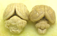
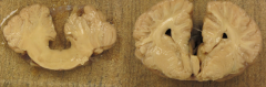
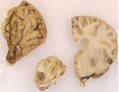
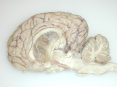
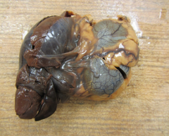
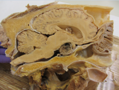
one brain is from a 3 week old female DSH kitten which had a wide base stance when standing & hypermetric gait. the other brain is normal. mdx? |
mdx: cerebellar hypoplasia
|
|
|
what is the likely pathogenesis of cerebellar hypoplasia in a kitten? |
fetal panleuk. infection --> tropism for rapidly dividing cells (ie cerebellum granular layer) --> destroys infected cells --> decreased growth and differentiation of purkinje neurons --> hypoplasia |
|
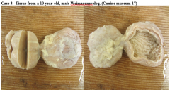
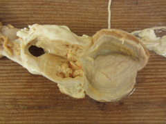
tissue from 8 mo MN weimaraner dog with ataxia and tetraparesis. mdx? |
mdx: spinal cord: syringomyelia |
|
|
what is hydromyelia? |
spinal cord cavitation lined by ependymal cell. |
|
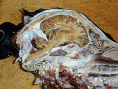
tissue from 1.5 yo QH with brief hx of neuro signs. signs started after horse fell over backwards. mdx? |
skull: basisphenoid/occipital fx or dislocation with extradural hematoma and brainstem compression. |
|
mdx? |
bilateral internal hydrocephalus, with cranial malformation |
|
tissue from QH colt unable to stand. mdx? common name? |
cerebrum, cerebellum and brain stem: aplasia of the cerebellar vermis and corpus callosum with internal hydrocephalus of lateral, 3rd, and 4th ventricles and mesencephalic aqueduct and polymicrogyria.
"dandy-walker syndrome" |
|
tissue from a heifer recently weaned and transported to a feedlot. |
multifocal acute hemorrhagic and necrotizing (or necropurulent) meningoencephalitis |
|
tissue from 4 mo MI lab that was HBC and presented with hind limb paralysis. mdx? |
focal acute complete vertebral body fx fo L5 with focal spinal cord compression adn hemorrhagic myelomalacia. |
|
tissue from 18 yo arab gelding with hx of colic. At presentation, he was seizuring, had severe nystagmus, dilated pupils, and increased RR. mdx? |
multifocal to coalescing, acute, severe necrohemorrhagic purulent meningoencephalitis and vasculitis |
|
tissue from 9 mo steer from a feedlot. molasses is fed as an energy source. this steer was found recumbent with opisthotonos & nystagmus. mdx? |
subacute multifocal cerebral polioencephalomalacia |
|
|
possible causes of polioencephalomalacia? |
- molasses is high in sulfur, which is converted by the rumen to hydrogen sulfide (a neurotoxin) - distiller's grains are high in sulfur and are frequently used as a feed additive - thiamine deficiency |
|
tissue from a 5 yo golden HBC. initial the dog was normal with minor injuries, but developed CNS signs (ataxia, opisthotonos & nystagmus). mdx? |
acute multifocal cerebral and mesencephalic hemorrhage |
|
tissue from a 15 yo oldenberg mare w/ a hx of 3 neuro episodes w/in the past year. mdx? ddx? |
right lateral ventricle: cholesterol granuloma (or cholesteatoma) with mild internal hydrocephalus.
ddx: ependymoma, choroid plexus carcinoma or choroid plexus papilloma. |
|
tissue from 9 yo SF pug presented for necropsy. mdx? |
vertebral column: disk protrusions, multiple |
|
tissue from a cat found as an incidental finding at necropsy. mdx? |
meningoma |
|
tissue from 10 yo k9 w/ pacing gait, truncal sway, anisocoria (r > left), positional nystagmus. change in behavior, eating/drinking habits. mdx? ddx? |
focal cerebral mass
ddx: oligodendroglioma, astrocytoma, ependymoma, granulomatous encephalitis. |
|
tissue from 8 yo lab with progressive HL weakness, ataxia, pyrexia, lethargy, anorexia and tachypnea. mdx? |
spinal cord: metastatic hemangiosarcoma |
|
tissue from dog w/ swelling of the distal limb and lameness. mdx? ddx? |
superficial digital flexor tendon- chronic focal mass
ddx: fibrosarc., synovial cell sarcoma of tendon sheath, granulomatous tendonitis, schwannoma |
|
tissue from 12 yo goat with 1 month hx of caudal cervical mass. mdx? |
intra-thoracic mass, cranial mediastinum (thymoma) |
|
tissue from an 8 yo samoyed. mdx? ddx? |
multifocal splenic nodules with hemosiderin plaques.
ddx: hemangioma, hemangiosarcoma, hematoma, nodular hyperplasia, histiocytic sarcoma, lymphoma. |
|
tissue from 9 yo lab with abdominal distention. mdx? |
spleen: locally extensive splenic mass |
|
tissue from 8 mo MI beagle. mdx? ddx? |
retropharyngeal lymph node: severe chronic diffuse suppurative bilateral lymphadenitis.
ddx= lymphoma, granulomatous lymphadenitis, metastatis neoplasia. |
|
tissue from 9 yo SF GSD. Mdx? Ddx? |
severe, diffuse splenomegaly
ddx: splenic congestion, extramedullary hematopoiesis, splenic macrophage hyperplasia, leukemia, or combination of the above. |
|
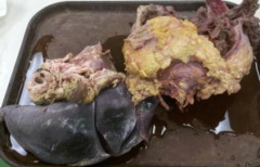
tissue from 4 yo holstein with chronic wt loss and poor condition adn palpable prescap. LN. mdx? |
lymph node: chronic diffuse severe lymphadenomegaly - lymphoma
abomasum: lymphosarcoma with multiple mucosal ulcers
|
|
tissue from 4 yo goat with enlarged LN of head and neck. mdx? |
lymph node: caseous lymphadenitis, diffuse, chronic |
|
tissue from newborn goat kid. mdx? |
thyroid: bilateral hypertrophy, severe, diffuse, chronic with prognathism and alopecia |
|
|
what is the pathogenesis of congenital thyroid hypertrophy? |
dam fed iodine deficient diet --> kid thyroid fails to produce T3/T4 --> lack of Hormone in circulation stimulates hypothalamus to produce TSHRH --> TSHRH stimulates thyroid to produce TSH --> TSH simulate thyroid epithelial hyperplasia. |
|
tissue from 7 yo Sf dog with lump in the ventral cervical region. mass made up of pleomorphic follicular epithelial cells invading thyroid capsule. mdx? |
mdx: thryoid carcinoma, unilateral |
|
|
what is the biologic behavior of a thyroid carcinoma? |
invasive, and can infiltrate local tissues such as blood vessels, esophagus, and trachea. metastasis is common and usually seen in the lungs. poor prognosis. |
|
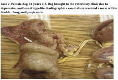
tissue from 8 yo MN poodle presenting with lethargy. AUS revealed a mass near the left kidney. mdx? |
adrenal gland: pheochromocytoma with invasion of the vena cava |
|
tissue from 13 yo SF lab euthanized due to metastatic hemangiosarcoma. mdx? |
adrenal gland: nodular cortical hyperplasia and focal adrenocortical adenoma |
|
tissue from 11 yo shetland pony euthanized due to severe laminitis. mdx? |
pituitary adenoma (likely a chromophobe adenoma of pars intermedia)
cholesteatoma of left lateral ventricle with mild hydrocephalus |
|
|
where do pituitary gland masses common originate from in horses? |
pars intermedia |
|
|
which clinical signs are associated with pituitary gland masses in horses? |
pu, pd, polyphagia, somnolence, hypertrichosis, hirsuitism, hyperhidrosis |
|
tissue from 11 yo lab with hx of recent D+. exploratory lead to left adrenalectomy, right was slightly enlarged. mdx? |
adrenal cortical ademona |
|
tissue from 9 yo rottweiler with pulmonary thromboembolism and hepatomegaly. mdx? |
adrenal glands: bilateral diffuse adrenal cortical hyperplasia. pheochromocytoma, left gland. |
|
tissue from 10 yo dog with hypercalemia |
unilateral parathyroid mass (adenoma)
multifocal parathyroid atrophy
|
|
tissue from a steer with dyspnea and a 'red nose'. mdx? |
trachea and larynx: severe, acute, diffuse, fibrinonecrotic (and ulcerative) laryngotracheitis |
|
8 yo SF lab with 3 month hx of chronic nasal discharge, that improved slightly after abx treatment, and epistaxis. the animal recently developled seizures. mdx? ddx? |
nasal cavity: locally invasive mass
ddx: neoplasia (adenocarcinoma, osteosarcoma, chondrosarc, fibrosarc), granulomatous rhinitis (aspergillosis, fb) |
|
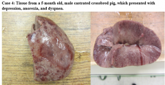
tissue from 2.5 week old pig with swollen left hock and swollen, red, inflamed, umbilicus. md? |
multifocal chronic pulmonary abscess |
|
tissue from 18 yo pony with hx of cough and respiratory distress. mdx? |
chronic bronchopneumonia with chronic pleural fibrosis (chronic, fibrosing, pleuritis) |
|
|
indicators of chronicity in lungs? |
- lesion is white vs red and inflamed - interlobular septa expanded by fibrous ct - pleural lesions cannot easily be removed with manual manipulation (fibrosing) |
|
tissue from 3 wk old Holstein calf. when it is born, it was found in the barn w/o farm staff observing the delivery. it was somewhat dull after birth, but suckled readily. for the past 2 day the calf has been down in lateral recumbency. mdx? |
severe chronic bronchopneumonia with fibrosis pleural adhesions (pleural fibrosis) |
|

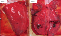
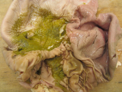
tissue from several different 12 wk old pigs. one had chronic cough and decreased growth, one had fever and acute resp. dz with cough, and 3rd had D+ with normal lungs. mdx? |
lung A: normal lung B: acute bronchopneumonia lung C: chronic bronchopneumonia |
|
|
ddx for acute bronchopneumonia in piglets? |
SIV + secondary bacterial infection (pasteurella multocida, strep. suis, H. parasuis) |
|
|
ddx for chronic bronchopneumonia in piglets? |
mycoplasma hyopneumonia + secondary bacterial infection (pasteurella multocida, strep. suis, H. parasuis) eg. enzootic pneumonia. |
|
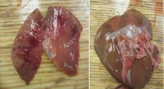
tissues from two 10 wk old pigs that are 'thumping'. mdx? |
lung 1: severe, diffuse interstitial pneumoniae lung 2: severe, diffuse interstitial pneumonia with mild/moderate bronchopneumoniae
liver: mild, multifocal hepatic fibrosis (milk spots) |
|
|
ddx for interstitial pneumonia in pigs? |
PRRSV, PCV2, S. choleraesuis, ascarid larval migration |
|
tissue from a 2 day old lamb. it was born into a clean, well-bedded stall. the umbilical cord was dipped and tied off. at necropsy, had intussussception of SI with venous infarct. mdx? |
moderate acute fibrinous pleuritis and moderate diffuse pulmonary edema |
|
tissue from a 7 day old holstein calf. Sunday morning the calf had D+/dehydration/recumbency, died on monday. mdx? |
severe fibrinous pericarditis |
|
tissue from a 12 y.o. feline with a cough that recently became bloody. mdx? |
lung: locally extensive multifocal pulmonary nodules
heart: hypertrophic cardiomyopathy |
|
tissue from a hunting dog that resided in the ohio-mississippi river valley and lived during the winter with her owner in arizona. the dog recently went into terminal respiratory distress. mdx? |
lung: chronic diffuse interstitial pneumonia with multifocal pulmonary nodules (pyogranulomas)
trachea: tracheal collapse, locally extensive mild-moderate
tracheobronchial lymph node: marked lymphadenomegaly |
|
tissue from a steer recently placed on lush pasture. mdx? |
interstitial pneumonia, diffuse acute with interlobular edema. |
|
|
what is the pathogenesis of bovine interstitial pneumonia secondary to lush pasture ingestion? |
ingest L-tryptophan on lush pasture --> rumen bugs convert l-tryptopha to 3 methyl indole --> clara cells convert 3MI to 3-methyleneindolenine --> toxic to type 1 cells and endothelial cells --> diffuse pneumocyte necrosis --> acute interstitial pneumonia |
|
tissue from pig that had been sneezing and coughing with nasal discharge. mdx? |
nasal cavity: chronic nasal conchae atrophy (or atrophic rhinitis) with septal deviation |
|
|
pathogenesis of atrophic rhinitis? |
bordetella bronchiseptica + pasteurella multocida colonize nasal cavity --> P. multocida cytotoxins inhibit osteoblastic activity and promote osteoclastic activity --> nasal bone resportion. |
|
tissue from a pig from a production facility undergoing an outbreak of respiratory dz with 12-15% of pigs effected. mdx? most likely cause? |
severe acute diffuse fibrinous adn necrohemorrhagic pleuropneumonia or necrohemorrhagic bronchopneumonia and fibrinous pleuritis.
most likely cause = actinobacillus pleuropneumonia (APP) |
|
tissue from two 80# pigs. one lung from animal has acute outbreak of rapidly spreading febrile dz w/ high morbidity and barking cough. one lung from a herd w/ slow progressive problem, decreased gain, and thumps. mdx |
lung A: severe diffuse interstitial pneumonia lung B: moderate, patchy (lobar) anteroventral consolidation |
|
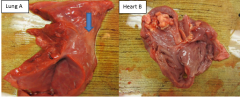
fresh tissue from two pigs. there has been an ongoing problem in the nursery. tissues from pig a are from an animal that was acutely anorexic and depressed w/ a high fever. tissues from pig B are from an animal that has stopped growing. mdx? |
lung A: severe acute fibrinous pleuritis
heart B: focally extensive epicardial fibrosis with chronic fibrous pericardia adhesions. severe chronic vegetative valvular endocarditis of the right AV valve and pulmonic valve |
|
|
how are parathyroid adenomas linked to hypercalcemia? |
parathyroid adenoma are functional --> produce PTH (without regards to signaling)--> hypercalcemia
at same time, the non-affected glands atrophy (down regulated by hypercalcemia) |
|
tissue from 400# bovine with wt loss and cough. mdx? |
severe chronic abscessing pneumonia (bronchopneumonia) with pleural and interlobular fibrosis. |
|
tissue from a cat w/ respiratory difficulty. rads demonstrated a ST opacity that obscured the heart. thoracocentesis showed PMN and bacteria. mdx? |
right lung: moderate acute fibrinous pleuritis w/ moderate diffuse atelectasis. |
|
tissue from a 10 yo FI golden that presented with hx of dyspnea, wt loss, weakness, and occasional collapse. mdx? ddx? |
lung: multifocal pulmonary nodules, chronic severe
ddx: metastatic neoplasm granulomatous pneumonia (fungal) |
|
tissue from 8 day old goat kid that has been growing poorly. developed resp distress and died. mdx? |
severe maxillary palatoschisis
severe acute hemorrhagic bronchopneumonia |
|
tissue from an 8 week old goat with fever, cough, and excessive salivation. mdx? |
severe, acute, fibrinonecrotic laryngitis |
|
tissue from 7 yo SF k9. developed progressive breathign problems, cough, and loss of appetite. of PE, there are decreased heart and sounds. afebrile. mdx? |
right middle lung lobe torsion with venous infarction and severe atelectasis |
|
|
what is the pathogenesis of serosanguinous thoracic effusion secondary to lung lobe torsion? |
torsion --> artery continues to pump while vein is collapsed --> venous congestion --> venous infarction --> fluid leaks from necrotic lung --> thoracic effusion --> respiratory distress |
|
tissue from 2 yo angus cow with 11 day hx of mild/moderate epistaxis (bilateral). mdx? ddx? |
focally extensive nasal mass
ddx= nasal carcinoma, nasal sarcoma, nasal lymphoma, hematoma, nasal granulmoa |
|
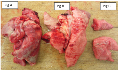
lung from three separate piglets. mdx? |
A: moderate, acute, diffuse, fibrinous pleuritis, pericarditis, & epicarditis B: moderate, chronic/active (hemorrhagic) bronchopneumonia C: normal lung |
|
tissue from race horse that was off feed. mdx? |
glandular and non-glandular stomach: acute multifocal to coalescing ulcerative gastritis (gastric ulcers) |
|
|
predisposing factors for equine gastric ulcers? |
infrequent, high CHO meals inadequate/infrequent access to pasture/forage heavy training schedules high stress excessive use of drugs, especially NSAIDS |
|
tissue from a pig that had respiratory signs a couple of days ago and was recently found dead. a few other pigs are pale and have melena. mdx? |
stomach: severe focal chronic ulceration of the pars esophagea. or, severe locally extensive chronic ulcerative gastritis of the pars esophagea. |
|
|
what are predisposing factors for pig gastric ulcers? |
stress respiratory tract disease small feed particle size irregular feeding patterns high unsaturated fat, low fiber, high energy diet |
|
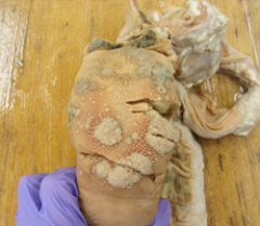
tissue from yearling replacement heifer in poor body condition. mdx? cause? |
severe disseminated chronic nodular abomasitis
cause: ostertagia ostertagii |
|
|
what is the pathogenesis of abomasitis secondary to ostertagia ostertagii? |
larvae destroy parietal cells of mucosal glands --> decreased HCL and pepsinogen production --> abomasal ph increases from 2-7 --> inable to digest protein --> bacterial overgrowth (pH and undigested feedstuff) --> D+, hypoproteinemia, wt loss |
|
tissue from a 9 day old piglet with severe D+. mdx? |
small intestine: severe acute locally extensive fibrinonecrotic enteritis |
|
|
what can cause fibrinonecrotic enteritis in piglets? |
necrotic coccidiosis subacute clostridium perfringens type C salmonella (not common in suckling pigs) |
|
|
pathogenesis of fibrinonecrotic enteritis in pigs? |
vili epithelial cell necrosis --> loss of superficial vili exceeds ability to replace and repair defect --> ulceration of vili tip --> fibrinous edudate |
|
tissue from 120# grow/finish pig that presented w/ a hx of D+ and reduced wt gain. mdx? common name? cause? |
small intestine and colon: severe, chronic, diffuse fibrinonecrotic and proliferative enterocolitis.
common name = PPE (porcine proliferative enteropathy)
cause: lawsonia intercellularis infection |
|
tissue from a canine with V+, wt loss and inappetence. mdx? likely dx? |
stomach: locally extensive, ulcerated, mural mass, chroncic severe
likely dx? gastric adenocarcinoma |
|
|
what is the anticipated behavior of a gastric adenocarcinoma? |
locally invasive advanced dz by time clinical signs present expected to met to regional LN peritoneal implantation likely liver and spleen likely met sites
|
|
tissue from 6 mo puppy. mdx? likely cause? |
small intestine: severe acute diffuse fibrinonecrotic enteritis with serosal hemorrhage
likely cause: parvovirus enteritis |
|
|
what is the pathogenesis of parvo virus enteritis? |
virus infects rapidly dividing crypt cells --> lack of replacement of normally sloughed epithelial cells --> vilous atrophy --> denudation of surface epithelium --> malabsorption, maldigestion, fluid loss into lumen --> dehydration, hypovolemia, electrolyte imbalance, secondary sepsis |
|
tissue from a 6 week old holstein calf with a hx of failure to thrive and gain weight, lethargy and fever. mdx? most likely pathogen? |
small intestine and colon: locally extensive, fibrinous (fibrinonecrotic) enterocolitis
likely cause: salmonella (typhimurium, newport, or dublin) |
|
tissue from a 1 yo bovine. mdx? likely etiologic agent? |
glossitis, pharyngitis, and esophagitis: proliferative multifocal chronic
cause: bovine papular stomatitis virus |
|
tissue from a 10 yo TB mare with hx of severe D+. she has now become progressively lame on forefeet. mdx? cause? |
severe diffuse fibrinonecrotic and multifocally ulcerative colitis with associated fibrinous peritonitis.
cause: salmonella |
|
tissue from a 4 yo SF doberman w/ 2 day hx of V+, lethargy, inappetence. owner reported 3 V+ episodes overnight. mdx? |
focal obstructing intestinal FB with segmental venous infarction of the small intestine. |
|
tissue from a 5 mo cat that died suddenly. mdx? likely cause? |
small intestine: focally extensive, segmental, acute necrohemorrhagic (or fibrinonecrotic and hemorrhagic) enteritis
cause: feline panleuk. virus |
|
tissue from a 6 month old foal with D+. mdx? |
small intestine: moderate to severe chronic diffuse proliferative enteropathy |
|
small intestine from a 2 yo cow w/ profuse watery D+ and progressive emaciation. mdx? likely cause? |
small intestine: severe, diffuse chronic granulomatous enteritis
cause: Mycobacterium avium ssp paratuberculosis |
|
|
pathogenesis of MAP in cow? |
fecal-oral infection route --> primary infection of SI at ileal M cells --> granulomatous enteritis --> infected macrophages release inflammatory mediators--> vascular permeability --> fluid, protein, electrolyte loss |
|
tissue from 6 mo dog that was anorexic and not passing feces. mdx? |
intussussception with associated fibrinonecrotic enteritis |
|
tissue from 10 yo gelding with hx of colic. mdx? |
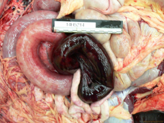
small intestine: intestinal volvulus with associated venous infarction, locally extensive, severe |
|
|
pathogenesis of intestinal volvulus in a horse? |
twisting of intestine around mesenteric root --> artery continues to pump blood in --> venous collapse causes congestion --> edema, hypoxia and subsequent necrosis of the bowel wall (venous infarction) |
|
tissues from 4 yo red angus bull. the animal was off feed for 3 days and had a fever of 106 F. mdx? |
severe chronic/active & fibrosing, granulating and fibrinous polyserositis (peritonitis and pleuritic) |
|
11 yo female cat w/ a 3 day hx of vomiting and anorexia. mdx? |
acute hemorrhagic pancreatitis |
|
|
pathogenesis of acute hemorrhagic pancreatitis? |
initiating event --> zymogen granules fuse wtih lysosomes --> trypsinogen activated to trypsin --> pancreatic secretory trypsin inhibitor overwhelmed --> trypsin activated additional digestive enzymes in pancreas--> autodigestion --> inflammation |
|
tissue from a 6 mo cat w/ hx of lethargy adn anorexia. mdx? |
intestinal serosa, chronic multifocal pyogranulomatous peritonitis (serositis) |
|
|
pathogenesis of FIP? |
exposure to FCoV --> internal mutation to FIP --> tropism for monocytes/macrophages --> systemic dissemination of virus --> weak CMI (dry FIP), absent CMI (wet FIP) --> vasocentric pyogranulomas |
|
tissue from 16 yo cat w/ cranial abdominal pain, V+, and weight loss. mdx? |
pancreas: focal, pancreatic mass with abscessation |
|
|
why is the pancreas prone to developing abscesses? |
autodigestion --> necrotic cavity --> pancreatic ducts connected to bacteria-laden intestine --> necrotic cavity + bacteria = abscess |
|
tissue from 16 yo cat that had died due to an 'accidental' gun shot wound to the chest. mdx? clinical significance? |
multifocal pancreatic nodules
sig = common incidental finding on old dogs and cats |
|
tissue from a cat that usually enjoys many meals, but has been hiding out under the stairs and has not eaten for several days. icteric. mdx? |
liver: severe hepatic lipidosis |
|
|
pathogenesis for hepatic lipidosis? |
obese cat --> anorexia (idiopathic, secondary to another dz process) --> mobilization of fatty acids --> anorexia = lack of protein for lipoprotein synthesis and an inability to remove excess lipid from liver --> lipid accumulates in liver --> hepatic lipidosis |
|
8 yo terrier-mix dog that presented with a distended abdomen. mdx? |
chronic diffuse hepatic fibrosis with nodular regeneration |
|
tissues from 15 yo mixbreed MN k9. mild abdominal pain and was slightly icteric. mdx? |
liver: multifocal hepatic nodules (likely nodular hyperplasia)
gallbladder: GB mucocele |
|
tissue from a big 6.5 yo MI dog. the animal had developed slowly progressive lethargy and depression. serum chem: alk. phos and AST elevated. BUN, TP and Alb were decreased. mdx? |
multifocal disseminated hepatic masses |
|
tissue from an aged, female lion that was euthanized due to metastatic mammary neoplasa. mdx? |
polycystic liver mass
multifocal chronic hepatic nodules |
|
tissues from a 9 mo. SF cat. slight icterus, 2 wk hx of anorexia and lethargy. mdx? ddx? |
liver: multifocal random nodules
ddx? systemic fungal infections, multifocal abscesses, metastatic neoplasia |
|
tissue from a pig that was raised on pasture. it was slaughtered for a pig roast adn at present did not have any clinical signs. mdx? common name? |
chronic multifocal and coalescing fibrosing hepatitis (hepatic fibrosis)
common name: milk spot liver |
|
tissue from an 11 yo MN cat w/ hx of lethargy, anorexia, icterus adn distended abd w/ elevated LE. mdx? dd? |
liver: biliary cystadenoma and hepatic cirrhosis
ddx: chronic toxicity, chronic cholangitis/obstruction, chronic congestion, inherited disorder of cu or fe metabolism, chronic hepatitis. |
|
tissue from a 8 yo male beagle. mdx? |
testis: focal testicular mass (based on gross appearance- interstitial/leydig cell tumor) |
|
tissue from a MI lab w/ unilateral cryptorchid testis. the dog w/ hx of gynecomastia, bilaterally symmetrical alopecia & unusual attractiveness to male dogs. mdx? |
testis: sertoli cell tumor |
|
|
bilateral, symmetrical prostatomegaly |
|
|
pathogenesis and resolution of protatomegaly? |
testis produces testosterone --> converted in prostatic epithelium to dihydrotestosterone (DTH) --> DTH regulates prostate gland growth. DTH receptors increase in number with age. androgen to estrogen ratio changes with age, makes DTH receptors more sensitive. --> glandular hyperplasia.
resolves with castration. |
|
tissue from 8 yo MN corgi with hx of urinary incontinence. mdx? ddx? |
prostate gland: malignant TCC
ddx: focal fibrinonecrotic prostatis locally extensive prostatic mass |
|
tissue from adult ram with swollen scrotum. mdx? causes? |
chronic, locally extensive abscessing or pyogranulomatous epididymitis w/ fibrous periorchitis.
causes: brucella ovis, corynebacterium pseudotuberculosis, actinobcillus seminis, trueperella pyogenes. |
|
tissue from a llama. mdx? |
penis and prepuce: chronic active locally extensive fibrinous balanoposthitis. |
|
tissue from a boar with only one testicle in the scrotum. mdx? |
unilateral testicular hypoplasia with contralateral testicular hypertrophy. |
|
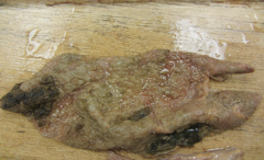
tissue from a 5 yo holstein cow. one quarter of the mammary gland is smaller and firm. the animal has reduced milk yield and high somatic cell count over the past 2 lactations. mdx? |
mammary gland: mastitis, purulent and fibrosing, chronic, diffuse, severe (or chronic abscessing mastitis) |
|
|
what are likely etiologic agents of chronic purulent mastitis? |
Trueperella pyogenes, Staphylococcus aureus |
|
tissue from a 12 yo FI feline with abdominal distention. mdx? |
focal ovarian mass hydrometra/mucometra multifocal uterine masses |
|
|
pathogenesis of muco/hydrometra? |
functional ovarian tumor --> hormonal stimulus led to increased uterine secretions --> functionally closed cervix --> fluid build up --> hydrometra/mucometra |
|
tissue fro a 14 yo FI canine with a firm mass on its ventrum. mdx? |
skin (mammary gland): mammary mass |
|
mammary gland from a 5 yo cow. appeared normal yesterday, but today is depressed, febrile, with firm hot swollen udder. treated with milk stripping, excenel and iv fluid, but died. mdx? |
mammary gland: severe locally extensive, acute hemorrhagic mastitis. |
|
|
pathogenesis of severe hemorrhagic mastitis? |
invasion of bacteria (S. aureus, E. coli) --> bacterial replication --> penetration of glandular tissue --> inflammatory response --> damage to secretory tissue and vessels from inflammation and bacterial toxins --> thrombosis and gangrenous mastitis |
|
tissue from a 10 yo FI golden. hx of V+ pu/pd. leukophilia. mdx? |
severe, chronic, diffuse, fibrinopurulent endometritis/metritis or pyometra
mild chronic, diffuse, cystic endometrial hyperplasia
multifocal paraovarian cysts |
|
|
what is the significance of paraovarian cysts? |
these are cystic distension of wolffian duct remnants. they are typically of no clinical signif. as they are not hormonally active. |
|
tissue taken at time of OHE from 7 yo mixbreed dog. mdx? |
uterus: cystic endometrial hyperplasia, chronic, diffuse. |
|
|
Pathogenesis of cystic endometrial hyperplasia? |
repeated estrogen priming (age) --> prolonged progesterone stimulation (60 day diestrus) --> progesterone increases growth of endometrial glands and secretion --> cysts develop following repeated cycles w/o pregnancy |
|
tissue from a newborn ewe lamb co-twinned to a male. mdx? |
uterine and ovarian hypoplasia, clitoral enlargement |
|
|
pathogenesis of freemartinism? |
fratenal twins --> male twin has testicular development before female twin ovarian development --> testosterone and anti-mullerian hormone reaches female via anastomoses --> inhibition of female mullarian duct development |
|
tissue from mare that had become aggressive and stud-like. tissue originated from expected location of right ovary. mdx? |
cystic ovarian mass (granulosa cell tumor of the ovary) |
|
tissue from 6 yo cow. animal died of hardware disease. mdx? |
placenta: multifocal adventitial placentation with amniotic plaques
uterus: adventitial caruncle formation |
|
tissue from cow that recently aborted. mdx? |
placenta: severe chronic diffuse placentitis with adventitious placentation. |
|
tissue from stillborn litter of puppies. mdx? most likely etiology? |
mild-moderate, acute multifocal and random, hepatic, pulmonary splenic and renal necrosis (necrotizing hepatitis, nephritis, pneumonia, and splenitis)
cause: canine herpesvirus |
|
tissue from late gestation aborted lamb. mdx? likely etiology? |
moderate, acute, multifocal necrosuppurative hepatitis
cause: campy (fetus fetus, jejuni), or flexispira rappini |
|
tissue from a 5 yo FI dog presenting for prolonged intervals between heat cycles. mdx? |
ovary: multiple follicular cysts uterus: mild, multifocal cystic endometrial hyperplasia |
|
tissue from 6 yo appy mare that aborted during 3rd trimester. mdx? |
placenta, chronic, multifocal placentitis |
|
tissue from one of several ewes that have aborted. mdx? |
moderate chronic placentitis |
|
placenta from an ewe that has aborted. mdx? cause? |
multiple foci of cotyledonary vilius necrosis (and mineralization)
cause: toxoplasmosis |