![]()
![]()
![]()
Use LEFT and RIGHT arrow keys to navigate between flashcards;
Use UP and DOWN arrow keys to flip the card;
H to show hint;
A reads text to speech;
30 Cards in this Set
- Front
- Back
|
Most common cause of acute pericarditis? |
idiopathic and viral; used interchangeably |
|
|
Most common virus associated with acute pericarditis? |
coxsackievirus B |
|
|
What are the other common causes of infective acute pericarditis |
-bacterial = pneumococcal spread most common -TB |
|
|
Noninfectious causes of acute pericarditis (8) |
1. Post MI 2. Uremia 3. Neoplastic disease 4. Radiation-induced 5. connective tissue diseases (SLE + RA) 6. Drug induced 7. Trauma 8. Post pericardiotomy |
|
|
Typical presentation of acute pericarditis in therms of: 1. Quality 2. Quantity 3. Onset 4. Duration 5. Location 6. Radiation 7. Aggravation 8. Alleviation 9. Associated symptoms |
1. Quality = Sharp pain, pleuritic 2. Quantity = Variable, often severe 3. Onset = Usually insidious 4. Duration = Hours/days 5. Location = restrosternal, precordial 6. Radiation = shoulder 7. Aggravation = breathing, lying down 8. Alleviation = Sitting up, NSAIDs 9. Associated symptoms = viral, constitutional |
|
|
What differentiates it from myocardial ischemia or infarction is that the pain of pericarditis is typically ? |
"typically sharp, pleuritic (it is aggravated by inspiration and coughing), and positional (e.g., sitting and leaning forward often lessen the discomfort)." |
|
|
Physical exam findings in acute pericarditis |
1. Tachycardia + often febrile. 2. Pericardial friction rub is pathognomonic |
|
|
1. How is the Pericardial friction rub described as sounding? 2. How is it best heard? |
1. scratching, grating, “velcro” sound |
|
|
How are WBC, CRP, and ESR levels effected in pericarditis |
All are increased |
|
|
Classic ECG finding in pericarditis? |
1. ST elevation in all leads with concave-up scooping 2. PR depression in inferior leads
|
|
|
What conduction disturbance and ventricular arrhythmias are a feature of pericarditis? |
Sinus tach is common; other atrial arrhythmias suggest other heart disease
Conduction disturbance, ventricular arrhythmias are not a feature of pericarditis |
|
|
Treatment of pericarditis |
Rest until pain free
Nonsteroidal anti-inflammatory drugs (NSAID) (indomethacin, ibuprofen, etc.) |
|
|
What is a pericardial tamponade |
Elevated intrapericardial pressure resulting from accumulation of pericardial fluid and 3. Diminished cardiac output resulting in
"A pericardial effusion that has gone bad" |
|
|
Cardiac tamponade should be suspected in any patient with |
known pericarditis, pericardial effusion, or chest trauma who develops signs and symptoms of systemic vascular congestion and decreased cardiac output |
|
|
Symptoms of pericardial tamponade are often associated with |
increased sympathetic tone (palpitations, anxiety, etc.) |
|
|
Halmark physical exam finding of pericardial tamponade |
Becks Triad: |
|
|
Other physical exam findings of pericardial tamponade |
1. Tachycardia |
|
|
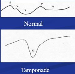
What does the pressure tracing of the pericardium normally look like? In tamponade? |
|
|
|
What is Increased pulsus paradoxus |
Normally the SBP decreases <10mmHg on inspiration due to increased venous return increasing RV size, which decreases LV size b/c of the septum bulging = decreased SV = decreased SBP
In tamponade the decrease is > 10-15mmHg because the space for the RV to expand is smaller, resulting in more of a bulge into the LV |
|
|
ECG findings in pericardial tamponade? |
1. low voltage, sinus tachycardia 2. electrical alternans (high spec, low sens) |
|
|
Echo findings in pericardial tamponade? |
1. shows RA or RV diastolic collapse, respiratory variation in transvalvular flow 2. usually a dilated inferior vena cava |
|
|
Treatment of pericardial tamponade |
1. Administer intravenous fluids and vasopressors (i.e., dopamine) to stabilize BP while awaiting definitive treatment |
|
|
Mild pericardial constriction symptoms? |
Related to elevated right heart pressures:
Ascites, edema, hepatic congestion |
|
|
More severe pericardial constriction symptoms? |
signs of left heart failure appear:
dyspnea, cough, PND, weight loss, fatigue |
|
|
Physical Exam findings of constrictive pericarditis (7) |
1. Elevated JVP 2. Kussmaul's sign 3. Pericardial knock 4. hepatosplenomegaly 5. ascites 6. edema 7. Pulsus paradoxus |
|
|
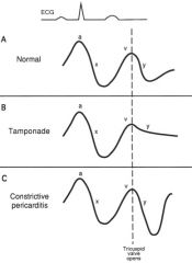
Jugular venous waveform changes with constrictive pericarditis compared to tamponade |
|
|
|
Kussumaul sign? |
Normally deep breath = decreased intrathoracic pressure = increased venous return = volume in R atria increases but CVP falls and Jugular veins collapse
Kussmauls = on inspiration veins dont fall or they can actually increase —> breathing increases venous return but it has nowhere to go so it just engorges the venous system |
|
|
How can the diagnosis of constrictive pericarditis be confirmed? findings (4)? |
by cardiac catheterization:
3. Prominent Y-descent 4. discordance between RV and LV systolic BP: RV SP rises with inspiration; LV decreases |
|
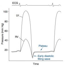
What does this pattern represent? |
This pattern reflects blood flow into the ventricles at the very onset of diastole, just after the tricuspid and mitral valves open, followed by sudden cessation of filling as further expansion of the ventricles is arrested by the surrounding rigid (constricted) pericardium.
Plateau = square root sign |
|
|
Treatment of constrictive pericarditis |
1. Treatment of right heart failure: |