Reading...
![]()
Play button
![]()
Play button
![]()
Use LEFT and RIGHT arrow keys to navigate between flashcards;
Use UP and DOWN arrow keys to flip the card;
H to show hint;
A reads text to speech;
28 Cards in this Set
- Front
- Back
|
Defining the foregut, midgut and hindgut
|
Foregut: esophagus, stomach, half of the duodenum
Midgut: most of the small intestine (second half of the duodenum, jejunum and ileum) and half of the large intestine (caecum, appendix, ascending and transvsere colon) Hindgut: Consists of the remainder of the transverse colon, descending and sigmoid colon, rectum, part of anal canal |
|
|
Arterial blood supply defines the foregut, midgut and hindgut
|
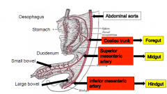
The blood supply, venous drainage and innervation to the gut is delivered from the posterior abdominal wall via the mesentery
|
|
|
Root of the Mesentery
|
The root of the mesentery is 15 cm long. It suspends the jejunun and the ileum from the post abdominal wall
|
|
|
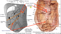
Peritoneum covering retroperitoneal structures
|
|
|
|
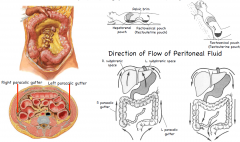
Paracolic Gutters
|
Paracolic gutters are important in flow of peritoneal fluid (and inflammatory material/cancer cells).
Greater absorption of peritoneal fluid by peritoneum in upper abdomen. Patients with intraperitoneal infection are placed in inclined position fluid gravitates to pelvic cavity where there is slower absorption of toxins. |
|
|
Peritoneal Spaces
|
|
|
|
The Small Intestine
|
Is the first part of the midgut. It consists of the second half of the duodenum, the jejunum and the ileum
|
|
|
Duodenum – 4 parts
|
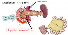
The embryonic foregut ends at the 2nd part of the
duodenum (Ampulla of Varter where the the common bile duct joins the chief pancreatic duct). The midgut starts at 3rd and 4th parts of the duodenum because they are supplied by the superior mesenteric artery. |
|
|
DDx Jejunum and Ileum
|
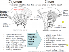
|
|
|
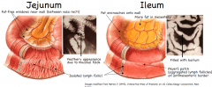
DDx Jejunum and Ileum on X-ray
|
|
|
|
Meckel’s diverticulum:
|
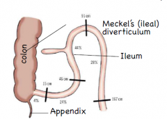
resembles the appendix
is usually within a metre of ileocaecal junction inflammation may mimic pain of appendicitis is the connection to the embryonic yolk sac |
|
|
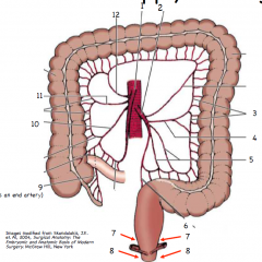
Blood Supply to Small Intestine
|
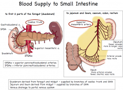
|
|
|
The Large Intestine Structures
|
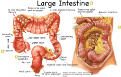
Caecum, appendix, colon (ascending,
transverse, descending, and sigmoid), rectum, first half of the anal canal Ascending and descending colon are typically retroperitoneal. |
|
|
The large Intestines;
Function |
Function: absorption of water & electrolytes, storage, elimination. Large intestine is 1.5 metres long,
extends from terminal ileum to anal canal |
|
|
Features that Distinguish the Colon
|
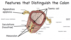
The colon is characterised by…..
1. taeniae coli start at base of appendix and extend to rectosigmoid junction 2. haustrations (sacculatrae) 3. epiploic appendages (appendices epiploicae) The colon also has a larger diameter than the small intestine but is shorter (1.5m) |
|
|
Caecum and Vermiform Appendix
|
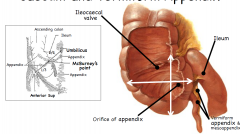
Caecum (L. blind) – 5-7cm long & wide, below level of ileocaecal valves. In contact with anterior abdominal
wall when full (distended with fluid/gas); taenia coli converge on base of appendix Vermiform appendix – variable (3-15 cm), narrow, blind-ended tube; suspended by mesoappendix; supplied by an end artery. Inflamed appendix starts as vague peri-umbilical pain shifts to pain in R. iliac fossa. |
|
|
Ileocacal Valve
|
makes like a flutter or farty sound when letting food through
|
|
|
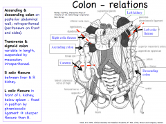
Colon - relations
|
|
|
|
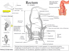
Rectum
|
Rectum from rectosigmoid junction (ant. to sacral segment 3) to anorectal jn (at pelvic diaphragm); located in pelvic cavity, anterior to sacrum & coccyx. Rectal ampulla is lower anterior dilatation.
No sacculations, appendices epiploicae or taeniae coli in rectum. |
|
|
Curvatures of the rectum
|
|
|
|
Rectum - Relationships
|
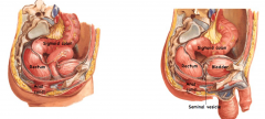
Rectum has peritoneum on front & sides of upper 1/3rd, on front of middle 1/3rd and no peritoneum
related to lower 1/3rd. Peritoneum reflects from rectum onto pelvic viscera forming rectouterine pouch in females & rectovesical pouch in males. Anterior – coils of intestine + cervix, vagina (female) or prostate, seminal vesicle (male) + vas deferens, bladder Posterior – sacrum, coccyx Lateral – coils of intestine (in pararectal fossa), pelvic diaphragm |
|
|
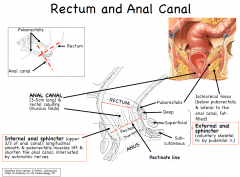
Anal Canal
|
|
|
|
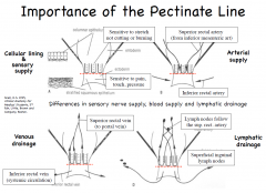
Importance of the Pectinate Line
|
|
|
|
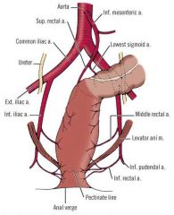
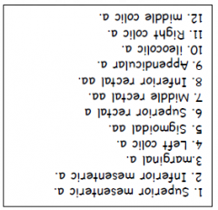
Large Intestines BS
|
Midgut supplied by superior mesenteric a; hindgut supplied by inferior mesenteric artery (to
pectinate line); rectal and anal canal walls also supplied by paired branches of internal iliac a. (middle & inferior rectal aa) |
|
|
Anal Canal BS
|
|
|
|
|
|
|
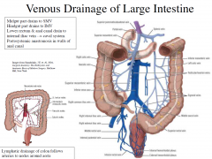
Venous Drainage of Large Intestine
|
|
|
|
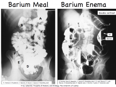
Barium Meal and Enema
|
|