Reading...
![]()
Play button
![]()
Play button
![]()
Use LEFT and RIGHT arrow keys to navigate between flashcards;
Use UP and DOWN arrow keys to flip the card;
H to show hint;
A reads text to speech;
35 Cards in this Set
- Front
- Back
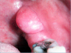
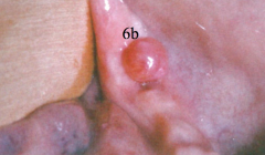
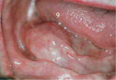
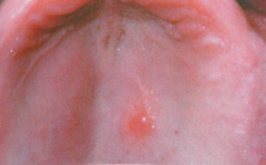
most common lesion of the oral cavity
|
Irritation Fibroma (traumatic Fibroma)
- sessile nonvascular soft, smooth mass made up of dense collagenous tissue with minimum inflammatory cells. - 1 to 2 cm. |
|
|
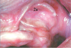
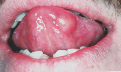
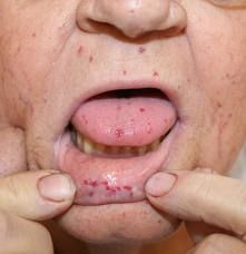
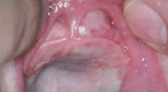
Epulis Fissuratum (Denture-Induced or Inflammatory Fibrous Hyperplasia, Denture Epulis)
- Long folds of dense connective tissue in the vestibule - Irritation by flange of loos denture over a long period of time. Often not inflamed, but may be ulcerated. - Similar lesion on hard palate beneath maxillary denture is called a FIBROEPITHELIAL POLYP or leaf-like denture fibroma. |
|
|
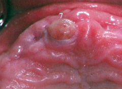
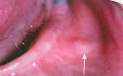
Visually looks like fibroma, the only difference is microscopic.
- Vascular fibrous tissue - Large stellate fibroblasts - usually less than 1 cm in diameter - first three decade of life |
Giant cell Fibroma.
- Does NOT appear to be associated with irritation. - 50% appear on gingiva (twice as common on mandible than maxilla) - |
|
numerous vertical projections each composed of orthokeratotic or parakeratotic squamous epithelium with connective tissue core.
|
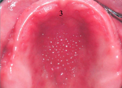
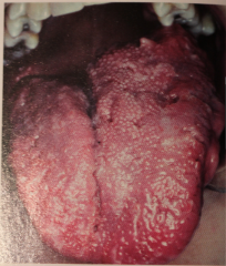
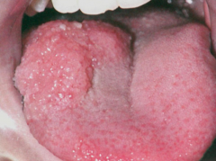
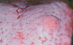
Papillary Hyperplasia (Inflammatory papillary hyperplasia, Palatal Papillomatosis)
- Often due to wearing denture or flipper 24 hrs a day. - Chronic atrophic candidiasis - poor denture hygiene For complete regression must do surgery or electosurgey - treat w/ scalpel, fluted bur, electro- or laser surgery. |
|
"3 p's"
|
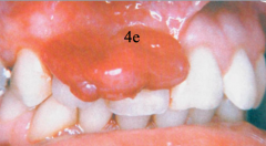
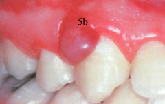
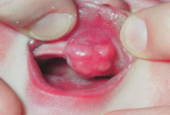
Pyogenic granuloma (pregnancy tumor)
- Usually on gingiva (interdental area), red, elevated adn pedunculated. Soft, bleeds easily, often ulcerated - exuberant tissue response to irritation (b/c of hormones) - excise, may have high rate of recurrence during pregnancy |
|
Probably arise from periodontal ligament or mucoperiosteum.
"3 p's" |
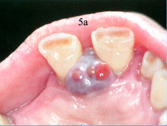
Peripheral Giant Cell Granuloma (Tumor)
-Occurs exclusively on gingiva - Dark and red usually or blue purple, but may be mucosal color (more pink than pyogenic granuloma, not as red) - more aggressive, can move teeth |
|
|
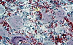
Peripheral Giant Cell Granuloma (Tumor) again
- Delicate C.T. stroma w/ multinucleated gian cells. |
|
|
Peripheral Giant Cell Granuloma (Tumor) again
- Delicate C.T. stroma w/ multinucleated gian cells. |
|
"3 p's"
|
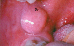
Peripheral (ossifying) Fibroma
- clinically can't distinguish from giant cell. - more common in young adults and children, twice as common in females - ONLY found on gingiva, usually anterior to molar region and less than 2 cm. - May be irritation or odontogenic in origin. - Dense connective tissue (very cellular like fibroma, but may have calcification or ossification) - may show radiopaque foci. |
|
|
Epulis granulomatosa
|
|
|
Which of the " 3 p's" is more aggressive and can move teeth?
|
Peripheral Giant Cell Granuloma (Tumor)
|
|
|
Which of the the "3 p's" occurs exclusively on the gingiva?
|
Peripheral Giant Cell Granuloma (Tumor)
& Peripheral (ossifying) Fibroma |
|
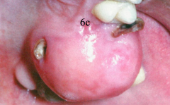
red vascular tissu growing out of a recent extraction site or socket ( may mimic a pyogenic granuloma)
|
Epulis Granulomatosa
- Made up of granulation tissue - metastatic carcinomas occasionally masquerade as this. Etiology = irritation - Calculus tooth fragment, bony sequestra in socket Treatment - excise |
|
|
Hyperplastic growth of granulation tissue that at times arise in healing extraction sockets.
- usually represent a granulation tissue reaction to bony sequestra in the socket |
Epulis granulomatosa.
|
|
Usually can see blood vessels on surface
|
Lipoma
- Benign neoplasm of Fat - uncommon orally - possible reaction of fat to trauma - 30+ years of age |
|
|
Most common mesenchymal neoplasm, but uncommon orally
|
Lipoma
|
|
|
Malignant neoplasm of fatty origin
|
LIposarcoma
- considered the most common soft tissue sarcoma and account for 20% of all soft tissue malignancies in adults. The most common sites are the thigh, retroperitoneum, and inguinal region. - They are rare in the head and neck regions. |
|
|
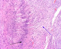
Neurilemoma (Schwannoma)
-Black arrow (left) = Antoni A; Blue arrow (right) = Antoni B - The Schwann cells of the Antoni A tissue form a palisaded arrangement around acellular zones known as Verocay Bodies |
|
slow growing nodular painless mass that can occur at any age.
|
Neurilemoma (Schwannoma)
- Uncommon orally but 25-50% occur in head and neck - Tongu is the most common intra-oral site - Can occur in bone (usually mandible) - Tumor of sheath of Schwann - Excise (well encapsulated) |
|
|
The most common type of peripheral nerve neoplasm.
|
Neurofibromas
- can arise as a solitary mass or be a component of neurofibromatosis. |
|
|
Neurofibroma
- skin is the most frequent location, but not uncommon orally. - Can arise centrally w/in bone. |
|
Probably arises from schwann cells, fibroblasts and perineural cells.
|
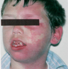
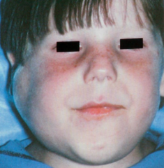
Neurofibromatosis type 1. AKA Von Recklinghausen's Disease of the skin.
Skin is the most common site, 72-92% have oral lesions majority of pt.s show cafe-au-lait spots may have small to large nodules to baggy pendulous masses (elephatitis neuromatosa) |
|
Not a true neoplasm
|
Traumatic (amputation) Neuroma
- attemped repair of damaged nerve. Often after tooth extraction - usually appears a small nodule less than .5 cm. - Mental nerve area common location (also tongu and lower lip) |
|
|
Proliferation of blood vessels that is often congenital and common in the head and neck regions (60%)
- 3:1 female predilection. |
Hemangioma
- will blanch, diascope. - common oral regions are lips, tongue, buccal mucosa and palate. - A unilateral hemangioma on the face following the division of the trigeminal nerve is called a port-wine stain. may also occur in bone. |
|
|
Methods of treatment for hemanioma?
|
Surgery
radiation cryosurgery steroids interferon-?-2A lasers NEVER do incisional biopsy |
|
|
Hereditary Hemorrhagic Telangiectasia (Rendu-Osler-Weber Disease)
- Autosomal dominant - tend to undergo repeated hemorrhage - Epistaxis may be an early sign - Pt. may suffer from anemia, but not usually life threatening. |
|
present at birth, but not hereditary.
|
Sturge-Weber Syndrome (Variant of hemangioma)
- "Portwine Nevi" Unilateral areas provided by trigeminal - May have vascular hyperplasia orally. - Neurological manifestations related to leptomeningeal angiomas and calcifications -a. may have convulsive disorders -b. may have mental retardations. |
|
|
Lymphangioma (most present at birth, 95% arise before age 10).
- commonly in the head and neck - Oral - most commonly occurs in tongue (may be papillary in appearance in superficial area) |
|
|
Lymphangioma
Histo - numerous spaces lined by endothelium containing lymph. Some may also have blood = mixed hemangiolymphanioma. - surgery only treatment of choice, tend to recur. |
|
|
Leiomyoma - benign neoplasm of SMOOTH muscle.
- uncommon orally - usually on poster of tongue - ecapsulated, painless, firm and may be multinodular. |
|
|
Rhabdomyoma - Benign lesion of SKELETAL muscle.
- Rare - most common location is tongue |
|
Occurs anywhere, especially tongue (50%+), all ages
|
Granular Cell Tumor
Benign soft tissue neoplasm, 2:1 female predilection asypmtomatic sessile nodule up to 2 cm in diameter. Controversial origin most likely Schwann cells or undifferentiated mesenchymal cell. Histo: large granular cells w/ eiosinophilic cytoplasm - may display pseudoepitheliomatous hyperplasia. |
|
|
Congenital Granular Cell Epulis (congenital epulis of the newborn)- present and birth...
Usually in Maxillary anterior gingiva 90% female predilections -excise - recurrence uncommon. |
|
|
What disease is similar to Granular cell tumor, but does NOT display pseudoepitheliomatous hyperplasia; and is seen in the new born?
|
Congenital Granular Cell Epulis (congenital epulis of the newborn)
|
|
Probably of neural crest origin
|
Melanotic Neuroectodermal Tumor of Infancy
- usually occurs in anterior maxilla -usually occurs as rapidly growing dark pigmented lesion - Pts. have high levels of vanilmadelic acid in urine. HISTO: infiltrating tumor mass of cells arranged in a patter of alveolus-like spaces lined by cuboidal cells. |