![]()
![]()
![]()
Use LEFT and RIGHT arrow keys to navigate between flashcards;
Use UP and DOWN arrow keys to flip the card;
H to show hint;
A reads text to speech;
64 Cards in this Set
- Front
- Back
|
chlamydoconidia |
|
|
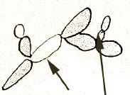
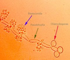
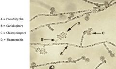
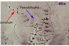
pseudohyphae &blastoconidia |
|
|
conidia |
spore |
|
|
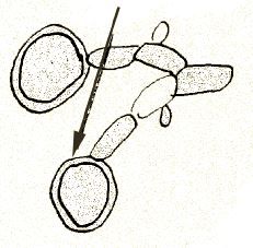
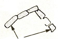
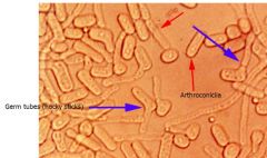
arthroconidia |
|
|
Colony Morphology |
Candida albicans |
|
|
Microscopic: – Pseudohyphae, some true hyphae – Clusters of round blastoconidia at the septa of pseudohyphae – Large, thick walled, usually single terminal chlamydospores
– Germ Tube positive – Urease negative (variable) |
Candida albicans
|
|
|
Candida albicans |
|
|
Candida albicans |
|
|
Colony Morphology |
Candida tropicalis |
|
|
Microscopic: – Pseudohyphae, some true hyphae – blastoconidia singly or in small groups
anywhere along pseudohyphae – Few tear drop shaped Chlamydospores may be present – Germ Tube negative – Urease negative |
Candida tropicalis
|
|
|
Candida tropicalis |
|
|
Candida tropicalis |
|
|
Candida tropicalis |
|
|
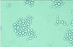
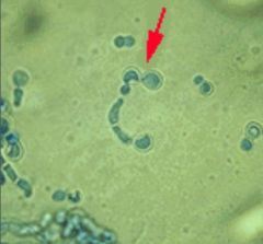
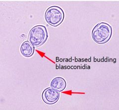
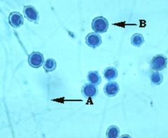
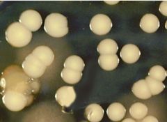
Infections in bloodstream,urogenital tract, normal flora• Colony Mor – Cream colored, sm yeast like -Microscopic: – Oval yeast cells w/ single terminal budding • No pseudohyphae
•Germ Tube neg – Urease neg |
Candida glabrata
|
|
|
Candida (Torulopsis) glabrata |
|
|
• normal flora |
Cryptococcus albidus |
|
|
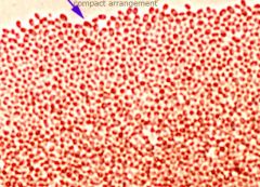
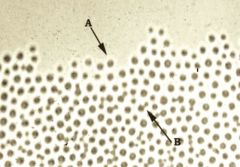
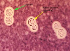
Cryptococcosis or normal flora • C: Flat or slightly heaped, shiny, moist, mucoid – blood agar, may show wrinkling at edges Micro – Yeast cells round, dark walled, budding – No hyphae
– Capsules may be – Bird seed agar / Caffeic Acid test Positive – Germ Tube negative – Urease pos– India ink for capsule |
Cryptococcus neoformans
|
|
|
Cryptococcus sp. |
|
|
Cryptococcus sp. |
|
|
Cryptococcus neoformans produces |
|
|
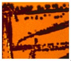
caffeic acid test for |
Cryptococcus neoformans |
|
|
The API 20-C strip |
carbohydrate yeast id tests |
|
|
– Disease in severely compromised hosts |
Geotrichum |
|
|
Geotrichum candidum |
|
|
Geotrichum sp. |
|
|
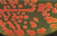
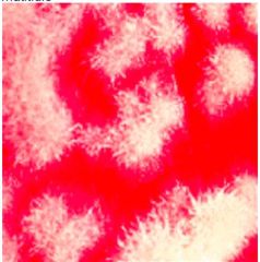
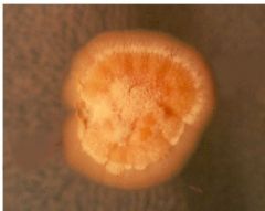
C:Usually pink or coral, orange to red Soft, moist, may be mucoid
• Micro Budding cells are round to oval Rare pseudohyphae– Faint capsule maybe |
Rhodoturola
|
|
|
Rhodotorula rubrum |
|
|
Rhodotorula rubrum |
|
|
– Disease in immuno-compromised hosts |
Trichosporon sp. |
|
|
Colony Morphology |
Trichosporon sp. |
|
|
Trichosporon sp. |
|
|
Trichosporon beigellii |
|
|
Trichosporon sp. |
|
|
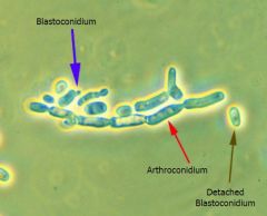
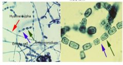
Colony Morph– Variable: “white fuzzy” – No hyphae Thick walled barrel shaped arthroconidia Arthroconidia alternate in the hyphae w/ |
Coccidioides immitis |
|
|
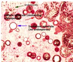
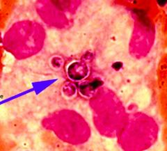
Spherules are lg, round, thick walled; contain endospores – DNA Probes are available – Immuno-diffusion 4 exoantigen
|
Coccidioides immitis
|
|
|
Coccidioides immitis |
|
|
Coccidioides immitis |
|
|
Coccidioides immitis |
|
|
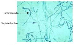
Colony Morph – Variable: white, beige,
orange, pinkish. Granular, powdery or woolly. – Light reverse • Microscopic: – Coarse Septate branched hyphae, no conidiophores – Arthroconidia not swollen or thick walled. – Arthroconidia alternate in the hyphae w/ empty cells that break away |
Malbranchea sp.
|
|
|
Malbranchea sp. |
|
|
Malbranchia sp. |
|
|
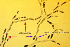
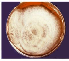
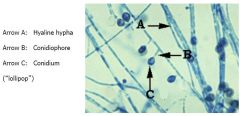
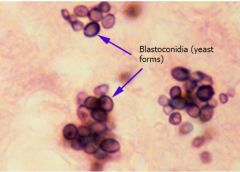
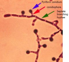
Colony Morph Initially yeast like, then white & colony, turning brown or tan w/ age – At 35 – 37, cream to tan, waxy yeast phase • Micro: – Septate hyphae w/ short or long conidiaphores Round or pear shaped conidia lollipop appearance Older cultures have thick walled chlamydoconidia Scedosporium apiospermum
|
Blastomyces dermatitidis
|
|
|
Blastomyces dermatitidis |
|
|
Blastomyces dermatitidis |
|
|
Blastomyces dermatitidis |
|
|
Blastomyces dermatitidis |
|
|
Blastomyces dermatitidis |
|
|
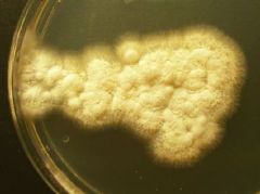
The colony is cottony and powdery with a pale brown color. Mature growth occurred in 6 days on SDA. There was no growth on media containing cycloheximide. The organism did not convert to a yeast form when cultured at 37 |
Chrysosoporium sp. |
|
|
Chrysosoporium sp. |
|
|
Chrysosoporium sp. |
|
|
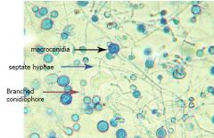
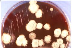
• Colony Morph – White- brown, fine dense several wks, lg thick walled macroconidia form |
Histoplasma capsulatum |
|
|
– Macroconidia can be confused w/ Sepedonium |
Histoplasma capsulatum |
|
|
Histoplasma capsulatum |
|
|
Histoplasma capsulatum |
|
|
Histoplasma capsulatum |
|
|
Histoplasma capsulatum |
|
|
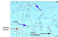
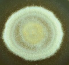
Colony Morph: White & waxy then becoming colony. Reverse is white – At 37, little or no growth. • Micro: Septate hyphae w/ single or branched conidiophores – Conidia are round, thick walled, & rough & knobby
– No microconidia formed, no yeast phase |
Sepedonium sp.
|
|
|
Sepedonium sp. |
|
|
Sepedonium sp. |
|
|
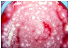
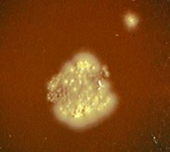
Colony Morph: White heaped compact, |
Paracoccidiodes brasilienses |
|
|
Paracoccidiodes brasilienses |
|
|
Paracoccidiodes brasilienses |
|
|
Paracoccidiodes immitis |
|
|
Paracoccidiodes immitis |