Reading...
![]()
Play button
![]()
Play button
![]()
Use LEFT and RIGHT arrow keys to navigate between flashcards;
Use UP and DOWN arrow keys to flip the card;
H to show hint;
A reads text to speech;
11 Cards in this Set
- Front
- Back
|
? מה מרכיב את פתח האף החיצוני
|
פתח בין 2 עצמות המאקסילה
|
|
|
? מה מרכיב את פתח האף הפנימי
|
medial pterygoid plate פתח בין 2 עצמות ה
|
|
|
? אילו עצמות מרכיבות את חלל האף
? ומהם חלקיהן |
ethmoid - יוצרת 3 חללי אוויר
1. Posterior Ethmoidal Air Cells. 2. Middle Ethmoidal Air Cells. 3. Anterior Ethmoidal Air Cells. Orbital Surface of Ethmoid Bone - the medial posterior wall of the orbit (posterir to the lacrimal bone) the bone consists of 2 plate : 1. cribriform plate (horizontal) crista gali is the part of the plate to which attaches the meninges 2.Perpendicular Plate (vertical) |
|
|
what are the concheas ?
|
3 curly shelves :
1. superior concha - O: from the midial air cells 2. middle concha - O: drom the midial air cells 3. inferior concha - O: attaches to the mexilla bone meatus - inferior to each concha there is a space draining the diffrent sinuses |
|
|
waht are the parts of the nasal septum ?
|
anterior part made of cartilage
posterior part made of 2 bones : 1. superior - ethmoid (Perpendicular Plate) 2. inferior - vomer bone the sepyum creates 2 distinguish spaces |
|
|
what spaces drain to the nasal cavity ?
|
1. sphenoid sinus - posterior to the superior concha
drains through the Sphenoethmoidal Recess 2. ethmois air cells - # posterior ethmoid air cells-> superior concha # middle cells -> middle meatus through the Ethmoidal Bulla # anterior cells -> Middle Meatus inferior to Ethmoidal Bulla through the Semilunar Hiatus 3. maxillary sinus - Opening of Maxillary Sinus in the inferior part of Semilunar Hiatus 4. drontal sinus - through thr Frontonasal Duct in the superior part of Semilunar Hiatus 5.lacrimal - through the Nasolacrimal Duct -> Inferior Nasal Meatus |
|
|
waht is the nasal tissue ?
|
made of mucusa - epithelial cells which consists of goblet cells. the goblet cells produces mucus . the mucus catches outside intuders
|
|
|
what is Kartagener's Syndrome ?
|
זוהי מחלה נפוצה הנקראת גם Primary Ciliary Dyskinesia, ופוגעת בתזוזה של תאי ה- Cilia. כתוצאה מהמחלה, הריר אינו מנוקז כמו שצריך. לכן, הבעיה תתבטא בסינוסיטיס חוזר ובדלקות ריאה. בנוסף, הבעיה תגרום לבעיה בפוריות כי תהיה בעיה בהסעה של הזרע בתוך צינור הזרע. בנוסף, החלבון של הסיליה משתתף גם בהנעה של האיברים של העובר לצדדים כאשר הוא מתפתח. כלומר, יש ביטוי גם ב"לטרליות" של האיברים. באנשים כאלו ניתן למצוא מצב שנקרא
Situs Inversus- האיברים ממוקמים הפוך ביחס למיקומם הרגיל בגוף. |
|
|
through which foramen the maxillary nerve & artery enter the nasal cavity ?
|
Sphenopalatine Foramen
|
|
|
who supplies blood to the nasal cavity ?
|
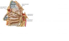
1. Sphenopalatine Artery ( branch of the maxillary ar.)
1. Lateral Posterior Nasal Branches: סעיפים שמספקים דם לקיר הלטרלי מאחור. 2. Septal Posterior Nasal Branches: סעיפים שמספקים דם לקיר הספטלי מאחור. 2. Anterior & Posterior Ethmoidal Arteries - branches of the ophtalmic ar. enters through the Anterior & Posterior Ethmoidal Foramina 1. ה- Ethmoidal Arteries Posterior מספקים דם בעיקר לקונכייה העליונה. 2. ה- Anterior מספק את המשטחים הלטרלי והספטלי מקדימה |
|
|
? מי מעצבב את חלל האף
|
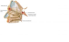
1. מאחור- עצבוב סנסורי (תחושה כללית) + פרה- סימפתטי:
בקיר הספטלי: ה- Nasopalatine Nerve המתפצל מה- Maxillary Nerve (V2). בקיר הלטרלי: סעיפים הנקראים Posterior Lateral Branches המתפצלים מה- Maxillary Nerve ומה- Greater Palatine Nerve. *העצבוב הפרה- סימפתטי המספק בלוטות להפרשת Mucosa בחלל האף נעשה ע"י Greater Petrosal (פיצול של Facial Nerve). הסעיפים הפרה- גנגליונים מגיעים ל- Pterygopalatine Ganglion, שם הם מבצעים סינפסה. הסעיפים הפוסט- גנגליונים מצטרפים ל- Maxillary Nerve ומגיעים איתו לחלל האף. 2. מקדימה- עצבוב סנסורי (תחושה כללית) + פרה-סימפתטי: Anterior Ethmoidal Nerve: זהו אחד הסעיפים של V1 (Ophthalmic Nerve) העובר דרך ה- Anterior Ethmoidal Foramen בארובת העין, מסתעף ומספק עצבוב לקיר הלטרלי ולקיר הספטלי בחלק הקדמי. (ל- Posterior Ethmoidal Nerve אין ייצוג בחלל האף). 3. החלק העליון של חלל האף מעוצבב (תחושה מיוחדת-ריח) ע"י Olfactory Nerves (1) |