![]()
![]()
![]()
Use LEFT and RIGHT arrow keys to navigate between flashcards;
Use UP and DOWN arrow keys to flip the card;
H to show hint;
A reads text to speech;
39 Cards in this Set
- Front
- Back
|
what is the primary function of the lungs? |
oxygenate blood by bringing inspired air close to venous blood in pulmonary capillaries |
|
|
describe the passage of inspired air through conducting airways |
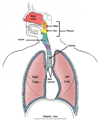
nasal cavity, nasal pharynx, oral pharynx, larynx, trachea, main bronchi |
|
|
describe how the lungs are separated |
right lung: 3 lobes (upper, middle and lower) separated by horizontal and oblique fissures. larger (higher diaphragm, no heart indent) left lung: 2 lobes (upper and lower) separated by oblique fissure. |
|
|
define the cardiac notch |
indentation in the left lung to create space for the heart |
|
|
define the lingula |
tongue-like lung process inferior to the cardiac notch |
|
|
describe the surface projections of the oblique fissure |
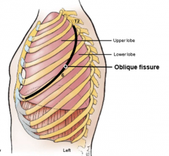
begins posteriorly at spinous process of T2, down to the anterior side where follows the 6th rib when scapula is abducted (arms raised), oblique fissures of both lungs follow medial border of scapula |
|
|
describe the surface projections of the horizontal fissure |
(right lung only) anteriorly parallels inferior border of 4th rib until it hits the oblique fissure |
|
|
describe the 3 lung surfaces |
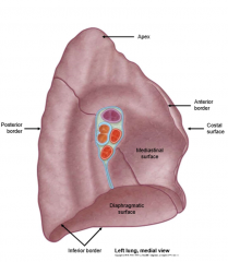
costal surface: posterior surface (ribs of back, costal cartilages and sternum) meets mediastinal surface at sternum mediastinal surface: facing mediastinum, anterior surface diaphragmatic surface: meets surface of diaphragm (inferior surface) |
|
|
describe the 3 lung borders |
anterior border (long, sharp, separates costal from mediastinal medially) posterior border (separates costal and mediastinal laterally) inferior border (separates diaphragmatic surface from costal and mediastinal surfaces) |
|
|
what is the lung apex |
superior point of lung, extending out of the thoracic cavity and into the neck |
|
|
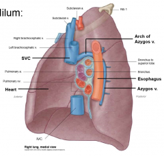
describe the components of the lung root in each lung |
Right lung: eparterial bronchus (most superior), pulmonary artery, main bronchus (surrounded by bronchiole vessels), pulmonary veins (anterior and inferior) left lung: pulmonary artery (most superior), main bronchus, pulmonary veins autonomic nerves, sensory nerves, lymphatics in mediastinal connective tissue in both surrounded by parietal pleura forming pulmonary sleeve and hangs down into pulmonary ligament |
|
|
define the hilum |
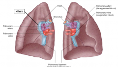
continuity of parietal pleura into the visceral pleura (where the pulmonary sleeve becomes the lung pleura) on mediastinal surface where everything enters/exits |
|
|
what structures enter/exit at the hilum |
enter: bronchus, pulmonary arteries, nerves exit: pulmonary veins, lymphatics |
|
|
define pulmonary artery and pulmonary vein |
artery: takes blood away from heart vein: takes blood to heart pulmonary artery is deoxygenated pulmonary vein is oxygenated |
|
|
describe the relationship of the hilum to other structures in the right lung |
anteriorly: superior vena cava and heart (cardiac impression) superiorly: arch of azygos vein posterior: esophagos and azygos vein |
|
|
describe the relationship of the hilum to other structures in the left lung
|
anteriorly: heart (cardiac impression) superiorly: aortic arch posteriorly: esophagos and thoracic aorta |
|
|
describe the main bronchi |
bifurcation of trachea at the sternal angle (transverse thoracic plane) surrounded by cartilagenous rings internally there is the carina |
|
|
what does a distorted carina indicate? |
possibly a metastasis of lung cancer (lymph nodes just under the carina) |
|
|
which bronchus do you aspirate foreign objects from and why |
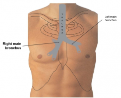
inhale a bite, goes down the right right bronchus is shorter, wider and more vertical |
|
|
describe bronchial branching |
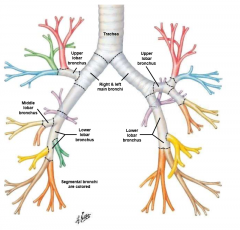
all occurs within the lung itself except for upper lobar bronchus of right lung which is early secondary upper, middle and lower lobar bronchus in right (each lobe) upper and lower lobar bronchus in left tertiary segmental branching (10 in right, 8-10 in left) |
|
|
what is the bronchopulmonary segment |
a wedge-shaped portion of the pulmonary artery supplying blood to the segmental branching of the lung (pulmonary artery follows branching of bronchi) |
|
|
what are the levels of the segmental branches |
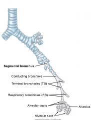
segmental bronchus, conducting bronchiole, terminal bronchiole (TB), respiratory bronchiole (RB), alveolar ducts, alveolar sacs (alveolus) gas exchange begins at RB level |
|
|
describe the intersegmental veins and their surgical relevance |
where the blood drains after gas exchange occurs located in connective tissue septa at periphery of bronchopulmonary segment tributaries of pulmonary veins landmarks for segmentectomy |
|
|
describe the dual blood supply to the lungs |
pulmonary arteries and veins (gas exchange, circulation between heart and lungs) bronchial arteries and veins (nutrition, vascular supply to lung tissue) |
|
|
describe the origins of the pulmonary vasculature |
pulmonary arteries (L+R) arise from pulmonary trunk (deoxygenated blood from right ventricle) behind aorta and vena cava (left has to go over bronchus) back to heart in pulmonary vein to left atrium |
|
|
describe the bronchiole arteries |
supply lung tissue, lung root and visceral pleura branches of the thoracic aorta 2 left 1 right bronchiole arteries right bronchiole artery arises with 3rd posterior intercostal artery |
|
|
describe the bronchiole veins |
only drain lung root near hilum rest of lung drains into pulmonary veins (reduces oxygen concentration ~3%) left bronchiole veins drain to accessory hemiazygos vein, cross midline into (right goes straight here) azygos vein right into superior vena cava |
|
|
describe the types of lymphatic drainage of the lungs |
superficial: just deep to visceral pleura at subpleural lymphatic plexus and drains towards hilum deep: lymph nodes in lung, parallel bronchiole tree |
|
|
describe the steps in lymphatic drainage in the lungs |
1. both superficial and deep drain to bronchopulmonary (hilar) nodes 2. inferior tracheobronchial (carinal) nodes 3. (L/R) superior tracheobronchial nodes 4. (L/R) paratracheal nodes 5. Bronchomediastinal trunk 6. Right lymphatic duct/ thoracic duct all drainage eventually reaches brachiocephalic veins |
|
|
how can cancer metastasize through lymphatic/venous/arterial methods |
lymphatic: enter lymph vessels and seed lymph nodes veins: enter bronchial vein and go to azygos system artery: (rare) enter pulmonary veins and pass to left side of heart |
|
|
describe basic lung innervation |
anterior and posterior pulmonary plexus (relative to bronchi, actually just a mess of nerves, hard to tell difference) both receive fibers from sympathetic trunks and vagus nerves on each side |
|
|
describe the different components of the pulmonary plexuses |
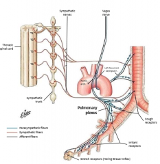
sympathetic (from chain ganglia of T1-T4)- post ganglionic fibers: bronchodilators, vasoconstrictors, decrease mucus secretion parasympathetic (directly from lateral horn) preganglionic fibers, ganglia and post ganglionic fibers: bronchoconstrictors, vasodilators and increase mucus secretion afferent (sensory): run in vagus nerve, reflexes for coughing and stretching (over inflation) |
|
|
describe the diaphragm |
dome shaped skeletal muscle separating thoracic and abdominal cavity superior meets fibrous pericardium of heart and diaphragmatic parietal pluera |
|
|
what are the functions of the diaphragm |
descent on contraction: increase vertical dimension of thoracic cavity, inter-abdominal pressure (with abdominal wall contraction= voiding responses-- defacation, micturition and childbirth) decrease pressure in lungs so air goes in |
|
|
describe the diaphragm attachments |
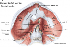
converge at central tendon sternal part (xiphoid process) R/L costal part (ribs) lumbar part-- lateral (arch over quadratus lumborum muscle) and medial arcuate (arch over psoas major muscle) ligaments, right and left crus (crua) forming from anterior longitudinal ligament (right is larger) meet at median arcuate ligament |
|
|
what are the 3 openings in the diaphragm and what passes through them
|
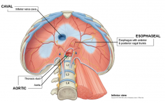
caval opening: vertebra T8, in central tendon (inferior vena cava) esophageal hiatus: T10 in right crus (esophagus and vagal trunks) aortic: T12, posterior to median arcuate ligament (aorta and thoracic ducts) |
|
|
describe the blood supply to the diaphragm |
thoracic side: superior phrenic artery (from thoracic aorta) abdominal side: inferior phrenic artery (from abdominal aorta veins mirror arteries, drain to inferior vena cava |
|
|
describe the diaphragm innervation |
from phrenic nerves (originate in neck from ventral primary rami on each side) C3,4,5 keep diaphragm alive into thoracic inlet and travel anteriorly to root of each lung between pericardium and mediastinal parietal pluera, supply hemidiaphragm on each side sensory in phrenic nerves as well |
|
|
what happens in a phrenic nerve lesion |
lesion= destruction normally, diaphragm contracts increasing vertical volume of lung if one side is paralyzed, it goes UP (increased abdominal pressure from one side going down)-- paradoxical movement detected with xray |