Reading...
![]()
Play button
![]()
Play button
![]()
Use LEFT and RIGHT arrow keys to navigate between flashcards;
Use UP and DOWN arrow keys to flip the card;
H to show hint;
A reads text to speech;
45 Cards in this Set
- Front
- Back
|
Boundries of the neck?
|
Superior boundries:
- mandible - mastoid process - Ionion # (superior occipital protuberence) Inferior: - sternum - clavicle - acromion of scapulae - 7th cervicle vertebrae |
|
|
structure coursing between the head and thorax are associated with which traingle?
|
anterior triangle
|
|
|
structure coursing through the head/neck and upper limbs are associated with which triangle?
|
posterior
|
|
|
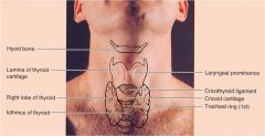
Know surface anatomy:
- hyoid bone - cricoid cartilage - thyroid cartilage - thyroid membrane - thyroid gland - trachea |
|
|
|
What are the 3 compartments of the neck?
|
viseral
vertebral vascular |
|
|
describe the viseral laminae of the neck.
|
- thin layer of areolar connective tissue
-lies between dermis and skin - contains platysma, cutaneous nerves (facial nerve, CN VII), blood vessles, lymphatic vessles - contains varying amounts of fat - continuous superficial facea elsewhere in the body |
|
|
Where is the external jugular vein found?
(know other superficial veins? - slide 21)? |
over the sternocleomastoid muscle
|
|
|
Describe the deep cervicle faciae.
|
- dense connective tissue deep to the superficial fasciae
- envelopes muscle, neurovascular structure, and certain vicera - serves as partitions, compartmentalizes the neck - clinical corralations - surgical cleavage plane and potential space (pathways for infections to spread) |
|
|
What are the 4 deep fasciae and where are the found?
|
Investing layer:
- just deep to superficial fasciae - surrounds muscle Prevertebral fasciae: a) axillary sheath (around axillary vessels) b)supraplural membrane ???? Pretracheal fasciae: - lies anterior to the trachea Carotid sheath: - contains corrotid arteries, internal jugular, vegas nerve etc... |
|
|
The investing layer of the deep fasciae covers which 2 glands?
|
submandibular and parotid
|
|
|
Where are the points of attachment for the investing fasciae?
Superior? Inferior? |
Superior:
- hyoid bone - inferior margin of mandible - zygomatic arch Inferior: - clavicle - acromion - spine of scapulae - manubrium of sternum |
|
|
superiorly the submandibular fasciae attaches where?
Inferiorly where does it attach? |
The myohyoid bone and the inferior border of the mandible
The hyoid bone |
|
|
The submandibular node is found in which fasciae?
|
the submandibular fasciae (part of investing layer)
|
|
|
describe the 2 layer of the parotid fasciae (part of deep investing fasciae)
|
superficial layer
- attaches to zygomatic arch superiorly - continuous with fasciae covering SCM behind and the masseter in front Deep parotid fasciae - extends along medial surface of gland and attaches to the skull - not as dense as superficial except for the Stylomandibular ligament |
|
|
What are the 2 divisions of the prevertebral membrane?
|
the suprapleural membrane and the axillary sheath.
|
|
|
axillary sheath (part of prevertebral division) surrounds what?
|
axillary vessles and brachial plexus
|
|
|
What does the carotid sheath contain?
|
common corotid, internal jugular and vagus nerve
|
|
|
The corotid fasciea is formed from what other fasciaes?
|
the pretracheal, prevertebral, and the investing/superficial fasciea.
|
|
|
the carotid sheath goes from where to where?
|
base of skull to root of neck
|
|
|
What is the posterior portion of the pretracheal fasciae called?
|
buccopharangeal fasciae (just anterior to alar fasciae)
|
|
|
What is the anterior portion of the prevertebral fasciae called?
|
alar fasciae (just posterior to buccopharangeal fasciae)
|
|
|
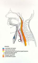
label the following fascial spaces:
Retroviseral Retropharangeal Danger Zone (space 4) Prevertebral Lateral pharangeal |
|
|
|
The investing layer encompasses what?
|
the entire neck and spits to surround SCM and trap. muscles
|
|
|
Pretracheal layer encloses what?
|
muscular portion incloses the infrahyoid muscle and the viseral portion encloses thyroid, larynx, trachea, pharynx and esophogus
|
|
|
the prevertebral layer surrounds what?
|
the cervical vertebral column and the associated muscles
|
|
|
The corotid sheath incloses what?
|
the common corotid, internal jugular and the vagus
|
|
|
What are the borders of the anterior triangle?
|
midline
inferior margin of mandible anterior margin of SCM muscle |
|
|
What are the borders of the posterior triangle?
|
Posterior margin of SCM
Anterior margin of trap middle 1/3rd of clavicle |
|
|
What are the 4 minor triangles of the anterior triangle?
|
muscular, submental, submandibular triangle, carotid.
|
|
|
What are the minor triagles of the posterior triangle?
|
supruclavicular (also called omoclavicular or subclavian) and occipital
|
|
|
What are the muscles of the posterior triangle?
|
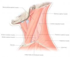
-semispinalis capitus muscle
-splenus capitus -levitator scapulae -posterior, anterior and middle scalene muscle |
|
|
What is the major vein in the posterior triangle?
|
external jugular
|
|
|
What are the important arteries in the posterior triangle?
|
First part of subclavian artery and its branches
Thyrocervical trunk a) tranverse cervical artery b) Suprascapular artery |
|
|
The superficial location of what nerve as it crosses the posterior triangle makes it suseptable to injury?
|
Accesory nerve XI
|
|
|
What is the hierarchy of peripheral nerves?
|
Dorsal and ventral ramus form plexuses of the Cervical, brachial, lumbar and sacral which form peripheral nerves
|
|
|
What makes up the cervical plexus?
|
Anterior Rami of cervical nerves (C1-C4)
|
|
|
What nerves make up the cervical plexus?
|
Muscular (deep) branches:
- phrenic and sensory nerves - Ansa cervicalis - direct motor branches to the prevertebral and lateral vertebral muscles Cutaneous (superficial branches) - Lesser occiptial - Greater Auricular - Transverse cervical nerve - Supraclavicular nerve |
|
|
What do the cutaneous branches of the cervical plexus arise from?
|
Erb's point
|
|
|
Brachial Plexus???
|
???? slide 63
|
|
|
How are the lymphatics in the neck arranged?
|
Horizontal Superficial ring
Horizontal deep ring Deep cervical (jugular) trunk (SEE POWER POINT!!!) slides 65-71 |
|
|
Why is the subclavian vein clinically important?
What other veins may be used for the same purposes? |
used for interveneous hyperalimentation, monitoring central venous pressure, obtaining central venous access and for chemotheropy, introducing intravenous pacemakers.
Internal jugular and chephalic veins |
|
|
Cervical rib and anterior scalene syndrome and hyperabduction are also preferentially called what?
|
Thoracic outlet syndrome.
|
|
|
in Cervical rib syndrome the anterior end of the cervical rib may articulate how?
|
articulate with the sternum,
(2) articulate or fuse with the first rib, (3) attach to the first rib by a fibrous band, or (4) present a free end. |
|
|
What is anterior scalene syndrome?
|
due to spasm or hypertrophy of anterior scalene muscle
|
|
|
The symtoms of thoracic outlet syndrome are due to what?
|
compression of the neurovascular
structures extending anywhere from the thoracic outlet to the insertion of the pectoralis minor muscle. They are characterized by neurological deficits and/or vascular (arterial or venous) changes in the upper extremity. (also see handout) |