![]()
![]()
![]()
Use LEFT and RIGHT arrow keys to navigate between flashcards;
Use UP and DOWN arrow keys to flip the card;
H to show hint;
A reads text to speech;
92 Cards in this Set
- Front
- Back
|
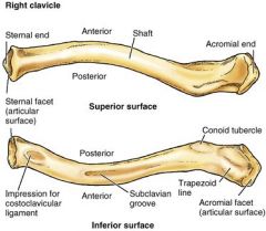
Clavicle (sternal end, acromial end) |
|
|
|
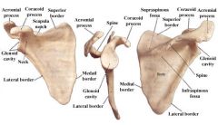
Scapula (body, superior, medial and lateral borders, glenoid cavity, subscapular fossa, coracoid process, acromion, scapular spine, supraspinous fossa, infraspinous fossa) |
|
|
|
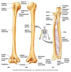
Humerus (head, greater tubercle, lesser tubercle, intertubercular groove, anatomical neck, surgical neck) |
|
|
|
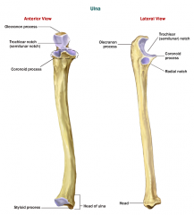
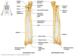
Ulna (olecranon, trochlear notch, radial notch, ulnar head, styloid process) |
|
|
|
Radius (radial head, radial tuberosity, ulnar notch, styloid process) |
|
|
|
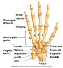
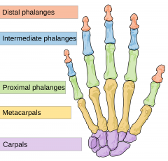
Carpal Bones (scaphoid, lunate, triquetum, pisiform) |
|
|
|
Metacarpals (1-5, beginning with pollex) |

|
|
|
Phalanges (proximal, middle, distal) |
|
|
|
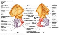
Coxal Bone (ilium, ischium, pubis, ala, body, acetabulum, greater sciatic notch, ischial spine, ischial tuberosity, obturator foramne, arcuate line, iliac crest, iliac fossa, pubic symphysis, auricular surface, superior and inferior rami of pubis, anterior superior and inferior iliac spine, posterior superior and inferior iliac spine) |
|
|
|
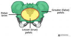
Pelvis (false vs true pelvis, pelvic brim, pelvic inlet/outlet) |
|
|
|
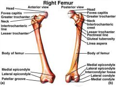
Femur (head, fovea capitis, neck, greater/lesser trochanters, intertrochanteric crest, shaft, linea aspera, medial and lateral epicondoyle, intercondylar fossa) |
|
|
|
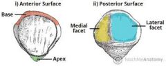
Patella (base, apex) |
|
|
|
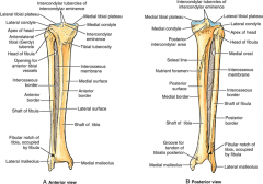
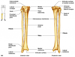
Tibia (medial/lateral condyles, intercondylar eminence, tibial tuberosity, anterior border, medial malleolus, fibular notch) |
|
|
|
Fibula (head, lateral malleolus) |
|
|
|
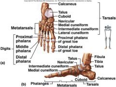
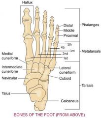
Tarsal Bones (Calcaneus, talus, cuboid, navicular, lateral/medial/intermediate cuneiforms) (Calvin Tasted Cubes of Nasty Limburger Cheese) |
|
|
|
Metatarsals (1-5) |
|
|
|
Phalanges (proximal, middle, distal) |

|
|
|
Sutures (lamboid, coronal, sagittal, squamos) |
|
|
|
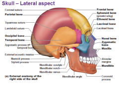
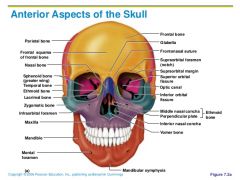
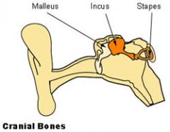
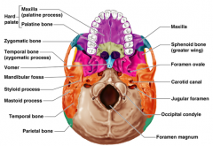
Cranial Bone (frontal, parietal, temporal, squamosal, zygomatic process, zygomatic arch, mandibullar fossa, mastoid process, petrous portion, malleus, incus and stapes, external and internal auditory meatus, occipital, occipital condoyles, foramen magnum, ethmoid, cribriform plate, crista galli, olfactory foramina, perpendicular plate, ethmoidal sinuses, sphenoid, sphenoid body, sella turnica, hypophyseal fossa, lesser/greater wings, pterygoid processes/plates) |
|
|
|
Facial Bone (nasal, external naris, maxilla, palantine process, zygomatic, zygomatic arch, temporal process, lacrimal, lacrimal fossa, palatine, horizontal plate, perpendicular plate, vomer, mandible, body, ramus, condylar process, coronoid process, Hyoid bone, body, greater horns, lesser horns) |
|
|
|
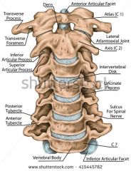
Cervical Vertebrae- n=7 (atlas, facet for dens of axis, axis, dens, vertebra prominens) |
|
|
|
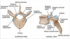
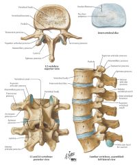
Thoracic Vertebrae- n=12 (facets, demifacets, transverse facets, T1- only vertebrae with superior whole facets and inferior demifacets, T2-8- superior and inferior demifacets, T9- only superior demifacets, T10-12- only whole facets) |
|
|
|
Lumbar Vertebrae- n=5 (massive oval body with flat faces; small, triangular, vertebral foramen, large transverse processes) |
|
|
|
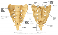
Sacral Vertebrae- n=5 (median sacral crest, sacral crest, sacral hiatus, sacral foramina, auricular surface, ala, coccyx- n=3-5) |
|
|
|
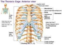
Sternum (Manubrium, body, xiphoid process) |
|
|
|
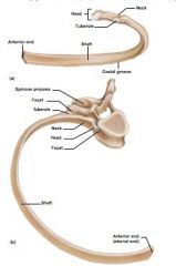
Ribs (head, nec, tubercle, articular facets, sternal end, body) |
Pair 1-7 (true ribs)
Pairs 8-10 (False ribs) Pairs 11-12 (floating ribs) |
|
|
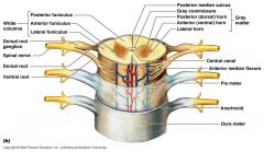
Spinal Cord (posterior median sulcus, anterior median fissure, cervical/lumbar enlargements, cnus medullaris, cauda equina, filum terminale, anterior/ventral (motor) root, posterior/dorsal (sensory) root) |
|
|
|
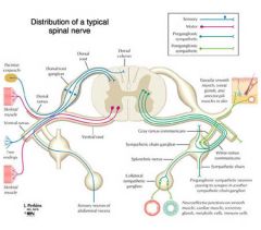
Spinal Nerves (central canal, white matter, gray matter, somatic & visceral sensory nuclei, somatic motor nuclei, visceral motor nuclei, anterior and posterior gray commissures, anterior white commissure) |
|
|
|
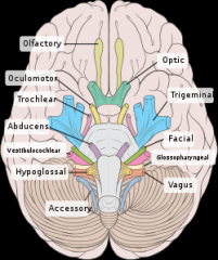
Cranial Nerves (Olfactory- I, Optic- II, Oculomotor- III, Trochlear- IV, Trigeminal- V, Abducens- VI, Facial- VII, Vestibulocochlear- VIII, Glossopharyngeal- IX, Vagus- X, Accessory- XI, Hypoglossal- XII) |
|
|
|
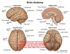
Brain (cerebral hemisphere, longitudinal fissure, gyrus, sulcus, corpus callosum, corpora quadrigemina (superior and inferior colliculi), cerebellum, vermis, medulla oblongata, dura mater) |
|
|
|
Why is it called the surgical neck (humerus)? What happens here? |
The anatomical neck is the portion that lies just below the head. As the neck continues along the humerus body, it is called the surgical neck (sonamed because this is the location of many fractures that require surgery |
|
|
What does trochlear refer to? |
motor/movement |
|
|
What are the names of the articulating surfaces between the humerus and ulna? |
trochlea of the humerus and the trochlear notch of the ulna |
|
|
What are the names of the articulating surfaces between the humerus and the radius? |
head of radius and capitulum of humerus |
|
|
what structure do you hit when you a) hit your butt cheek hard. b) bump the bony part of your groin. c) get a "hip-pointer" |
a) b) c) |
|
|
What are the real names of the bulges people call ankles? |
malleolus |
|
|
Synarthroid joints |
joints lacking a joint cavity and not movable. these types of joints are used to connect bones rather than provide a surface for movement relative to each other. Types: coronal suture, sagittal suture, lamboidal suture, squamos suture |
|
|
Diarthrotic joints |
joints where there is a synovial cavity and the bones move freely relative to each other. Synovial cavity is oddly filled with synovial fluid, which acts to cushion and lubricate the joint. |
|
|
Hinge joint |
flexion/extension as seen at knee or elbow |
|
|
Pivot joint |
rotation as seen at atlas/axis joint |
|
|
Spheroidal joint |
(ball and socket) flexion/extension as seen in shoulder; abduction/adduction, external rotation, internal rotation and circumduction as seen at hip |
|
|
Saddle joint |
flexion/extension, abduction/adduction, and circumduction of thumb between carpals and metacarpals |
|
|
Ellipsoidal joint |
flexion/extension, abduction/adduction, and circumduction as seen at wrist or ankle |
|
|
explain names of lamboid, sagittal, and coronal sutures |
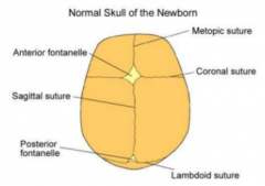
sagittal- joins 2 parietal bones coronal- frontal bone to two parietal bones lamboid- joins parietal bone to occipital bone |
|
|
what is the function of the mandibular fossa? |
The mandibular fossa is the cavity in the temporal bone that enables interaction with the mandibular condyle |
|
|
What is the function of the crista galli? |
The crista galli (Latin: "crest of the rooster") is a median ridge of bone that projects from the cribriform plate of the ethmoid bone. It is where the falx cerebri attaches anteriorly to the skull. The olfactory bulbs lie on either side of the crista galli on top of the cribriform plate |
|
|
What passes through the olfactory foramen? |
olfactory nerve 1 |
|
|
What does sella turcica mean? |
The sella turcica (Latin for Turkish seat) is a saddle-shaped depression in the body of the sphenoid bone of the human skull and of It serves as a cephalometric landmark. The seat of the saddle, the deepest part of the sella turcica known as the hypophyseal fossa, holds the pituitary gland |
|
|
What sits on the hypophyseal fossa? |
the deepest part of the sella turcica known as thehypophyseal fossa holds the pituitary gland |
|
|
what four bones make up the hard plate? |
Maxillary (left and right), palatine process of maxilla bone, an palatine |
|
|
what does the hard plate separate? |
separate the oral cavity from the nasal cavity |
|
|
In general, what passes through most of the foramina? |
cranial nerves |
|
|
what is the difference between the vertebral foramen and invertebral foramen? |
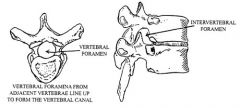
INTERVERTEBRAL FORAMINA- spaces between adjacent vertebrae which givespassage for spinal nerves to exit. VERTEBRAL FORAMINA forms vertebral canal. |
|
|
How do facets differ from demifacets? |
A facet is a flat or nearly flat surface on a bone. A demifacet is actually half of a facet |
|
|
What does facet/demifacet articulate? |
the head of the rib or part of the costal cartilage articulates and these are usually found on ribs 2 through 9. The superior demifacet of the body of the vertebra receives the head of the rib with the same number as the vertebra of interest. |
|
|
What does transverse facet articulate? |
The transverse costal facet (or transverse costal fovea) is a site where a rib forms a joint with the transverse process of athoracic vertebra. |
|
|
sacral foramina are serially homologous to what structures? |
intervertebral foramina? |
|
|
Why is the auricular surface so named? |
because its shape resembles an ear |
|
|
With what does the auricular surface articulate with? |
The auricular surface of the ileum articulates with the auricular surface of the sacrum to form the sacroiliac joint. The auricular surface of the sacrum articulates with the auricular surface of the ilium to form the sacroiliac joint. |
|
|
what is a sulcus? |
a groove or furrow, especially one on the surface of the brain |
|
|
why does the spinal cord enlarge slightly in the cervical and lumbar regions? |
The cervical and lumbar enlargements of the spinal cord result from enlargement of the gray matter that contains the neural machinery necessary to operate the limbs. |
|
|
what is a commisure? |
the joint between two bones. a band of nerve tissue connecting the hemispheres of the brain, the two sides of the spinal cord, etc. |
|
|
why are lateral grey horns located in the thoracic and lumbar regions only? |
The lateral grey column is primarily involved with activity in the sympathetic division of the autonomic motor system. It projects to the side as a triangular field in the thoracic and upper lumbar[1] regions (specifically T2-L1) of the postero-lateral part of the anterior grey column. sympathetic begins in thoracic. |
|
|
why is the sympathetic division referred to as the thoracolumbar system? |
Relating to the thoracic and lumbar portions of the vertebral column. Relating to the origins of the sympathetic division of the autonomic nervous system. |
|
|
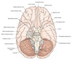
how would you id the optic nerve distinct from the optic tract? |
The optic tract is actually comprised of two separate tracts: the left optic tract and the right optic tract. It begins at the area where information from the left eye and right eye cross |
|
|
why would you need to label the optic nerve, optic tract, and optic chasm seperately? |
The optic nerve , cranial nerve II, transmits visual information to occipital cortex. Axons caudal to the intersection, called the optic chiasm, are optic tract fibers. |
|
|
which of the cranial nerves innervate most (4) of the extrinsic eye muscles? |
oculomotor (nerves III) |
|
|
4 of the cranial nerves comprise the cranial portion of the parasympathetic division of the ANS. Which 4 nerves? |
the oculomotor, facial, glossopharyngeal and vagus nerves, which are also known as cranial nerves III, VII, IX and X. An easy way to remember these is with this mnemonic: "Faeries occupy glimmering valleys," with the first 2 letters in each word matching those of the corresponding cranial nerve. |
|
|
which three of the cranial nerves are involved in taste? |
The Facial Nerve carries information from the front 2/3 of the tongue. (Cranial Nerve VII) The Glossopharyngeal Nerve carries information from the back 1/3 of the tongure (Cranial Nerve IX) The Vagus Nerve also carries some taste information, but it's a minor component (Cranial Nerve X). |
|
|
which additional nerve innervates the tongue, but is not involved in taste? what does it do? |
Motor supply for all intrinsic and extrinsic muscles of the tongue is supplied by efferent motor nerve fibers from the hypoglossal nerve (CN XII) |
|
|
Why do you think the facial nerve is so named? |
It controls the muscles of facial expression, and functions in the conveyance of taste sensations from the anterior two-thirds of the tongue and oral cavity. It also supplies preganglionic parasympathetic fibers to several head and neck ganglia. |
|
|
what is a gyrus? |
a ridge or fold between two clefts on the cerebral surface in the brain |
|
|
what is the difference between a sulcus and fissure? |
The sulci and fissures are both grooves in the cortex but they are differentiated by size. A sulcus is a shallower groove that surrounds a gyrus. A fissure is a large furrow that divides the brain into lobes, and also into the two hemispheres as the medial longitudinal fissure does. |
|
|
Auditory |
|
|
|
male vs female pelvis |
|
|
|
Cranial Bone 2 (frontal, parietal, temporal, squamosal, zygomatic process, zygomatic arch, mandibullar fossa, mastoid process, petrous portion, malleus, incus and stapes, external and internal auditory meatus, occipital, occipital condoyles, foramen magnum, ethmoid, cribriform plate, crista galli, olfactory foramina, perpendicular plate, ethmoidal sinuses, sphenoid, sphenoid body, sella turnica, hypophyseal fossa, lesser/greater wings, pterygoid processes/plates) |
|
|
|
Brain (olfactory bulb and tract, optic nerve, optic chiasm and optic tract, infundibulum and pituitary, mammillary body, pons, medulla oblongata) |
|
|
|
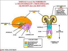
Brain (telencephalon- cerebral hemispheres, longitudinal fissure, gyrus, sulcus, frontal/temporal/parietal/occipital lobes of cerebrum, corpus callosum, olfactory bulb and tract. Sagittal- central sulcus, corpus callosum, anterior commissure, lateral ventricles. Diencephalon- optic chasm, optic tract, infundibulum and pituitary, hypothalmus, mammillary body. Sagittal- thalamus, pineal gland, optic chiasm, mammillary body, 3rd ventricle) |
|
|
|
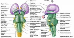
Brain (Brainstem-mesencephalon, pons, medulla oblongata, L & R superior and inferior colliculi, cerebellar peduncles, pons, 4th ventricle, medulla oblongata, central canal, Sagittal section- tectum, tegmentum, mesencephalic aquaduct, 4th ventricle) |
|
|
|
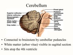
Brain (Cerebellum- peduncles, vermis, cerebellar cortex, sagittal- arbor vitae) |
|
|
|
I Olfactory |
Smell |
|
|
XI accessory |
Moves head |
|
|
II optic |
Sight |
|
|
III oculomotor |
Moves eye, pupil |
|
|
IV trochlear |
Moves eye |
|
|
V trigeminal |
Face sensation |
|
|
VI abducens |
Moves eye |
|
|
VII facial |
Moves face, salivate |
|
|
VIII vestibulococear |
Hearing, balance |
|
|
IX glossopharyngeal |
Taste, swallow |
|
|
X vagus |
Heart rate, digestion |
|
|
XII hypoglossal |
Moves tongue |