Reading...
![]()
Play button
![]()
Play button
![]()
Use LEFT and RIGHT arrow keys to navigate between flashcards;
Use UP and DOWN arrow keys to flip the card;
H to show hint;
A reads text to speech;
29 Cards in this Set
- Front
- Back
|
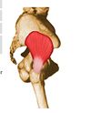
Origin Anterior surface of lateral process of sacrum and gluteal surface of ilium at the margin of the greater sciatic notch
Insertion Superior border of greater trochanter Action: laterally rotates thigh |

Piriformis
|
|
|
Origin Anterior part of the popliteal groove on lateral surface of lateral femoral condyle
Insertion Posterior surface of tibia in a fan-like fashion, just superior to the popliteal line Action Felexes and medially rotates leg |
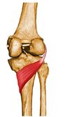
Popliteus
|
|
|
Origin Internal surface of obturator membrane and posterior bony margins of obturator foramen
Insertion Medial surface of greater trochanter of femur, in common with superior and inferior gemelli Action Laterally rotates thigh |
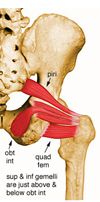
Obturator Internus
|
|
|
Origin Anterior surface of inferior pubic ramus, inferior to origin of adductor longus
Insertion Pectineal line and superior part of medial lip of linea aspera Action Abducts and medially rotates thigh |
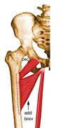
Abductor Brevis
|
|
|
Origin: From lateral lip of linea aspera, and lateral supracondylar ridge of femur.
Insertion: Primarily on fibular head; also on lateral collateral ligament and lateral tibial condyle Action: Flexes leg |
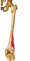
Biceps Femoris Short Head
|
|
|
Origin Dorsal ilium between inferior and anterior gluteal lines; also from edge of greater sciatic notch
Insertion Anterior surface of greater trochanter Action Abducts and medially rotates the hip joint |
Gluteus Minimus
|
|
|
Origin Anterior surface of the fibula and the adjacent interosseous membrane
Insertion Base and dorsal center of distal phalanx of great toe Action Extends great toe and dorsiflexes and inverts foot |
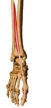
Extensor Hallucis Longus
|
|
|
Origin Inferior 2/3 of posterior surface of fibula, lower part of interosseous membrane
Insertion Plantar surface of base of distal phalanx of great toe Action Flexes Hallux, plantarflexes and inverts foot |
Flexor Hallucis Longus
|
|
|
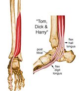
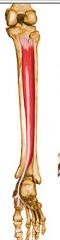
Origin Posterior surface of tibia distal to popliteal line
Insertion Splits into four slips after passing through medial intermuscular septum of plantar surface of foot; these slips then insert on plantar surface of bases of 2nd - 5th distal phalanges Action Flexes toes 2 - 5; also helps in plantar flexion and inversion of foot |
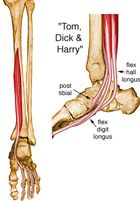
Flexor Digitorum Longus
|
|
|
Origin: Posterior aspect of interosseous membrane, superior 2/3 of medial posterior surface of fibula, superior aspect of posterior surface of tibia,
Insertion Splits into two slips after passing inferior to plantar calcaneonavicular ligament; superficial slip inserts on the tuberosity of the navicular bone deeper slip divides again into slips inserting on plantar sufraces of metatarsals 2 - 4 and second cuneiform Action Principal invertor of foot; plantar flexes ankle, |
Tibialis Posterior
|
|
|
Origin Inferior 2/3 of lateral fibular surface; also anterior and posterior intermuscular septa of leg
Insertion Lateral surface of styloid process of 5th metatarsal base Action Everts foot and plantar flexes ankle |
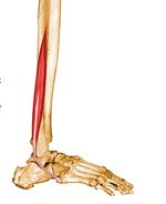
Fibularis Brevis
|
|
|
Origin Medial third of superior nuchal line; external occipital protruberance, nuchal ligament, and spinous processes of C7 - T12 vertebrae
Insertion Lateral third of clavicle, acromion, and spine of scapula Action Elevates, depresses, rotates, and retracts scapula |
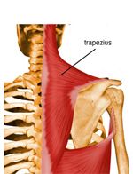
Trapezius
|
|
|
Origin Spinous processes of inferior 6 thoracic vertebrae, thoracolumbar fascia, iliac crest, and inferior 3 or 4 ribs
Insertion Floor of intertubercular groove of humerus Action Extends, adducts, and medially rotates humerus; |
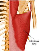
Latissimus Dorsi
|
|
|
Orign: spinous processes of T2 - T5 vertebrae
Insertion: Medial border of scapula from level of spine to inferior angle Action: Rotates downward and retracts scapula |
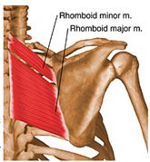
Rhomboid Major
|
|
|
Origin: Nuchal ligament and spinous processes of C7 and T1 vertebrae
Insertion Medial border of scapula from level of spine to inferior angle Action: Rotates downward and retracts scapula |

Rhomboid Minor
|
|
|
Origin Posterior tubercles of transverse processes of C1 - C4 vertebrae
Insertion Superior part of medial border of scapula Action Elevates scapula and tilts its glenoid cavity inferiorly by rotating scapula |
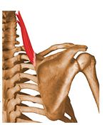
Levator Scapulae
|
|
|
Origin 3rd to 5th ribs near their costal cartilages
Insertion Medial border and superior surface of coracoid process of scapula Action: Depresses, protracts and rotates scapula downward |
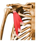
Pectoralis Minor
|
|
|
Origin: Anterior and superior margins of ribs 1-8 or 1-9
Insertion: Anteriro surface of vertebral border of scapula. Action: Protracts and rotates scapula upwards. |
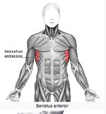
Serratus Anterior
|
|
|
Muscles of the Pectoral Girdle
|
Trapezius
Rhomboid major Rhomboid Minor Levator Scapulae Pectoralis Minor Serratus Anterior * Actions of these muscles involve the movement of the scapula.** |
|
|
Muscles of the Shoulder Joint
|
Pectoralis Major
Latissimus Dorsi Deltoid Supraspinatus Infraspinatus Subscapularis Teres minor Teres major Coracobrachialis *Actions of these muscles involve the movement of the arm** |
|
|
Muscles of the Elbow Joint
|
Biceps brachii long head
Biceps brachii short head Brachialis Tricepts brachii long head Tricepts brachii lateral head Tricepts brachii medial head Brachioradialis Pronator Teres Supinator Action of these muscles involve the movement of the forearm. |
|
|
Muscles of the Wrist Joint
|
Wrist Flexors:
Flexor carpi radialis Flexor carpi ulnaris Palmaris longus Wrist Extensors: Extensor Digitorum Extensor Carpi Radialis |
|
|
Muscles of the Hip Joint
|
Psoas major
Iliacus Gluteus maximus Gluteus medius Gluteus minimus Tensor fascae latae Piriformis Obturator internus Adductor magnus Adductor longus Adductor brevis Pectinueus |
|
|
Muscles of the hip and knee Joint
|
Gracilis
Sartorius Rectus Femoris Semitendinosus Semimembranosus Biceps femoris long head |
|
|
Muscles of the knee joint
|
Biceps femoris short head
Vastus lateralis Vastus medialis Vastus intermedius Popliteus |
|
|
Muscles of the knee and ankle joint
|
Gastrocnemius: medial head and
lateral head. |
|
|
Muscles of the ankle joint
|
Tibialis anterior
Tibialis posterior Extensor hallucis longus Extensor digitorum longus Fibularis longus Fibularis brevis Soleus Flexor digitorum longus |
|
|
Muscles of Massication
|
Muscles that move the mandible:
Masseter Temporalis Playsma Platysma and Masseter are antagonistic pairs. |
|
|
Steps that result in the contraction of Skeletal Muscle (Sliding filament theory)
|
1. The motor neuron at the neuormuscular juntion releases acetylcholine (Ach) to bind to the sarcolemma of the skeletal muscle fiber.
2. The signal spreads throughout sarcolemma of skeletal muscle fiber 3. The sginal is spread down T-tubules to center of cell. 4. T-tubules spread signal to cell interior and pass signal to the Sarcoplasmic Recticulum (SR). 5. Calcium is relesed from the SR 6. Calcium combines with the troponin-tropomysonsin complex on actin exposing binding sites for myosin. 7. Myosin crossbridge heads with ATP go from a low affinity state to a high affinity state by splitting ATP into ADP + Pi causing heads to attach to binding sites on actin. 8. Attachement of myosin to actin results in the release of Pi and actin "slides" past myosin (sarcomeres shorten/ muscle cell shortens/ z lines pull together) 10. ATP molecule binds to myosin heads resulting in the release of crossbridge attachment to actin. |