![]()
![]()
![]()
Use LEFT and RIGHT arrow keys to navigate between flashcards;
Use UP and DOWN arrow keys to flip the card;
H to show hint;
A reads text to speech;
43 Cards in this Set
- Front
- Back
|
What are the main functions of the thorax?
The internal aspect of the thoracic wall is covered by the ____ which separates the internal muscles from what pluera? |
to protect the contents of the thoracic cavity but also provide the mechanical function of breathing -endothoracic fascia -parietal fascia |
|
|
the ___ is the superior part of the trunk between the neck and the abdomen. The ___ surrounded by the thoracic wall contains the thymus, heart , lungs, distal part of the trachea, and most of the esophagus The ___ is an osteocartilagenous structure that protects the thoracic organs and some abdominal organs (eg liver) |
thorax (chest) thoracic cavity thoracic cage |
|
|
the bony thorax includes what? (3)
what are the two distinct apertures in the thoracic cage? |
12 thoracic vertebrae, 12 ribs and costal cartilage and the sternum ( manubrium, body, and xiphoid process)
superior thoracic aperture (inlet) and (outlet) inferior thoracic aperture |
|
|
what are the bones of the thoracic inlet (superior)?
What are the contents of the thoracic inlet? |
first pair of ribs and cartilage, manubrium sterni (superior border), and first thoracic vertebrae
trachea, esophagus, vagus nerve (CN X), phrenic nerve (C3,4,5), and apex of the lungs |
|
|
What are the bones the thoracic outlet? |
the 12th thoracic vertebrae, the 12th pair of ribs , and the costal margin (costal cartilage 7-10) |
|
|
Important landmarks: ____ at T10 or Tt11 ____ at the lower border of T4 ____ corresponds directly to the manubriosternal joint the ___ is the angle formed between the manubrium and the body of the sternum (manubriosternal joint) |
Xiphoid sternal angle costal cartilage of 2nd rib angle of louis |
|
|
The angle of louis marks the level of the ___ costal cartilage. it also denotes the level of the what? (3) |
2nd aortic arh, bifurication of the trachea (carina), T4/T5 intervertebral disc |
|
|
Rib __ is the largest Rib __ is the shortest Ribs _ are the typical ribs Ribs __ are the atypical ribs Ribs ___ articulate directly with the sternum through the costal cartilages and are the true ribs Ribs ___ share a common costal cartilage and are the false ribs |
7 12 3-9 1,2,10,11,12 1-7 8,9,10 |
|
|
Ribs __ do not articulate anteriorly , they end among the muscles of the body wall so they are the floating ribs _ is located at the inferior aspect of the ribs, shelters the intercostal neurovascular bundle (vein, artery, nerve- VAN) The first rib is short and broad, forms part of the thoracic inlet and it has a groove for the __ artery and vein |
11, 12 Costal Groove sublclavian |
|
|
Deepest muscle of the rib cage is what? What are the main functions of the thoracic cage? |
-innermost intercostal muscle -protection of internal organs, framework of attachment of muscles and assitance in respiration |
|
|
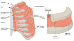
The Innermost intercostal muscle consist of what 3 parts? ___ are between the ribs ___ muscle is at an angle and is variable in size and shape. It is more developed inferiorly, and it covers one or two intercostal space ___ muscle are continuous with transversus abdominus muscle. (posterior aspect of sternum) |
innermost proper, subcostalis muscle, and sternocostalis (transversus thoracis)
innermost proper subcostalis sternocostalis (transversus thoracis)
|
|
|
What forms the deepest muscular layer of the thoracic cage? |
innermost proper, subcostalis, and transversus thoracis |
|
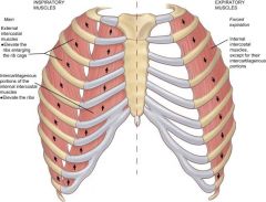
Name the muscle, innervation and function |
Internal and external intercostal muscle fibers corresponding intercostal nerve External will elevate the ribs and help with inspiration. Internal will help with inspiration |
|
|
Innermost intercostal muscle (subcostalis, transversus thoracis (sternocostalis), innermost costalis) -corresponding intercostal nerve -Expiration |
|
|
If the long thoracic nerve is lacerated what muscle is damaged and what are the clinical considerations of this situation? |
Serratus anterior, wing scapula, loss of elevation of arm, and problems with respiration |
|
|
What are the three origins of the diaphragm? What are the three openings of the diaphragm? __ opening- thoracic aorta, thoracic duct, greater splachnic nervers (t5-9), and azygos veins __opening- esophagus, right and left vagus nerve __opening- phrenic nerves (C3,C4,C5), IVC, Lymphatics
|
sternal, costal, vertebral Aortic (T12), esophageal (T10), caval (T8) aortic esophageal Caval |
|
|
What is a direct branch off the subclavian artery? What is a indirect branch off the subclavian artery? What are the arteries of the thoracic wall? |
internal thoracic artery highest intercostal artery (from costocervical trunk) internal thoracic, highest intercostal, posterior intercostal, and subcostal arteries |
|
|
The anterior aspect of the thoracic wall is supplied by what artery? The posterior aspect of the thoracic wall is supplied by the ___ in ICS1, ____ artery in ICS 2-11 and ___ artery in ICS 12 |
internal thoracic artery posterior intercostal artery or highest intercostal artery posterior intercostal artery (thoracic aorta) subcostal artery (thoracic aorta) |
|
|
In the anterior aspect of the wall ICS 1-6 are supplied by the __ artery. ICS 7,8,9 are supplied by the ___ artery. And ICS 10 and 11 are by what? |
internal costal artery musculophrenic (from internal thoracic) no blood supply |
|
|
The anterior aspect of the thoracic is drained by the what? the posterior aspect of the thoracic is drained by the what? On the right side- the 1st drains into ___, 2nd and 3rd into ___ vein, 4th through 11th drain into __ vein and 12th drains into __ vein |
internal thoracic into the brachiocephalic vein azygos system into the SVC highest intercostal vein (drains into brachiocephalic) higher intercostal vein ( azygos vein) azygos vein subcostal vein (drains into azygos) |
|
|
the left side of the thoracic wall, the ICS1 drain into ___ vein, the 2nd, 3rd, and 4th drain into __ vein. the 5th through 11th and 12th drain into the __ vein |
highest drains into brachiocephalic vein accessory hemiazygos hemiazygos |
|
|
The Right upper extremity or the area to the right drains into the ___ duct. The rest of the body drains into the ___ duct. |
lymphatic thoracic |
|
|
When penetrating the Mid Axillary line the ___ layer is where you wanna stop. The endothoracic fascia is the CT layer that functions to prevent the ___ from rubbing agaisnt the innermost intercostal muscle. This fascia is thickened over the apex of the lungs which is called ____ |
Endothoracic fascia parietal pleura Sibsons Fascia |
|
|
___ is when rib rising from C7 (0.5%) which means there is brachial plexus compression, and subclavian compression (subclavian artery is not on the clavicle __ is displacement of the costal cartilage from the sternum (2-7) (contact sports) ___ is dislocation pf the costochondral joint between the rib and its costal cartilage |
cervical rib rib dislocation rib separation |
|
|
___ aka funnel chest, body of the sternum projects inferiorly and posteriorly pressing on the heart, which widens it making it look large on AP radiographs ___ aka pigeon chest, sternum projects anteriorly |
Pectus Excavatum Pectus Carinatum |
|
|
____ is surgical puncture of the chest wall using a tap ___ can use a needle (medial or lateral approach) (thoracentesis) or a tube (ICS 4 or ICS 5 at the anterior axillary line) ___ surgical incision of the chest wall. Over ICS 4 or ICS 5, extending from the lateral margin of the sternum to the anterior axillary line. use to expose heart for cardiac massage |
Thoracentesis Thoracostomy Thoracotomy |
|
|
The ___ are the covering membranes of the lungs and with them occupy the lateral parts of the thoracic cavity The parietal layer of pleura, ___ forms the its external wall. And ___ invests the lungs |
pleurae pleural sac parietal pleura Visceral pleura |
|
|
The ___ lines the thoracic wall and covers the thoracic surface of the diaphragm and the lateral surface of the mediastinum The ___ covers the outer surface of the lungs and extends into the interlobar fissures The pareital pluera becomes continuous with the visceral pleura at the ___ of each lung |
parietal pleura visceral pleura hilum |
|
|
The __ cavity is a slit like space that separates the parietal and visceral pleurae. __ fluid lubricates the apposing pleural surfaces and reduces friction Cervical Pleura or ___ The ___ is the area of parietal pleura relected over the apex of the lungs |
pleura pleura fluid Cupula Cupula |
|
|
A ___ is a potential pleural space that is not occupied by the lungs during quiet respiration. what are the 3 recesses of the pleural recesses? __ recess is lowest area of the plueral cavity into which the lungs expand during inspiration . A tap can be performed here at ICS_ |
Pleural Recess Costodiaphragmatic, costomediastinal, and costovertenbral recesses Costodiaphragmatic 9 |
|
|
What is the blood supply of the parietal pleura? what is the blood supply of the visceral pleura?
What is the innervation of the parietal pleura? what is the innervation of the visceral pleura? Is the parietal pleura sensitive to pain? what about the visceral pleura? |
intercostal arteries, and internal thoracic artery Bronchial arteries (thoracic aorta)
intercostal nerves (1-11), subcostal nerve (Tn12), Phrenic nerve (Cn 3,4,5) -no sensory innervation -Yes -no
|
|
|
What lies between the lungs in the mediastinum? How are the lungs attached to the medaistinum? How are the lungs attached to the pericardium? The ___ is about 1 inch or 2.5 cm above the clavicle. The concave base sits on the ___, a convex ___ surface that corresponds to the concave chest wall, and a concave __ surface that is molded into the pericardium . |
heart and great vessels roots (pulmonary arteries and veins, and main bronchi) pulmonary ligaments apex diaphragm costal mediastinal
|
|
|
The anterior border of the lung, it is notched on the left lung=___ Oblique fissure is at __ costal cartilage Horizontal fissure is at __ costal cartilage The left lung has a modified middle lobe called ___ |
cardiac notch (costal cartilage 4,5,6) 6 4 lingula |
|
|
the __ lung has a tracheal and esophageal areas, a groove for the SVC and IVC, cardiac depression, a groove for the azygos vein, and a concave diaphragmatic depression. the __ lung has large cardiac depression, a groove for the aortic arch, and a groove for the descending aorta, as well as a concave diaphragmatic depression |
right left |
|
|
What is the arterial supply of the lungs? What is the venous supply of the lungs? What is the lymphatic drainage of the lungs? |
-bronchial arteries (thoracic aorta), and pulmonary arteries -bronchial veins drain into the azygos system and into the pulmonary veins -from the pulmonary nodes to the tracheobronchial nodes |
|
|
Innervation: __ nerve will cause bronchoconstriction and vasodilation, and increase glandular secretion. also does the senosry of the lungs ___ nerve has a vasomotor innervation of T2-T5 and produce bronchial dilation and vasoconstriction and inhibits glands of the bronchial tree |
Vagus (parasympathetic) Sympathetic |
|
|
The ___ is a mobile fibrocartilagenous tube, the superior half of which is located in the cervical region Where does the trachea begin? What is it anterior to? What does it enter into? At the angle of louis it deviates into what plane before it divideds? How and where does it end? |
Trachea lower border of cricoid cartilage (c6) esophagus superior mediastinum slightly right to the median plane By dividing into the right and left main bronchi at the level of the sternal angle (T4 or T5)
|
|
|
The trachea is kept patent by a series of what? What is anterior to trachea? What is posterior to trachea? |
series of 15-20 C shaped cartilage -sternum, thymus, arch of aorta, origins of brachiocephalic trunk and left common carotid -esophagus, Azygos vein and right vagus and pluera (on the right); aortic arch, left common carotid, left subclavian artery and left vagus, left phrenic and pluera (on the left) |
|
|
What is the blood supply of the trachea? What is the nerve supply of the trachea? |
Superior thyroid artery (external carotid artery), inferior thyroid artery (thyrocervical trunk [subclavian]), bronchial arteries (thoracic arteries), internal thoracic artery (subclavian artery) -Vagus (CN X) and sympathetic chain |
|
|
The __ bronchi is wider, shorter and more vertical than the __ bronchi. Before entering the hilum it gives off the ___ bronchus. When it enters the hilum it divides into what? The ___ bronchi is narrower, longer and more horizontal the the right bronchi. it passes below what? What is it in front of? When it enters the hilum what doe it divide into what? |
right left superior lobar bronchus middle and inferior lobar bronchus left arch of aorta espophagus superior and inferior lobar bronchus
|
|
|
What are the main blood supply of the bronchi? What is the venous drainage of the bronchi?
Bronchial arteries: two on the left from the __ and one on the right from the ___ artery |
The bronchial arteries (supply the conductive airways, non respiratory), pulmonary arteries to the terminal portion of the bronchioles -bronchial veins to azygos and hemiazygos veins -thoracic aorta -first posterior intercostal artery |
|
|
Innervation: __ nerve leads to vasoconstriction and mucus secretion. __ nerve leads to bronchodilation |
Vagus (parasympathetic) Sympathetic (T2-T5) |
|
|
___ is inflammation of the pleurae. Pain is referred to the thoracic/abdominal wall. hear a "pleural rub" on ascultation To prevent recurrent spontaneous pneumothorax, the pleurae are obliterated surgically called ___. ___ is lung collapse, the pleurae sacs are separated |
Pleuracy/Pleuritis Pleural poundrage (type of Pleurectomy) Atelectasis |