![]()
![]()
![]()
Use LEFT and RIGHT arrow keys to navigate between flashcards;
Use UP and DOWN arrow keys to flip the card;
H to show hint;
A reads text to speech;
104 Cards in this Set
- Front
- Back
|
Small bones found in tendons are termed __________. |
Sesamoid |
|
|
The shaft of a long bone is mostly made of __________ type tissue, while the extremities of long bones are typically made of __________ tissue. |
Compact/cortical Spongy/cancellous |
|
|
The hollow central portion of a long bone is termed the __________ and is filled with __________. |
Medullary cavity Bone marrow |
|
|
What are at least two examples of flat bones? |
Calvarium Sternum Ribs Scapulae |
|
|
What are at least two examples of irregular bones? |
Facial bones Vertebrae Pelvis |
|
|
The membranous covering of a bone is termed __________. |
Periosteum |
|
|
The membranous covering of cartilage is termed __________. |
Perichondrium |
|
|
The type of cartilage found at the extremities of long bones and at their articulating surfaces is called __________. |
Hyaline cartilage |
|
|
The term used to describe secondary centers of ossification is _________; the term for primary ossification centers is _________. |
Epiphysis Diaphysis |
|
|
What is the name of the somewhat wider portion of a long bone adjacent to the epiphyseal plate? |
Metaphysis |
|
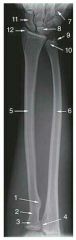
Identify the labeled parts 1 through 12 seen in this image. |
1. Radial tuberosity 2. Neck of radius 3. Head of radius 4. Proximal radioulnar joint 5. Radius 6. Ulna 7. Base of 5th metacarpal 8. Lunate 9. Styloid process of ulna 10. Head of ulna 11. Scaphoid 12. Radial styloid process |
|
|
What is the most lateral carpal of the distal carpal row? |
Trapezium |
|
|
The radiocarpal joint is the articulation between the radius and which carpal? |
Scaphoid |
|
|
What articulation is formed by the ulnar notch and radial head? |
Proximal radioulnar articulation |
|
|
The lateral aspect of the distal humerus presents a raised smooth surface called the _________, which articulates with the superior surface of the __________. |
Capitulum Radial head |
|
|
Tennis elbow is a painful condition that affect what bony area of the elbow? |
Lateral epicondyle Sometimes radial head |
|
|
In which projection of the elbow should the coronoid process be viewed in profile? |
Internal oblique |
|
|
How and where should the CR enter for the PA projection of the hand? |
Perpendicular to the third MCPJ |
|
|
How and where should the CR enter for the AP projection of the shoulder? |
Perpendicular to 1" inferior to the coracoid process |
|
|
How and where should the CR enter for the PA projection of the second digit? |
Perpendicular to the second PIPJ |
|
|
Which projection on the digits will best demonstrate forward/backward fracture displacement? |
Lateral |
|
|
The common fracture located at the base of the first metacarpal is called a/an __________ fracture. |
Bennett fx |
|
|
The type of fracture common to the neck of the fourth or fifth metacarpal is the __________ fracture. |
Boxer |
|
|
A fracture of the distal radius characterized by anterior displacement of the fragments is termed a __________ fracture. |
Smith fx |
|
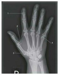
Identify the numbered structures in this image; what bone does number 3 articulate? |
1. DIPJ of second digit 2. Sesamoid bone 3. Base of first metacarpal 4. Head of 5th metacarpal
Trapezium |
|
|
The bones that form the palm of the hand are called the __________; that form the digits are called the __________. |
Metacarpals Phalanges |
|
|
If the AP diameter of the concave carpal arrangment is diminished, the __________ nerve is impinged upon; this condition is called __________. |
Median nerve Carpal tunnel syndrome |
|
|
What is the most commonly fractured carpal? |
Scaphoid |
|
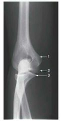
The image seen here is a __________ projection of the elbow. Identify the labeled parts. |
AP internal oblique 1. Medial epicondyle 2. Trochlea 3. Coronoid process |
|
|
Fracture of the distal radius, accompanied by posterior displacement and fracture of the ulnar styloid process, is typical of what type of fracture? |
Colles fx |
|
|
The __________ portion of the radius and ulna are superimposed when the hand is __________. |
Proximal Pronated |
|
|
What elbow fat pad, usually unseen in normal and accurately positioned lateral elbow, is often visible in the presence of pathology? |
Posterior fat pad |
|
|
What position will best demonstrate medial/lateral fracture displacement? |
AP |
|
|
What bones form the shoulder girdle? |
Two scapulae and clavicles |
|
|
What structure is composed of the supraspinatus, infraspinatus, teres minor, deltoid, and subscapularis muscles? |
Rotator cuff |
|
|
An oblique hand image demonstrates foreshortening of the digits in poor visibility of the interphalangeal joints how can this be corrected? |
Extend the fingers and make them parallel to the IR |
|
|
The PA wrist with ulnar deviation is to better demonstrate which carpal? |
Scaphoid |
|
|
Where should the CR enter for an AP projection of the first digit? |
Perpendicular to the first MCP |
|
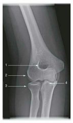
Identify the labeled structures in this image. |
1. Olecranon fossa 2. Capitulum 3. Radial head 4. Coronoid process |
|
|
The PA projection of the hand provides a/an __________ projection of the first digit. |
Oblique |
|
|
The carpal canal, tangential inferosuperior, projection of the wrist requires a CR angulation of how many degrees and in what direction? |
25-30° to the long axis of the hand |
|
|
If a lateral projection of the forearm demonstrates epicondyles not super imposed how should that positioning be corrected? |
Place the humerus and forearm on the same plane |
|
|
When the elbow cannot be extended for an AP projection, what projections can be taken to demonstrate the required AP anatomy? |
AP with forearm parallel to the IR AP with humerus parallel to the IR |
|
|
AP lateral oblique, external rotation, projection of the elbow should demonstrate what structure free of superimposition? |
Radial head |
|
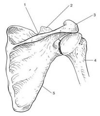
Identify the labeled and atomic parts seen in this image. |
1. Clavicle 2. Head of humerus 3. Scapula |
|
|
The AP medial oblique, internal rotation, projection of the elbow should demonstrate what structure free of superimposition? |
Coronoid process |
|
|
Images made of the elbow in the lateral position with the hand in external rotation, lateral position, pronation, and internal rotation are done to demonstrate what structure? |
Radial head |
|
|
What is the long, curved process that extends laterally above the head of the humerus? |
Acromion process |
|
|
What is the only articulation between the upper extremity and thorax? |
Sternoclavicular joint |
|
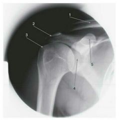
Identify the labeled anatomic parts seen in this image. |
1. Clavicle 2. Acromion process 3. Greater tubercle 4. Glonoid fossa 5. Coracoid process |
|
|
A true AP projection of the shoulder is obtained in the __________ rotation position; the __________ should be seen in profile laterally. |
External Greater tubercle |
|
|
How can you obtain a lateral projection of the humerus in the case of trauma? |
Transthoracic |
|
|
The inferosuperior axial projection of the shoulder requires that the arm be abducted 90° from the body and should be in the __________ rotation position. |
External |
|
|
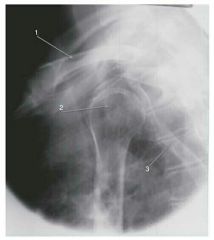
The posterior oblique position, Grashey method, of the shoulder requires that the part be rotated so that the __________ of the affected side is parallel to the IR. |
Scapula |
|
Identify the numbered anatomical parts in the illustration seen here. |
1. Scapular notch 2. Coronoid process 3. Acromion process 4. Humerus 5. Axillary/lateral border |
|
|
An internal rotation projection of the shoulder should demonstrate the __________ in profile medially. |
Lesser tubercle |
|
|
In a Y-view, where is an anterior humeral dislocation seen? |
Beneath the coracoid process |
|
|
How are AC joints usually examined in order to demonstrate small separations? |
With and without weights |
|
|
In order to visualize a larger portion of the scapula in the AP projection, how should the arm be positioned? |
Abducted |
|
|
The vertebral and axillary borders should be superimposed in the __________ projection of the scapula. |
Lateral |
|
|
Which metatarsal has a large tuberosity, commonly subject to trauma? |
5th |
|
|
Which tarsal lies immediately anterior to the talus? To the calcaneus? |
Navicular Cuboid |
|
|
Bones formed in tendons, often near articulations, are called __________. |
Sesamoids |
|
|
What bones form the ankle joint? |
Tibia Fibula Talus |
|
|
The lateral malleolus is the distal expanded end of which bone? |
Fibula |
|
|
What position best demonstrates visualization of the entire ankle mortise? |
15° internal oblique |
|
|
Osgood-Schlatter disease affects what bony part? |
Tibial tuberosity |
|
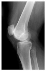
What positioning change(s) should be made to improve this image? |
Rotate posteriorly |
|
|
What are the names of the crescent-shaped fibrocartilage discs located on the tibial plateau? |
Lateral and medial menisci |
|
|
Which projection of the toes should demonstrate no overlapping of soft tissues? |
AP |
|
|
The dorsoplantar projection of the foot requires that the CR be directed how much, in what direction, and to what point? |
10° posteriorly to the base of the third metatarsal |
|
|
Cuboidal articulations and the sinus tarsi are well visualized in which projection of the foot? |
AP medial oblique |
|
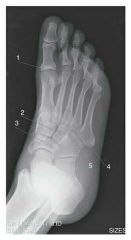
Identify the numbered anatomical structures in this image. |
1. 1st MCPJ 2. Lateral cuneiform 3. Navicular 4. Base of 4th metatarsal 5. Cuboid |
|
|
The AP medial oblique projection of the foot requires what degree of obliquity? |
30° |
|
|
How should the foot be examined in order to demonstrate the longitudinal arches and ligament injuries? |
Weight-bearing |
|
|
The dorsoplantar axial projection of the calcaneus requires that the CR be directed how many degrees and in what direction to the plantar surface? |
40° caudad |
|
|
Where should the CR be directed for the AP projection of the ankle? |
Midway between the malleoli |
|
|
For the AP projection to demonstrate congenital clubfoot, Kite method, how should the foot be aligned on the IR? |
In its natural AP position; no attempt should be made to straighten the foot |
|
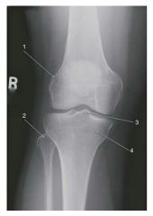
Identify the numbered anatomical structures in this image. |
1. Lateral femoral epicondyle 2. Head of fibula 3. Intercondylar eminence 4. Medial tibial condyle |
|
|
What is the relationship between the plantar surface and the IR in the plantodorsal axial projection of the calcaneus? |
Perpendicular |
|
|
The AP oblique, medial rotation, of the ankle requires what degree of obliquity? |
45° |
|
|
A 15° medial oblique ankle is performed to demonstrate the __________. |
Mortise |
|
|
In order to demonstrate both the ankle and knee joints on an examination of the lower leg, the IR is often placed __________ to the long axis of the lower leg. |
Diagonal |
|
|
An AP projection of the knee should be centered at what point? |
1/2" distal to the patellar apex |
|
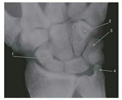
Identify the numbered anatomical structures in this image. |
1. Scaphoid 2. Hamate 3. Triquetrum 4. Ulnar styloid process |
|
|
If the distance between the ASIS and tabletop is less than 19 cm, what tube angle should be employed for the AP projection of the knee? |
5° caudad |
|
|
What are two evaluation criteria for correct positioning of the lateral knee? |
Femoral condyles superimposed Patellofemoral joint space open |
|
|
To evaluate knees for arthritis, it is recommended that they be imaged in the __________ position. |
Weight-bearing |
|
|
To visualize the proximal tibiofibular articulation, the AP knee should be obliqued how many degrees in which direction? |
45° medially |
|
|
The PA axial projection of the intercondyloid fossa requires that the CR be directed __________ to the long access of the tibia. |
Perpendicular |
|
|
The PA tangential projection (Hughston method; "sunrise") of the patella and the patellofemoral joint requires that the knee may be flexed about 50° and the CR directed how many degrees and in what direction? |
45° cephalad |
|
|
What image is usually made to determine pediatric bone age? |
PA (left) wrist and hand |
|
|
Long bone measurement examinations are usually performed to detect ___________. |
Limb length differences between left and right sides |
|
|
Punched-out radiolucent lesions are representative of the malignant condition called __________, and the x-ray examination most frequently requested to diagnose this condition is __________. |
Multiple myeloma Skeletal/bone survey |
|
|
X-ray Imaging of bony articulations, and their soft tissue structures, using positive and/or negative contrast agents is termed __________. |
Arthrography |
|
|
Fluoroscopic and radiographic examination of the spinal cord and its meninges using positive contrast agents is termed __________. |
Myelography |
|
|
What is the type of fracture that vertebral bodies are subject to, especially in cases of osteoporosis? |
Compression fx |
|
|
What is the name of the final stage of healing/repair of a bone fracture? |
Remodeling |
|
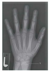
The study/projection shown here is most likely performed for __________. Identify the part indicated as #1. |
Bone age study Epiphyseal plate |
|
|
What is the type of bone fracture where bone appears shattered or broken into several fragments? |
Comminuted |
|
|
What is the type of fracture that can be common to the patella or cranium, radiating from a central point, and being star-shaped? |
Stellate |
|
|
What is the term used to describe movement of fractured ends of bones away from each other? Name the term used to describe the gap between fractured ends of bones. |
Displacement Distraction |
|
|
A break in the bony cortex on one side of the shaft/body of a long bone, especially in children, is termed __________. |
Greenstick |
|
|
A small chip of bone that breaks away when a joint is dislocated, or when a tendon is pulled, is termed __________. |
Avulsion |