Reading...
![]()
Play button
![]()
Play button
![]()
Use LEFT and RIGHT arrow keys to navigate between flashcards;
Use UP and DOWN arrow keys to flip the card;
H to show hint;
A reads text to speech;
32 Cards in this Set
- Front
- Back
|
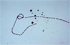
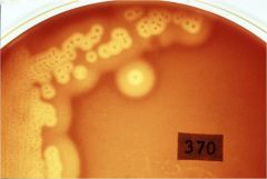
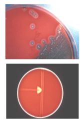
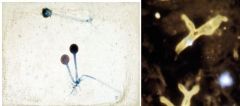
A positive result for CAMP test is indicated by ?
|
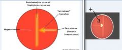
A positive result is indicated by an "arrowhead"-shaped enhanced zone of b-hemolysis
|
|

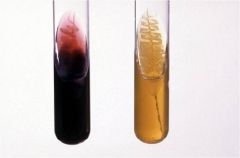
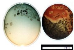
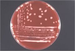
Triple Sugar Iron agar
Which is Salmonella and which is E.coli? Which examples that might yield these results? proteus, kleibsiella, psuedom.? |
Which is which?
left: Salmonella (red or purple; not fermenting) right: E. coli (yellow due to low pH) fyi: proteus, kleibsiella also produce yellow color with TSI; while pseudomonas retains red color |
|
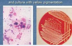
Identify from following information:
Gram + Bacillus Catalase test Pos. CAMP + Glucose fermentation + |
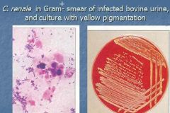
Corynebacterium renale
Gram + Bacillus Catalase test Pos. CAMP + Glucose fermentation + |
|
Identify from following information:
Gram + Bacillus Catalase test Pos. CAMP + Glucose fermentation - |
Rhodococcus equi
Gram + Bacillus Catalase test Pos. CAMP + Glucose fermentation - |
|
|
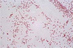
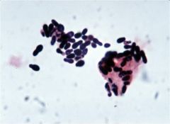
Which forms chains strep or staph?
|
Streptococcus
|
|
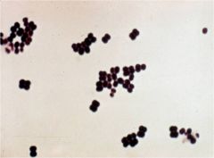
Strep or staph?
|

Staphylococcus
|
|
Which staph have dbl. zone hemolysis?
|

Staphylococcus aureus & S. intermedius
|
|
Catalase positive strep or staph?
|

Staphylococcus
Catalase test + |
|
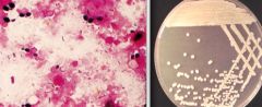
Staph. which forms non-hemolytic colonies on blood agar
|

Staphylococcus epidermidis – forms non-hemolytic colonies on blood agar
|
|

type hemolysis?
|
: alpha hemolytic streptococci. (note greening/ incomplete hemolysis)
|
|
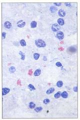
Which Strep. exclusively associated with mammary gland. Recurring mastitis
Catalase negative, mostly B hemolytic, CAMP +ve (arrow-head pattern) |

S. agalactiae
|
|
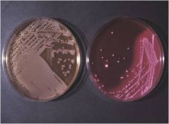
Lactose fermenters (LF) appear what color on plate?
(which plate above?) Give 2 example of gram negative lactose fermenters: |
Lactose fermenters (LF) (Pink on Mac)(right)
Escherichia coli, Klebsiella |
|

Examples of Non-Lactose fermenters (NLF)
|

Non-Lactose fermenters (NLF)
(no color on MAC) Salmonella, Proteus note: these all Gram-ve rods |
|

Mucoid colonies on blood agar
Lactose fermenting pink colonies on MacConkey agar |
Klebsiella pneumoniae: mucoid colonies on blood agar
Lactose fermenting pink colonies on MacConkey agar |
|

T/F
E. coli: Gram smear(Klebsiella, Salmonella, Proteus: cannot be distinguished in smear) |
True
|
|
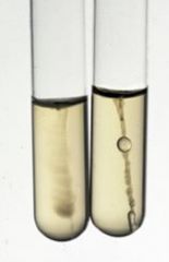
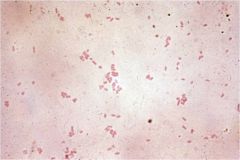
E. Coli and Klebsiella inoculated into soft agar ; urease test
Results? |

E. Coli and Klebsiella inoculated into soft agar (SIM: sulfide-indole-motility medium):
E. coli: motile; urease – negative (clear/yellow) Klebsiella: non-motile; urease – positive (not shown but should appear red) |
|

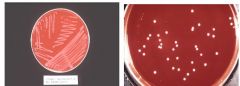
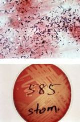
swarming growth on blood agar plate
|
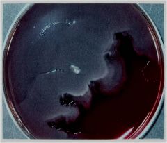
Proteus mirabilis : swarming growth on blood agar plate
|
|

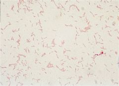
Gram - Rod
Non-lactose-fermenter Most strains produce green pigment Colorless to greenish colonies on MacConkey medium |
Pseudomonas aeruginosa : Gram smear (G-ve rods )
Non-lactose-fermenter Colorless to greenish colonies on MacConkey medium |
|
|
Is Pseudomonas aeruginosa: positive oxidase test?
|
Yes
Pseudomonas aeruginosa: greenish colonies and a positive oxidase test FYI: some strains hemolytic, some not! |
|
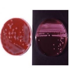
Culture : non hemol colonies, no growth- MacConkey
Culture exudates, lung or transtracheal wash Culture nasal swabs from pigs (atrophic rhinitis), rabbits (snuffles) |

PASTEURELLOSIS (P.multocida)Coccobacilli
|
|
(Gram-negative, curvy rods)
hint: incubated in a microaerophilic atmosphere |

Campylobacter
|
|
Terminal spores (drumstick)
|

Terminal spores (drumstick)
Tetanus (lockjaw) in animals, humans Habitat: soil |
|

(acid fast stain) on rectal scraping -> Fig: clumps of bacilli (pink)
Johne’s disease: diagnosis |
Mycobacterium paratuberculosis
|
|

Unicellular fungi (yeasts)
|
Malassezia in dogs’s ear: Unicellular fungi (yeasts)
|
|
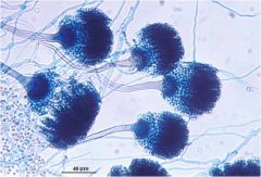
on Sabouraud agar, 5 days
|

Candida albicans on Sabouraud agar, 5 days
|
|
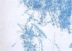
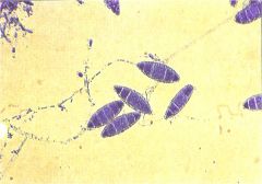
spindle shaped macroconidia (lactophenol cotton blue wet mount from culture)
|

Microspoum canis : spindle shaped macroconidia (lactophenol cotton blue wet mount from culture)
|
|

: boat shaped macroconidia
|
Microsporum gypseum : boat shaped macroconidia
|
|
wet mount from culture (lactophenol cotton blue stain)
|

Aspergillus fumigatus : wet mount from culture (lactophenol cotton blue stain)
|
|

What closed sporangium (left)Aseptate hyphae in tissue
|
Zygomycosis Rhizopus : closed sporangium (left)Aseptate hyphae in tissue
|
|

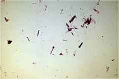
Gram stain of pus (Gr+ Bacillus)
Catalase Negative CAMP - Culture on BA” Penicillin effective |
Arcanobacterium pyogenes
|
|
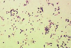
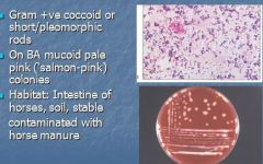
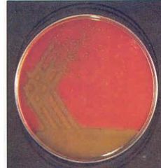
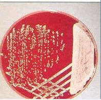
identify
colonies that tend to become salmon pink on BA |

Rhodococcus equi (formerly Corynebacterium equi): colonies that tend to become salmon pink
|
|
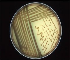
identify culture on BA
Gram-positive, catalase-positive, rod-shaped bacteria. It forms partially acid-fast beaded branching filaments (acting as fungi, but being truly bacteria). |
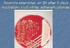
Nocardia
Gram-positive, catalase-positive, rod-shaped bacteria. It forms partially acid-fast beaded branching filaments (acting as fungi, but being truly bacteria). |