Reading...
![]()
Play button
![]()
Play button
![]()
Use LEFT and RIGHT arrow keys to navigate between flashcards;
Use UP and DOWN arrow keys to flip the card;
H to show hint;
A reads text to speech;
111 Cards in this Set
- Front
- Back
|
Microbiology
|
Study of an organism which must be seen with the use of a microscope.
|
|
|
Bacteriology
|
Study of bacteria.
|
|
|
Mycology
|
Study of Fungi (molds & yeasts)
|
|
|
Protozoology
|
Study of protozoa
|
|
|
Virology
|
Study of viruses
|
|
|
Parasitology
|
Study of parasites
|
|
|
Infectious Disease
|
invasion of the body by mo which will result in an abnormal state of health in which the body doesn't function properly.
|
|
|
Zaccharias Janseen
|
1590- Invented 1st microscpe
|
|
|
Robert Hook
|
1660-1665- Writes micrographie, use of cork, & coined the word "cell"
|
|
|
Francisco Reidi
|
1668- Disputed spontaneous generation. Meat experiment- meat uncovered and covered. meat=magets
|
|
|
Spontaneous Generation (SG)
|
Some form of life could arise from non-living matter
|
|
|
Antone Von Leuwenhoek
|
1676- Provided 1st microscopic details & descriptions of bacteria, algae, fungi, & protazoans. Coined the word "animalcules". Known as the Father of Bacteriology & Protozology
|
|
|
John Needam
|
1745- Agreed on SG. Used heated broth.
|
|
|
Lazzaro Spallanzani
|
1776- Disputed SG. Used broth heated & selaed the container, preventing vital force from entering.
|
|
|
Edward Jenner
|
1796- Introduced a vaccination for small pox. Developed from cow pox.
|
|
|
Golden age of Microbiology
|
1857-1910- Civil war victims lost limbs and died due to having no medical facilities and using dirty medical instruments.
|
|
|
Rudolf Virchow
|
1858- Presents Theory of Biogenisis- the concept that all cells originatre from preexisting cells.
|
|
|
Louis Pasteur
|
1858-1861- Developed pasteurization, developed the Germ Theory of Disease- a specific mo causes a specific disease.
|
|
|
Joseph Lister
|
1867- Ist to introduce aseptic technique. Disenfecting hands by washing and using chemicals Phenol & Carbolic Acid. Used heat for sterilization.
|
|
|
Robert Koch
|
1881- Developed vaccine for Anthrax- caused by bacterium-bacillus anthracis( came from animls).
1882- Identified the causitive agent for TB. 1884- Established Koch's Postulates 1885- created Rabies vaccine |
|
|
Koch's Postulates
|
A series of proofs that verified the germ theory & established if the organism was pathogenic & what disease it caused.
|
|
|
Koch's procedure for Determining Postulates
|
1. Remove the disease causing organism from the patient.
2. Propogate it (grow it) in the lab animals. 3. Remove the live organism from the animal. 4. Put it in another healthy animal. |
|
|
Paul Erhlich
|
1908- Developed chemotherapy drug Salvarsan- arsenic/sulfur based. Nicknamed the "The Magic Bullet". Was 1st used to treat syphilis.
|
|
|
Alexander Fleming
|
1929- Developed penicillin.
|
|
|
Ability to see viruses
|
1940's
|
|
|
Watson & Crick
|
1953- Identified the structure of DNA.
|
|
|
Jonas Salk
|
1954- Developed polio vaccine
|
|
|
Infantile Meningitus Vaccine
|
1990
|
|
|
Genome of Haemophilus Influenzae was published in Science Magazine.
|
1995
|
|
|
Human Genome was maped
|
2000
|
|
|
Taxonomy
|
Formal system for organizing, classifying, & naming living organisms.
|
|
|
Carl Linnaeus
|
1739- 7 Classification levels
|
|
|
7 Classifaction Levels
|
1. Species: sapiens
2. Genus: homo 3. Family: hominoidea 4. Order: primates 5. Class: mammalia 6. Phylum: chordata 7. Kingdom: animalia |
|
|
Nomenclature
|
Process of assigning names to taxonomic groups.
|
|
|
Rules for writing nomenclature
|
Genus (capitalized) species (lowercase)
Underline both words, but NOT the space in between!! Binomial- 2 groups named Italicize only in published documents. |
|
|
Identification
|
Discover & record particula parts & traits of organism. Enable placement in taxonomic scheme.
|
|
|
Whittaker's 5 Kingdom System
|
1. Procaryotae (Monera) - Bacteria
2. Protista- Protozoans & Algae 3. Fungi (Mycetae)- Yeasts & Molds 4. Plantae- Plants 5. Animalia- Animals |
|
|
Woese- Fox
|
1970's created 3 super kingdoms known as domains.
Utilized ribosomal ribonucleic acid (rRNA) to develope the system |
|
|
Woese- Fox System
|
1. Domain Bacteria- Clinical bacteria (Gram+ & Gram-)
2. Domain Achaea- Prokaryotes & methane producers 3. Domain Eukarya- Protista, Fungi, Plantae, & Animalia |
|
|
Prokaryotes
|
Bacteria has no true nucleus
Has no structural features Has ribosomes Reproduces by binary fission Bacteria have cell wall |
|
|
Eukaryats
|
Plants, animals, fungi, & protista
Have a nucleus Have ribosomes Reproduce by sexual & asexual reproduction Some have cell walls ( Humans do not) |
|
|
Positive Staining
|
Cells are colored by dye
Background not stained Basic dyes: Crystal violet, Methylene blue, Safranin,& Malachite green Basic dyes are positive and attract to negative charged cells. |
|
|
Negative Staining
|
Cells remain clear and colorless
background stains Acidic dyes: Nigrosin, India ink, Congo red, Acidic dyes are negative and repel negative charged cells. |
|
|
Gram Staining
|
Gram + :
Stain purple, cell walls stain, dye crystals trap in cells with grams iodine, with alcohol- crystals remain in cells, Red dye has no effect. Gram - : Stain red, Cell wall stains with dye, no effect with Grams iodine, Alcohol- cell looses dye, Red dye stains colorless cells. |
|
|
Pathway of Light
|
Passes through the condenser, gathered into a light beam that's focused on the specimen. Light leaves the specimen, enters the objective lens, and is refracted to form an enlarged image, the real image.
|
|
|
Resolution
|
Longer waves are too large to penetrate between the finer spaces and produce a fuzzy, undetailed image.
Shorter waves are small enough to enter small spaces & produce a much more detailed image. |
|
|
Compound Light Microscope
|
Simple to use. Brightfield; more than 1 lens, what you see is upside down & backwards. All specimens are stained & use visible light.
|
|
|
Darkfield Microscope
|
Uses opaque disc & light is blocked.
Typically do not stain specimens. Organisms are still alive. |
|
|
Phase-Contrast Microscope
|
Defraction plates that move back & forth, cause the light to move out of phase & results in contrast. Because of light, organism is seen.
|
|
|
Fluorescence Microscope
|
Uses fluorochrome (fluorescent dye) to locate microbes
|
|
|
Electron Microscope
|
Uses electrons to see specimen & stains.
|
|
|
Scanning Electron Microscope (SEM)
|
100,000 tmag
3D view of image |
|
|
Transmission Electron Microscope (TEM)
|
1,000,000 tmag
view of image is in slices |
|
|
Resolving Power
|
Ability of lens to distinguish fine details.
|
|
|
Refractive Index (RI)
|
The amount the light bends.
|
|
|
Total Magnification (tmag)
|
Ocular Power x Objective Power= tmag
|
|
|
Oil immersion ( bending light)
|
A continuous cone of light from the condenser to the objective, which increases the amount of light. WIth immersion oil it decreases the scattering of light & enhances resolution. WIthout it, some of the peripheral light that passes through the specimen is scattered (bent) through the air or onto the slide, which decrease resolution.
|
|
|
Morphololgy
|
Shape
|
|
|
Spherical
|
One, perfectly round; cocci- more than 1, in cluster.
|
|
|
Bacilli
|
Singular shape, rods-elongated shape
|
|
|
Spiral
|
Corkscrew, squiggly
spirochete- more than one spirillum- long, spiral, wormlike |
|
|
Pleomorphic
|
Can chage shape, have more than one shape
|
|
|
Glococalyx
(Capsule, slime layer) |
Gelatnus layer outside the cell wall. Helps retain moisture. Attaches to surfaces.
Protection from dehydration & phagocytes. |
|
|
Flagellum
|
Typically rods. Movement, Attached mainly by gram negatives.
|
|
|
Pilus
|
2 Types:
Common- point of attachment Sex pili |
|
|
Fimbriae
|
Another point of attachment
|
|
|
Cell wall
|
Domain- Bacteria, & Kingdom-Monera,
Identification- by shape & cell walls 2 types of cell walls: Gram + & Gram - |
|
|
Gram +
|
Mainly peptidoglycan
Thicker |
|
|
Gram -
|
Thinner
Lipopolysaccharide (LPS) Lipoprotein peptidoglycan |
|
|
Function of cell wall
|
Determines ridged shape & protects the cell.
|
|
|
Cell Membrane
|
Semi-permeable, Controls what enters or exits.
|
|
|
Cytoplasm/Protoplasm
|
Gelatenous pool that everything floats around in. 70-80% water.
|
|
|
Nuclear Region
|
Consists of:
Nucleoid and Plasmid |
|
|
Nucleoid
|
Contains chromosome region} contains chromatin body} contains a circular, single strand of DNA
|
|
|
Plasmid
|
non-essential DNA, allows bacteria to invade the body. They carry Rfactors (R-resistance)
|
|
|
Ribosomes
|
Free floating in cytoplasm. Involved in protein synthesis.
|
|
|
Inclusion Bodies
|
Granule- store nutrients
Gas Vacuoles- Store gas, like to float |
|
|
Endospore
|
Immortality. Part of the DNA is walled off, eventually seperates & can survive forever, regenerate & start all over.
|
|
|
Bergey's Manuel
|
Characteristics of prokaryotes
|
|
|
How the Bergey's Manuel is organized
|
1. Mophologcal characteristics
2. Differential staining 3. Nutrition- seking biochemical reactions & tests 4. Metabolism |
|
|
Metabolism physical aspects:
|
pH, temperature, osmotic pressure
|
|
|
Psychrophiles
|
cold temperature-: 0C- 25 C ( 32F-77F)
|
|
|
Mesophiles
|
Moderate temp.: 20C - 45 C (68F- 113F)
|
|
|
Thermophiles
|
Heat loving: 40C-70C, 90C - 100C (104F-158F) (100F- 212F)
|
|
|
pH
|
6.5 - 7.5
|
|
|
Osmotic pressure
|
Hypotonic
hypertonic salt-loving: extreme halophiles 30% Facultative halophiles 2% |
|
|
Chemical requirements
|
H2O, C, N, minerals
|
|
|
Oxygen
|
Obligate Aerobes- with O2
Facultative Anaerobes- with or without O2/( E.Coli. & Yeasts) Obligate (strict) Anaerobes- Killed in O2 ( Botulism) Aerotolerant anaerobes- can't use O2, tolerates O2 Microaerophilic- aerobic, requires less O2 than in air |
|
|
Binary Fission
|
Cell elongates, developing cell #2
Significant growing inward of cell wall & cell membrane cell forms two different cell walls cells divide completly into two different cells |
|
|
4 Phases of Growth Curve
|
1. Lag- Metabolizing nutrients in it's growth place.
|
|
|
Compare, draw, & label each type. Cell wall essay
|
|
|
Monitoring Tuberculosis Indicator Project ( NTIP )
(My Paragraph ) |
The National Tuberculosis Indicator Project is an indicator monitoring system that uses routine surveillance data to measure TB program performance.
|
|
|
According to NTIP results, TB program performance was mixed for the recent 5yrs, with general improvement for indictors related to
|
TB case management, but lower performance for indicators related to contact investigations of pt's with infectious TB.
|
|
|
Program should ensure that all pt's with TB promptly begin & then complete a full course of treatment, & that contacts to infectious pt are
|
identified, evaluated, & if infected given a full course of treatment; progress should be monitore using NTIP, & better understanding & overcome barriers & challenges.
|
|
|
1590
|
Zaccharias Janseen
|
|
|
1660-1665
|
Robert Hook
|
|
|
1668
|
Francisco Reidi
|
|
|
1676
|
Antone Von Leuwenhoek
|
|
|
1745
|
John Needam
|
|
|
1776
|
Lazzaro Spallanzani
|
|
|
1796
|
Edward Jenner
|
|
|
1857-1910
|
Golden age of microbiology
|
|
|
1858
|
Rudolf Virchow
|
|
|
1858-1861
|
Louis Pasteur
|
|
|
1867
|
Joseph Lister
|
|
|
1881-1885
|
Robert Koch
81- anthrax 82- causitive agent of TB 84- Kochs' Postulates 85- rabbies vaccine |
|
|
1908
|
Paul Erhlich
|
|
|
1929
|
Alexander Fleming
|
|
|
1953
|
Watson & Crick
|
|
|
1954
|
Jonas Salk
|
|
|
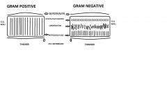
Cell Wall Essay
|
There are two types of cell walls: Gram Positive and Gram Negative. Gram positive cell walls consist mainly of peptidoglycan. The outer layer of both cells is called glycocalyx. Glycocalyx is a gelatinous layer outside the cells the aids in protection of phagocytes and from dehydration by retaining moisture. It also aids in attachment to surfaces. Gram positive cells are thicker than gram negative cells that are thinner. They consist of mainly three layers; lipopolysaccharides, lipoprotein, and peptidoglycan. They also contain flagellum. Flagellums are typically rods, & provide movement. Another structure is pilus. There are 2 types of Pilus; common- which are another point of attachment, & sex pili. The outside of the cell have hair-like structures fimbriae which also aid in attachment. The function of the cell wall is to determine the ridged shape & protects the cell. Inside the cell wall is the cell membrane, which is semi-permeable and controls what enters and exits the cell. Inside there is also a gelatinous pool that everything floats in called cytoplasm or protoplasm. It consists of 70% - 80% water. Towards the center region is the nuclear region. It contains the nucleoid and plasmid. The nucleoid contains chromosomes that secrete chromatin, which contains a circular, single strand of DNA. The plasmid has non-essential DNA, allows bacteria to invade the body, and carry Rfactors (R –resistance). Both Types of cells contain ribosomes, which are free floating in cytoplasm and aid in protein synthesis. Another structure is the inclusion bodies, which are granules, that store nutrients and gas vacuoles that store gas. Also, there are endospores, which are immortal. Part of the DNA is walled off, eventually separating & can survive forever, regenerate, & start all over.
|