Reading...
![]()
Play button
![]()
Play button
![]()
Use LEFT and RIGHT arrow keys to navigate between flashcards;
Use UP and DOWN arrow keys to flip the card;
H to show hint;
A reads text to speech;
7 Cards in this Set
- Front
- Back
|
CCB kinetics
|
metab by cyp3a sytem
only active metabolite is norverapamil from n-demethylation of verapamil |
|
|
Diltizem
|
deacetylated to deacetyldiltiazem )minimally active) excreted biliary tract
Vd large = (5.3 L/kg) inhibit cyp 3a/p-glycoprotein (elevation of cyclosporine and digoxin from p-glyco) minimally active met deacetyldiltiazem which is elim in biliary tract |
|
|
Verapamil
|
Vd =large (5.5)
inhibit cyp 3a/p-glycoprotein (elevation of cyclosporine and digoxin from p-glyco) Active metab norverapamil from n-demethylation |
|
|
Nifedipine
|
Vd small (0.8 L/kg)
|
|
|
CCB Toxicity Figure
|
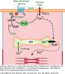
Myocardial toxicity of calcium channel blockers and use of antidotal therapies. Calcium channel blockers reduce calcium ion influx through the L-type calcium channel (Cav-L) and thus reduce contractility. Mechanisms to increase intracellular calcium include recruitment of new or dormant calcium channels by increasing cyclic adenosine monophosphate (cAMP) either by stimulating its formation by adenyl cyclase (AC) with catecholamines or glucagon (see text), or by inhibiting its degradation to 5′-monophosphate with phosphodiesterase inhibitors (PDEI) such as amrinone. Increasing the calcium concentration gradient across the cellular membrane to further its influx may improve contractility. The mechanism by which insulin therapy enhances inotropy is not fully known. PKA, protein kinase A; RyR = ryanodyne receptor.
|
|
|
CCB Toxicity figure - Vascular
|
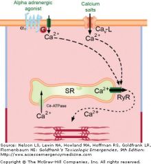
Vascular toxicity of calcium channel blockers and antidotal therapies. Calcium's entry via voltage-sensitive channels (Cav-L) initiates a cascade of events that result in actin-myosin coupling and contraction; this is inhibited by calcium channel blockers. Mechanisms to increase intracellular calcium include activation of receptor-operated calcium channels with 1-adrenergic agonists or increasing the calcium ion gradient across the cellular membrane to further its influx; RyR = ryanodyne receptor.
|
|
|
Ca receptor type affected
|
All CCB's antagonize L-type voltage sensitive Ca ch.
CCB's are classified into 3 groups. slightly different region of the Ca Ch affinitiy and differ affinities for myocardial and peripherla vascualture. |