Reading...
![]()
Play button
![]()
Play button
![]()
Use LEFT and RIGHT arrow keys to navigate between flashcards;
Use UP and DOWN arrow keys to flip the card;
H to show hint;
A reads text to speech;
25 Cards in this Set
- Front
- Back
|
HEART
|
hollow organ with thick muscular tissues that propels the blood to different parts of the body by rhythmically contracting
|
|
|
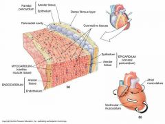
PERICARDIUM LAYERS
|
Fibrous Pericardium
Serous Pericardium - Inner mesothelial layer which in turn has two layers |
|
|
SEROUS PERICARDIAL LAYERS
|
Parietal Layer - lines the external wall of the pericardial sac
Visceral Layer - direct covering of the heart (epicardium) |
|

LAYERS OF THE HEART:
EPICARDIUM |
outermost covered by mesothelial cells, immediately below mesothelium are blood vessels, elastic fibers, nerves and adipose tissue
|
|

LAYERS OF THE HEART:
MYOCARDIUM |
Middle layer, thickest and contains cardiac muscles arrangen in a complex spiral manner
|
|

LAYERS OF THE HEART:
ENDOCARDIUM (3 layers) |
innermost layer exposed to the blood and participates in the formation of heart valves (determines direction of blood flow)
|
|
3 LAYERS OF ENDOCARDIUM:
SUBENDOCARDIAL LAYER |
biggest part, loose connective tissue (areolar) that connects the myocardium and the endocardium.
contains blood vessels, nerves, Purkinje fibers |
|

3 LAYERS OF THE ENDOCARDIUM:
SUBENDOTHELIAL LAYER ( Lamina Propria) |
thin layer of connective tissue containing fibroblast, collagen and elastic fibers
|
|

3 LAYERS OF ENDOCARDIUM:
ENDOTHELIUM ( Epithelium) |
simple squamous epithelium
|
|
|
CARDIAC SKELETON
|
Central support of the heart. muscle fibers and heart valves connect
|
|
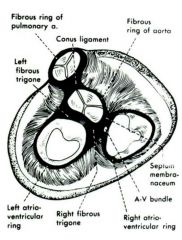
CARDIAC SKELETON PARTS:
ANNULAR FIBROSA |
dense fibrous rings of the AV node and Atrial foramina
|
|

CARDIAC SKELETON PARTS:
TRIGONA FIBROSA |
triangular area between AV opening, islands of cartilage like tissue or chondroid tissue
|
|

CARDIAC SKELETON PARTS:
SEPTUM MEMBRANACEUM |
regualar orientation of collagen bundles in layers superior to interventricular septum, connects muscle septum to trigona fibrosa
|
|
|
CARDIAC VALVES
|
reduplication of endocardium (2 layers), leaflets that control bloodflow by covering openings of the AV, Aortic and Pulmonary valves
supported by dense fibroelastic tissue and chondroid tissue |
|
|
IMPULSE CONDUCTION SYSTEM
|
specialized cardiac system that is responsible for the generation of the heart beat and the conduction of the impulse to the different parts of the myocardium, and ensure that the atria and the ventricles contract in succession
|
|
|
INTERCALATED DISCS', DESMOSOMES' AND GAP JUNCTIONS' ROLES IN CONTRACTION
|
intercalated discs consists of desmosomes and gap junctions.
desmosomes are responsible for keeping your cardiac cells together during contraction gap junctions (protein lined tunnels) are responsible for the direct transmission of depolarizing current from cell to cell |
|
|
IMPULSE CONDUCTION SYSTEM CONSISTS OF...
|
SA node, AV node, Bundle of his/Atrio-ventricular bundle/Truncus-Atriosos-Ventriculosos, right and left bundle, Purkinje fibers
|
|
|
LOCATION OF THE IMPULSE CONDUCTION SYSTEM PARTS
|
SA node - below your epicardium, right atrial wall , near superior vena cava
AV node - beneath the endocardium, floor of the right atrium Bundle of His/ Atrio-Ventricular Bundle/ Truncus-Atriosos-Ventriculosos - short bundles that originate from your AV node separating into.... right and left bundle that will further branch and anastomose... Purkinje fibers |
|
|
What is a Purkinje fiber?
|
specialized cardiac muscle
|
|
|
BLOOD VESSELS
|
hollow organs
|
|
|
ENUMERATE THE LAYERS OF THE ARTERY
|
Tunica Intima
-epicardium -subendothelial layer -internal elastic layer Tunica Media - thickest External Elastic Layer Tunica Adventitia - with vasa vasorum |
|
|
VASA VASORUM
|
blood vessel that supply nutrient to artery
|
|
|
VASA NERVORUM
|
Blood vessel that supply nutrient to nerve
|
|
|
NERVI VASORUM
|
nerve that supply nutrient to artery
|
|
|
NERVI NERVORUM
|
nerve that supply nutrient to vasa nervorum
|