Reading...
![]()
Play button
![]()
Play button
![]()
Use LEFT and RIGHT arrow keys to navigate between flashcards;
Use UP and DOWN arrow keys to flip the card;
H to show hint;
A reads text to speech;
123 Cards in this Set
- Front
- Back
|
sensitivity
|
- ability to distinguish among stimuli of different intensity
|
|
|
specificity
|
- ability to distinguish among stimuli of different types
|
|
|
4 basic components of sensory pathways
|
- sensory reception
- transduction - transmission - integration |
|
|
sensory reception
|
- detection of stimuli by sensory receptors
- neuron ending or specialized receptor cells in close contact with neurons - specialized to respond to stimuli, changes in environment - absorb energy from a stimulus and transduce that energy into electrochemical energy - produce graded receptor potentials = more or less neurotransmitters - generate action potential in sensory neuron - senstation (awareness) and perception (interpretation) occur in brain - sense organs consist of sensory receptors and accessory cells - cover large amounts of membrane to accommodate need to detect - membrane has cell specialization = microvilli and cilia |
|
|
cell specialization
|
- vertebrate = cilia
- protosomes = extensions of membrane such as microvilli |
|
|
transduction
|
- conversion of stimulus energy into a change in membrane potential of a sensory receptor
- process of converting stimulus energy into an electrical signal - requires sensory receptor molecules - initiate transduction of stimulus to produce receptor potential - action potential = release NT = produce receptor potential - initiation of receptor potential caused by a stimulus - receptor potential = depolarization of sensory cell |
|
|
transmission
|
- sensory cells transmit an action potentials to CNS
- work alone = specialized neurons to produce action potentials * axons extend into CNS - others are specialized that regulate neurons * specialized cells into CNS |
|
|
perception
|
- brain's construction of stimuli
- stimuli from different sensory receptors travel as action potentials along dedicated neural pathways - brain distinguishes stimuli from different receptors based on area in brain where action potentials arrive |
|
|
sensory receptor cell
|
- cell that is specialized to transform the energy of a stimulus into electrical signal
- stimulus = chemical, mechanical, electromagnetic |
|
|
stimulus
|
- form of external energy to which a sensory receptor cell can respond
|
|
|
sense organs
|
- anatomical structures that are specialized for the reception of particular stimuli
- many similar receptor cells and nonneural tissues |
|
|
sensory systems
|
- sense organs and all of their associated central processing areas
|
|
|
sensory receptor molecules
|
- initiate transduction of stimulus
- produce receptor potential - receptor molecules = membrane proteins - increased surface area = cilia and microvilli |
|
|
4 classifications of sensory receptor cells
|
- sensory modality
- form of stimulus energy - mechanism of transduction - location |
|
|
sensory modality
|
- subjective nature of sensory stimulus
- thing sensed |
|
|
mechanism of transduction
|
- receptor type
- ionotropic transduction = sensory stimulus is received and then transduced into an electrical signal - metabotropic transduction = sensory receptor molecules act like NT or GPCR in activating metabotropic cascade |
|
|
location
|
- exteroceptors
- interoceptors |
|
|
interoceptors
|
- respond to internal stimuli
- visceroreceptors = stimuli from viscera and blood vessels - detect changes in pH, osmotic pressure, body temperature, chemical composition of blood, tissue stretch |
|
|
exteroceptors
|
- respond to stimulus outside the body
- receive stimuli from outside - touch, pressure, pain, temperature, most receptors of sense organs |
|
|
2 functions of sensory receptor cells
|
- transduce
- encodes information about stimulus |
|
|
transduce
|
- energy = receptor potential
- receptor potential = depolarization of sensory cell |
|
|
encodes information about stimulus
|
- carried via action potentials to CNS
- keeping wiring straight = segregation of axons - labeled lines = sensory modality or quality of sensation associated with stimulus depends solely on which receptor cells are stimulated, rather than how - afferent axons mirrors geometric arrangement of receptor cells |
|
|
mechanoreceptors
|
- specialized to respond to different types of mechanical stimuli
- mediate touch, pressure, equilibrium, hearing, osmotic simulation - ionotropic - NOMPC = non mechanoreceptors C - transient receptor potential family - open in response to deformation = stretch - deformation allows cations to flow through channel - creates receptor potential - found in muscle - 6 membrane spanning segments - formed from nerve endings of dorsal root ganglia cells |
|
|
sensillum
|
- miniature sense organs that cover insects hard exoskeleton
- mechonsensory = hollow with sensory neuron endings - stretch activated channels = open in response to stretch and allow cations to flow through channels |
|
|
receptor potential
|
- primary electrical response of sensory receptor cell to stimulation
- output of sensory transduction - reversal potential near zero - frequency of action potential denotes strength of stimulus - stronger stimulus = greater receptor potential = higher frequency of action potentials |
|
|
stretch activated channels
|
- nonselective cation channels
- permeable to Na and K |
|
|
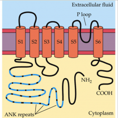
no mechanoreceptor potential C (NOMPC)
|
- ion channel responsible for stretch activated channels
- 6 transmembrane sequences and a P loop - part of transient receptor potential (TRP) channel family - ankyrin repeats = protein structural motif used to link proteins to elements of cytoskeleton |
|
|
4 kinds of mechanoreceptors in skin
|
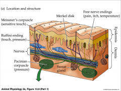
- merkel disc = sensitive to touch
- Meisner's corpuscle - Ruffini endings = touch pressure - Pacinain corpuscles = deep pressure and vibration |
|
|
unencapsulated mechanoreceptors
|
- free nerve endings of sensory neurons = superficial light touch, pain, itch, and temperature
- merkel disc hair follicle = superficial light touch |
|
|
encapsulated mechanoreceptors
|
- meisner's corpuscles
- pacinian corpuscles - ruffini's corpuscle - muscle spindle = stretch - golgi tendon organ = stretch |
|
|
merkel disc
|
- tactile sensile
- produces long trains of action potential in response to sustained deformation - merkel cell with nerve ending - merkel cell contain and release NT |
|
|
meisner's corpuscle
|
- wrapped in myelin and collagen
- 2 to 6 sensory neurons ending together - associated with Schwann cells and collagen |
|
|
pacinian corpuscle
|
- unmyelinated sensory neuron terminal
- concentric lamellar layers of membrane and connective tissue - layers separated by fluid - sudden vibrations/deep pressure distorts sensory ending producing a receptor potential - steady stimulus allows fluid to redistribute relieving distortion |
|
|
touch receptor cells
|
- association of epithelial cells with distal endings of neurons that soma in dorsal root ganglia adjacent to spinal cord
- dorsal root ganglion (DRG) = send disal processes into skin and their central axons into dorsal or sensory part of spinal cord |
|
|
sensory adaptation
|
- frequency of action potential in response to continuous and constant stimulation decreases over time
- tonic = slowly adapting * merkel discs, ruffini endings - phasic = rapidly adapting * messiner corpuscles - decrease in responsiveness due to continued stimulation |
|
|
lamellae
|
- thin, concentric, accessory cells
- encase pacinian corpuscles - responsible for phasic nature - absorb energy |
|
|
proprioceptors
|
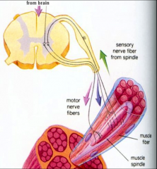
- internal mechanoreceptors that monitor movement, position, tension/stretch, and mechanical stress within the body
- musculoskeletal system = mostly associated with skeletal muscles - provide most info to brain about joint angle, muscle length, tension which is integrated to give information about position of limb in space - muscle spindle - golgi tendon organ |
|
|
muscle spindle
|
- most well known proprioceptor
- provides information about muscle length |
|
|
golgi tendon organ
|
- provides information about changes in muscle tension
|
|
|
vestibular organs in invertebrates
|
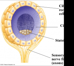
- sense organ that detects acceleration and direction of gravitational force
- ionotropic - statoliths = mechanoreceptors ciliated receptor cells that detect movement of granules - grain of sand or calcium carbonate that move the cilia around as position changes |
|
|
statocyst
|
- mechanoreceptor organ for orientation
- contains grains of sand or secretion of calcium carbonate - found in jellyfish - detect acceleration and direction of gravitational force - most invertebrates maintain equilibrium |
|
|
tympanal organ
|
- thin cuticular tympanum (eardrum)
- common form of auditory organ in insects - mechanosensory cells attached to typanum are stimulated - sensitive detectors and encoders of sound intensity - thin eardrum displaced by sound - located on thorax, legs, or other body locations |
|
|
lateral line organs
|
- mechanoreceptors of fish
- sensitive to minute, local, water displacements - sense-hillock or neuromast consist of cluster of pear-shaped sensory cells surrounded by long, slender supporting cells - sense hairs on top of sensory cells project into jellylike substance (cupula) that bends in response to water displacement |
|
|
vertebrate acoustico-lateralis system
|
- semicircular canals
- sensory organs for hearing and equilibrium are closely associated in the ear |
|
|
vestibular organs of humans
|
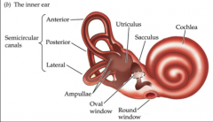
- 3 semicircular canals which detect angular acceleration of head
- 2 otolith organs detect linear movement and acceleration = sacculus, utriculus - semicircular canals filled with fluid and are oriented at right angles to each other - at base of each canal is ampulla that contains a cluster of hair cells in structure - lie adjacent to auditory organs - labyrinth = vestibular chambers and neighboring chambers of the cochlea - subserve sensory functions of acceleration and balance |
|
|
anatomy of mammalian ear
|
- change in acceleration and position move the fluid, endolymph against hair cells
|
|
|
hair cells
|
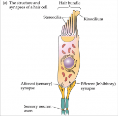
- mechanoreceptor cells in acoustico-lateralis system of vertebrates
- epithelial cell with apical surface that faces overlying lumen and basal surface that faces underlying tissue - hair bundle at apical end consisting of sterocilia of increasing height - don't posses axons and don't generate action potentials - bending of sterocilia transduced into receptor potential by release of transmitter substance onto an afferent/sensory neuron - joined by filamentous tip links |
|
|
sterocilia
|
- tuft of microvilli
- may have single true cilia = kinocilium - narrow at apical end - length is held rigid by actin filaments - pivots at base producing a shearing force - directionally sensitive = short to long arrangement - displacement toward tallest increase depolarization and amount of NT release - displacement toward shortest decreases NT release |
|
|
tip links
|
- directly involved in producing hair-cell response
- directly gate channel opening - when stretched open ion channels that permit ion influx and depolarization or closes ion channels |
|
|
macula
|
- hair cells in utriculus and sacculus
- horizontal for utriculus - vertical for sacculus - covered by otolithic membrane |
|
|
ear consist of 3 parts
|
- external ear = distal to eardrum
- middle ear = air filled - inner ear = liquid filled |
|
|
ossicles
|
- transmit sound wave vibrations from eardrum to oval window of inner ear
- transmit enough force/unit area at oval window to vibrate endolymph in vestibular canal - vibration of endolymph causes vibration of basilar membrane on which hair cells rest = 20,000 times/sec - because basilar membrane varies in thickness, rigidity, along its length = different frequencies cause basilar membrane to vibrate maximally at different points along its length - low frequencies at apical, wider, thinner, more flexible end - high frequencies at basal, thicker, narrower, stiffer end |
|
|
3 middle ear ossicles
|
- malleus
- incus - stapes |
|
|
tensor tympani and stapedius
|
- contact to clamp the movement of ossicles
- protect auditory membranes - middle ear muscles |
|
|
eustachian tube
|
- connects middle ear with pharynx
- equalize pressure with environmental pressure |
|
|
cochlea
|
- coiled tube containing chambers filled with fluid
- chambers are separated by basilar membrane - hair cells rest on basilar membrane in organ of corti |
|
|
basilar membrane
|
- separated cochlea into upper and lower chambers
- oval window stimulates movement - varies in width and thickness along length - every frequency has different place of max amplitude - movement bends sterocilia of hair cells whose tips are embedded in overlying tectorial membrane - sterocilia of hair cells project into scala media or cochlear duct -- a bath hight in K |
|
|
cochlear amplifier
|
- active component of basilar membrane that contributes to sound localization
|
|
|
organ of corti
|
- hair cells in region of cochlea
- sit on basilar membrane and vibrate with it - 3 rows of outer hair cells and 1 row of inner hair cells - only have stereocilia - tectorial membrane = flap of tissue covering hair cells - inner air cell major source of auditory input to brain |
|
|
outer hair cells change length
|
- amplify local movement of basilar membrane
- prestin = responsible fro shortening and lengthening - change length in response to changes in membrane potential/frequencies |
|
|
time difference
|
- arriving at 2 ears at slightly different times
|
|
|
intensity difference
|
- louder in ear that more directly faces sound source
- sound shadow = shielded from sound by head - head affective to high frequency better than low frequency |
|
|
cochlear hair cells
|
- bend in response to movement of basilar membrane
- brush tectorial membrane - displacement toward tallest sterocilia increases the depolarization and amount of NT released - displacement toward shortest decreases NT release by hyperpolarization - bending depolarizes membrane of mechanoreceptors cuaring the release of NT and send action potentials to brain via auditory nerve - K flows in = depolarization causes release of NT - stimulates afferent sensory neuron - organ of corti has 3 rows of hair cells - 80% - 95% of these synapses are with inner hair cells - outer hair cells maybe important in amplification |
|
|
hearing
|
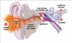
- outer ear directs sound waves distal to eardrum sound pressure waves vibrate tympanum
- wave transmitted through middle ear to oval window by ear ossicles - eustachian tube of middle ear equalizes pressure in middle ear with environmental pressure - middle ear ossicles transfer sound from air to liquid by pushing against oval window of cochlea of inner ear |
|
|
chemoreceptors
|
- transmit information about total solute concentration of solution
- specific ones respond to individual kinds of molecules - when stimulus molecules binds to a chemoreceptor, the chemoreceptor becomes more or less permeable to ions - sensory response to chemical stimulus - emerged very early in evolution = bacteria - taste = gustatory sense - olfaction = sense of smell |
|
|
taste
|
- chemical sense
- gustation is sensation of taste - results from action of chemicals on taste buds |
|
|
taste buds
|
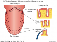
- confined to lingual papillae
- filiform (no taste buds) = important for texture - foliate (taste buds) - fungiform taste buds = tips and sides - vallate (circumvallate) = rear and half of taste buds - taste cells grouped together on tongue |
|
|
vertebrate taste receptor cells
|
- epithelial sensory cells
- taste buds - papillae = small swelling confining taste buds - 50 to 150 slender, elongated cells of at least 4 types - lifetime of 5 to 10 days |
|
|
physiology of taste
|
- molecules must dissolve in saliva
- 5 primary sensations of taste - influenced by food texture, aroma, temperature, appearance - mouthfeel = detected by lingual nerve in papillae - hot pepper stimulates free nerve endings |
|
|
5 primary sensations
|
- throughout tongue
- sweet = tip - salty = lateral - sour = lateral - bitter = posterior - umami = tast of AA |
|
|
mechanisms of action
|
- activate 2nd messenger systems = sugars, alkaloids, glutamates bind to receptors
- depolarize cells directly = sodium and acids penetrate cells |
|
|
taste cells
|
- apical microvilli serve as receptor surface
- synapse with sensory nerve fibers at their base |
|
|
basal cells
|
- precursor to new taste receptor cells
|
|
|
taste transduction mechanism
|
- different for every taste
- taste receptor cells have channels permeable to Na - Na concentration inside mouth increases = membrane potential of receptor cell depolarizes - channels not voltage gated - fairly large increase in [NA] required to open channels extracellular domains-amino termini = provide binding site for sugars - loop between 5 and 6 transmembrane domain interacts with G protein |
|
|
salty
|
- simplest transduction
- channels permeable to Na - [Na] increases = salt receptor cells depolarize - ionotropic permeability - taste is sensitive |
|
|
sour
|
- channel mediated
- polycystic kidney disease = like ion channel subfamily - PKD1L3 and TRPP3 - H+ modulate permeability of channels |
|
|
sweet, bitter, umami
|
- metabotropic GPCRs
- sweet is a dimer of T1R1 and T1R3 - umami sensed by dimer of T1R3 and T1R1 bitter receptors form T2R family - all use similar G proteins - activate phospholipase C = IP3 and diacylglyerol - IP3 = release Ca from intracellular store - opens another TRP channel - binding of receptor activates G protein - G protein activate phopholipase C - produce IP3 and DAG finally opening transient receptor potential channel |
|
|
olfactory epithelium
|
- contain receptor cells with cilia for olfaction = in layer of mucus
- olfactory receptor cells is bipolar neuron - highly sensitive = up to 10000 odors - 5cm^2 of superior concha and nasal septum - olfactory receptor surface - lines part of internal nasal cavity - area of nasal mucosa = varies greatly among species |
|
|
olfactory receptor cell
|
- bipolar neuron with cell body in olfactory epithelium
- single, narrow dendrite extends from cell body to mucus covered epithelial surface and ends in a dendritic knob which projects into layer of mucus - 20 to 30 olfactory cilia extend and intermesh within the mucous layer - bind odor molecules dissolved in thin layer of mucus - membranes of cilia sites of olfactory transduction - parykaryon in olfactory epithelium - axons pass through cribiform plate - cell survive for about 60 days - molecules bind to receptor on olfactory hair - hydrophilic = diffuse through mucus - hydrophobic = transport by odorant binding protein - send their axons to neighboring olfactory bulb in CNS - golmerus = globular cluster - all particular receptor molecule terminate in same glomeruli |
|
|
physiology of smell
|
- activate G protein and cAMP system
- opens ion channels for Na and Ca - creates receptor potential - Ca binds to calcium activated Cl channels augmenting the depolarization - receptors adapt quickly due to synaptic inhibition in olfactory bulbs |
|
|
olfactory receptor proteins
|
- 7 transmembrane domains
- G protein coupled receptors - active GPCRs leads to opening cyclic nucleotide-gated channels = receptor potential |
|
|
vomeronasal organ
|
- located below main olfactory epithelium
- detect pheromones and other chemical signals - self enclosed pouch normally isolated from air breathed - receptor cells have microvilli - GPCRs - respond specifically to one or only a few compounds with high sensitivity - open TRP channels to depolarize membrane |
|
|
electromagnetic receptors
|
- detect electromagnetic energy such as light, electricity, magnetism
- some snakes very sensitive infrared receptors that detect body heat of prey against colder background - many animals migrate using earth's magnetic field orient - visual receptors of diverse animals depend on light absorbing pigment - animals use diverse set of organs for vision but underlying mechanism for capturing light is same = common evolutionary origin |
|
|
vertebrate eye
|
- detects color and light, but brain assembles the information and perceives the image
- camera eye - cornea and lens focus an inverted image on retina - macula lulea - optic disc is blind spot where ganglion cell axon pass through - retina contains rods and cones photoreceptors and network of neurons = disc, body, and synapse - horizontal cells, bipolar cells, ganglion cells, amacrine cells |
|
|
macula lulea
|
- cells on visual axis of eye
- fovea centalis = center of macula - finely detailed images due to packed receptor cells |
|
|
photoreceptors
|
- sensory receptor cells sensitive to light
- rhodopsin first GPCR to be studied - pigments epithelium = absorb stray light and prevent reflections |
|
|
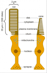
rod cells = rhodopsin
|

- night = scotopic vision
- outer segment = stack of coin like membranous discs studded with rhodopsin pigment molecules - absorption peak at 500nm - 2 major parts of molecule * opsin = vitamin A derivative * retinal = vitamin A derivative - absent in central part of fovea but greatly outnumber cones elsewhere - used in dim light - more sensitive - nocturnal animals |
|
|
cone cells = photopsin (iodopsin)
|
- color = photopic vision
- outer segment tapers to a point - used in brighter light for color vision and high acuity - opsin molecules contain different amino acids that determine wavelength of light absorbed - 3 kinds of cones absorbing different wavelengths of light produce color vision - animals and humans having fovea - diurnal animals |
|
|
photoreception
|
- response of sensory cell to light
- photopigment = absorbs light - detect light using pigment - all photoreceptor cells have greatly increased membrane surface areas that increase light sensitivity |
|
|
photoreceptor cells subdivided
|
- ciliary photoreceptor
- rhabdomeric photoreceptor |
|
|
ciliary photoreceptor
|
- modified cilia contain rhopsin
|
|
|
rhabdomeric photoreceptor
|
- collections of microvilli
- vertebrate = ciliary - arthropod = rhabotomeric |
|
|
2 major kinds image forming eyes
|
- camera eye
- compound eye |
|
|
camera eye
|
- lens forms an inverted image on an array of photoreceptors at back of eye
- cornea and lens focus an inverted image on retina - retina = photoreceptor containing layer at back of eye - light refracted at surfaces where materials differ in density - lens refraction focus image by changing shape of lens - rod and cone photoreceptor cells - network of neurons : horizontal, bipolar, amacrine, ganglion * perform first stages of visual integration - pigmented epithelium absorbs light not captures by photoreceptors and performs many metabolic functions photoreceptors and performs many metabolic functions - retina inverted with photoreceptors in outermost layer - fovea = central high acuity region in which intervening cell layers and blood vessels are displaced to the side - optic disc = points at which axons exit retina producing blind spot in visual field |
|
|
compound eye
|
- ommatidia each with its own lens, together produce mosaic image
- each ommatidium conveys info about one part of visual world, and nervous system constructs image as a mosaic of tiles of individual ommatidial responses |
|
|
rhodopsin
|
- consists of protein containing associated nonpeptide organic molecule = chromophore
- chromophore is retinal and bound to integral membrane protein opsin = rhodopsin - photochemical reaction = twists the aldehyde tail of chromophore around one of its double bonds and produces all trans retinal - conformation changes in opsin = activation rhodopsin - activates G protein signal transduction cascade |
|
|
reticular cell
|
- photoreceptors in drosophila
- 8 or more reticular cells arranged in a circle - transduction cascade is localized to membranes of its microvilli |
|
|
rhabdomere
|
- array of microvilli along edge of reticular
- contain rhodopsin photopigment - G proteins and associated proteins - channes that produce electrical response to light |
|
|
phototransduction
|
- absorption photon causes change in conformation of rhodopsin = activation of G protein
- activated G protein activates phospholipase C = produces second messengers IP3 and diacylglycerol (DAG) - DAG opens 2 cation channels = TRP channels - produces depolarization = triggers synaptic transmitter release - doesn't generate action potentials - proteins bound together by cytoplasmic scaffolding protein |
|
|
light induced change in rhodopsin
|
- activated series of reactions at disc membrane
- result in enzymatic degradation of cAMP - transduction = G protein activated by rhodopsin - cGMP phosphodiesterase (PDE) = enzyme in disc membrane that hydrolyzes cGMP to 5'-GMP - activated PDE decrease cytoplasmic [cGMP] = cation channel close = decrease Na influx = hyperpolarization |
|
|
dark current
|
- produced by constant flow of Na
- keeps rod relatively depolarized - detectable change from absorption of single photon |
|
|
receptors recover slowly from bright light
|
- dark adaption = slow adjustment to darkness
- regeneration = photochemically or enzymatically - insects = photochemically - vertebrates = enzymatically - slow enzymatic process - partly occurs in pigment epithelium |
|
|
transduction of visual information to nerves system
|
- begins when light induces conversion of cis-retinal to trans-retinal
- trans-retinal activates rhodopsin = activates G protein transduction = eventually leading to hydrolysis of cyclic GMP |
|
|
vertebrate photoreceptors
|
- light activates rhodopsin
- activate rhodopsin activates transduction - activated transduction = activates cGMP phosphodiesterase - enzyme decreases concentration of cGMP by converting it to 5' GMP - decrease of cGMP closes cGMP gated ion channels - Na decreases and photoreceptor is hyperpolarized |
|
|
light hyperpolarizes
|
- dark current enters rod outer segment in dark carried largely by Na influx
- light acts to decrease dark current by closing cGMP gated Na - brighter the light = greater the hyperpolarization |
|
|
humans perception of color based on 3 types of cones
|
- each with different visual pigment: red, green, blue
- pigments called photospins and are formed when retinal binds to 3 distinct opsin proteins |
|
|
light and dark
|
- in dark, rods and cones release the NT glutamate into synapses with neurons called bipolar cells
- in light, rods and cones become hyperpolarized, shutting off release of glutamate - bipolar cells are then either depolarized or hyperpolarized |
|
|
3 other neurons contribute to information processing in retina
|
- ganglia cells transmit signals from bipolar cells to brain
- horizontal and amacrine cells help integrate visual information before its sent to brain |
|
|
interaction among different cells
|
- result in lateral inhibition
- enhanced contrast in image |
|
|
optic nerves
|
- meet at optic chasm near cerebral cortex
- senstation from left visual field of both eyes are transmitter to right side of brain - senstation from right visual field are transmitted to left side of brain |
|
|
ganglion cell axons
|
- lead to lateral geniculate nuclei
- lateral geniculate nuclei relay information to primary visual cortex in cerebrum - at least 30% of cerebral cortex, in dozens of integrating centers are active in creating visual perceptions |
|
|
receptive light fields
|
- divided into 2 parts
- center - surrond |
|
|
cell response
|
- on center cell response = increase rate of impulse discharge when center of receptive field is illuminated
- off center cell response = decrease rate of impulse discharge when center of receptive field is illuminated = inhibited by light at center |
|
|
retinal pathways
|
- straight through pathway
- lateral pathways |
|
|
lateral pathways
|
- horizontal cells from a lateral pathway in outer plexiform layer and amacrine cells from a lateral pathway in inner plexiform layer
|
|
|
straight pathways
|
- photoreceptors to bipolar cell to ganglion cell
- produce center of ganglions cells contrast receptive field |
|
|
photoreceptor excited by light
|
- hyperpolarizes off center bipolar cell
- inhibiting generation of action potential - depolarizes an on center bipolar cell - increasing probability of generating action potential |
|
|
light in surrond hyperpolarizes horizontal cell
|
- depolarizes other cones they have synapse with = opposes light
- hyperpolarizes on center bipolar cells and inhibits on center ganglion cell - depolarizes off center bipolar cell and excites off center ganglion cell |
|
|
vertebrate brain integrates visual information
|
- through parallel pathways
- axons of ganglion cells relay information to brain = all axons form optic nerve - axons from right side of brain cross to left side in corpus colosseum - where they cross form optic chiasm - respond to light as well as pattern - contrast or changes in light level and color over time |
|
|
color vision
|
- accomplished by cones that contain different photopigments
- depends on ratio of three classes of photoreceptors sensitive to different wavelengths of light - retinal circuitry integrates color contrasts based on red, green, blue, yellow |
|
|
blue, red, green
|
- 16% blue
- 17% red - 10% green - each has different core of opsin sensitive to different wavelengths |
|
|
ganglia cells
|
-output of retina
- receptive field of ganglion cell is are of retina in which neuron can be influenced by light |
|
|
ganglion cell
|
- axons synapse with lateral geniculate nuclei
- send their axons to primary visual cortex |