Reading...
![]()
Play button
![]()
Play button
![]()
Use LEFT and RIGHT arrow keys to navigate between flashcards;
Use UP and DOWN arrow keys to flip the card;
H to show hint;
A reads text to speech;
52 Cards in this Set
- Front
- Back
|
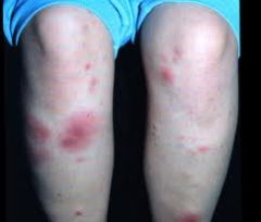
Erythema Nodosum:
• Red lumps form on the shins, and less commonly the thighs and forearms. • Peak occurrence is between 20-30. • Is generally idiopathic, although the most common identifiable cause is streptococcal pharyngitis. • May be the first sign of systemic disease such as TB, sarcoidosis, IBD or cancer. • The hallmark of this condition is tender, erythematous, subcutaneous nodules that are typically located symmetrically on the anterior surface of the lower extremities. • Self-limiting and usually resolves itself around 3-6 weeks. |
|
|
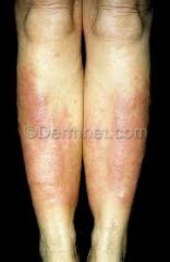
Pretibial Myxedema
|
|
|
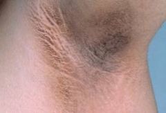
Acanthosis Nigricans
|
|
|
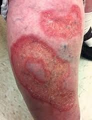
Necrobiosis Lipoidica (Diabetica):
|
|
|
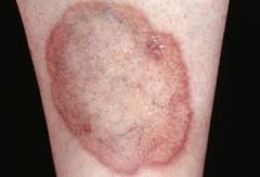
Necrobiosis Lipoidica (Diabetica):
• Rare skin disorder which can affect the SHIN of insulin dependent diabetics, although it may occur in non-diabetics as well. • Typically, one or more tender yellowish brown patches develop slowly on the lower legs over several months. They may persist for years. • The center of the path becomes shiny, pale, thinned, with prominent blood vessels. • Often painless. • Skin biopsy needed for diagnosis. • Topical steroids for treatment. |
|
|
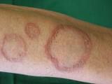
Granuloma Annulare:
|
|
|
Granuloma Annulare:
• Common condition of unknown cause, which affects the skin of children, teenagers or young adults. Commonly seen in DIABETICS. • Can occur on any site of the body. • Only affects the skin. • Areas are tender when knocked. • Often disappear after a few weeks or months without leaving a scar, but may recur at the same site. • Management is not really needed because patches disappear by themselves. If they do persist, potent topical corticosteroids should be trialed. |
|
|
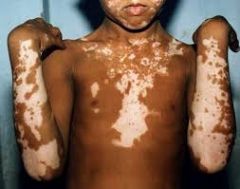
Vitiligo:
|
|
|
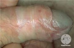
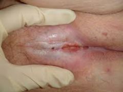
lichens sclerosis
|
|
|
lichens sclerosis
|
|
|
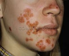
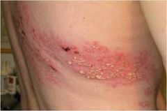
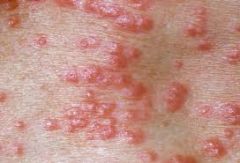
Impetigo:
• Bacterial skin infection, usually strep pyogenes or staph aureus. • Looks like pustules and round, oozing patches that grow larger day by day. • There may be clear blisters (bullous impetigo) or golden yellow crusts. • Managed via moistening crusted areas, antibiotic ointment and oral antibiotics if infection is extensive. |
|
|
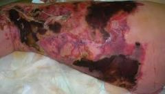
Necrotizing Fasciitis:
• Bacterial infection of the soft tissue and fascia. • May occur in anyone almost half of all known cases of streptococcal necrotizing fasciitis have occurs in young and previously heathy adults. • Disease occurs if there is an opening in the skin that allows bacteria to enter the body. May only need to be a small cut or graze. • Immunosuppressed individuals are at increased risk. • Symptoms appear within 24 hours of the minor injury. • There is pain in the general area of the injury. • There are flu-like symptoms such as nausea, fever, diarrhea, dizziness and general malaise. • Within 3-4 days the affected area starts to swell and dark marks appear. • Necrosis occurs and the wound becomes blackened. • At this point there is severe pain. • At 4-5 days the patient can die from toxic shock. • Management is hospitalization in ICU, culture of causative organisms and high dose IV antibiotics. This is followed by removal of all dead tissue. • 25% of pati |
|
|
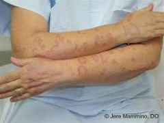
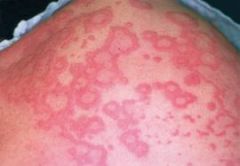
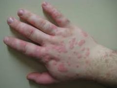
Erythema Multiforme:
|
|
|
Erythema Multiforme:
• Hypersensitivity reaction triggered by infections, most commonly HSV, but also commonly triggered by drugs. • Presents as skin eruption characterised by a typical target (iris) lesion. • It is acute and self limiting, usually resolving without complication. • It classically presents with a few to hundreds of skin lesion erupting within a 24 hour period, upper limbs more commonly than lower. • For the majority of cases, no treatment is required as the rash settles by itself over several weeks. • Supportive treatment may be necessary. |
|
|
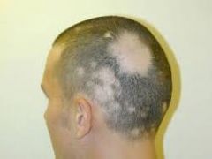
Alopecia Areata:
|
|
|
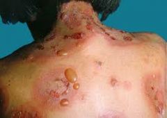
Bullous Pemphigoid:
|
|
|
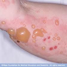
Bullous Pemphigoid:
• Blistering skin disease that is due to an immune reaction within the skin. Rare in children – more common in middle aged or elderly. • Presents as crops of tense, fluid-filled blisters. • Raise from normal looking or inflamed skin. • May be localized or widespread. • Managed via oral prednisolone and antibiotics. |
|
|
Herpes Zoster (Shingles):
|
|
|
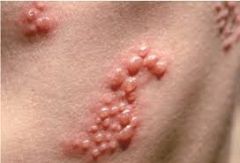
• Painful blistering rash caused by reactivation of varicella (herpes zoster).
• Reactivation typically occurs in the elderly and immunocompromised. • Must have had chickenpox to develop shingles. • Patient may also feel unwell. • Pain precedes blistering rash by 1-3 days. • Mx via rest a relief, as well as a bland protective cream. • Oral antivirals (acyclovir) are recommended in the immunocompromised, the elderly and those with facial shingles. |
|
|
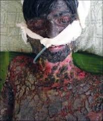
Stevens Johnson Syndrome & Toxic Epidermal Necrolysis:
• Potentially fatal skin reaction where there is sheet-like skin and mucosal loss. • Nearly always caused by medications >200 medications identified. • Involves a prodromal flu-like illness of several days, then an abrupt onset of a tender/painful red skin rash starting on the trunk and rapidly spreading over hours to days onto the face and limbs. • May be macular, targetoid or bullous. • The blisters then merge to form sheets of skin detachment, exposing red, oozing dermis. The skin sloughs off when rubbed gently. • Mx involves cessation of causative drug, hospital admission and resuscitation therapy. |
|
|
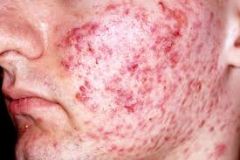
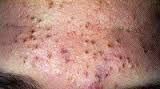
Acne Vulgaris
|
|
|
comedones - open = blackheads, closed = white
|
|
|
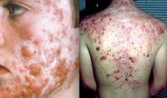
Cystic acne
|
|
|
Treatment for acne
|
Appropriate Investigations:
None needed for classic cases. May investigate for PCOS if suspected. CAH 17-OH progesterone. Management: 1. Benzoyl peroxide (BPO). 2. Topical antibiotics (to reduce inflammation). 3. OCP. 4. Oral Isoretinoin (Roacataine). |
|
|
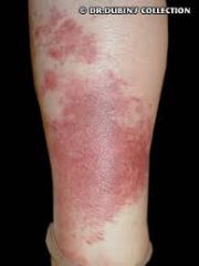
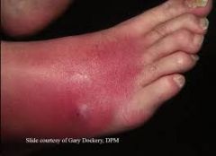
Erysipelas
Strep pyogenes. Affects SUPERFICAL dermis. Well demarcated. Hot, painful, swollen. Fever, malaise. Oral antibiotics for 2 weeks. Rest and elevate leg. |
|
|
Cellulitis
Strep progenies or Staph aureus. Affects DEEP dermis. Ill-demarcated erythema. Hot, pain, swollen, indurated. Swab. Oral antibiotics for 10 days. Treat portal of entry. |
|
|
Varicella - Chicken Pox
|
|
|
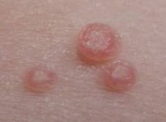
molluscum contagiosum - umbilicated papules
|
|
|
Scabies
|
|
|
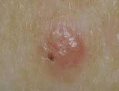
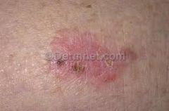
Basal Cell Carcinoma:
Slow growing Small shiny Pearly |
|
|
Basal Cell Carcinoma:
|
|
|
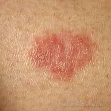
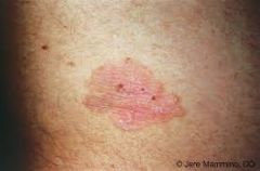
Bowens Disease
Intraepidermal SCC presents as one or more irregular, flat, red and scaly patches of up to several centimetres in diameter. Cryotherapy, 5-FU cream, Imiquimod, superfifical skin surgery. |
|
|
Bowens
|
|
|
Bowens
|
|
|
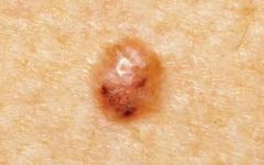
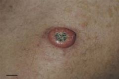
Keratoacanthoma:
Keratoacanthoma is a skin lesion that erupts in sun damaged skin, rather like a little volcano. It grows for a few months, then may shrink and resolve by itself. |
|
|
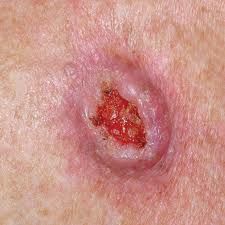
Squamous cell carcinoma
|
|
|
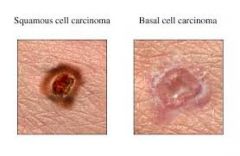
SCC vs BCC
|
|
|
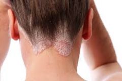
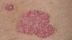
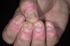
Psoriasis
|
|
|
Psoriasis
Histology: Histology is rarely required for a psoriasis diagno sis, but can be used to determine rarer diseases that can look like psorias is such as Mycosis Fungoidies. Morphology: Well demarcated. Erythematous. Silvery scale. |
|
|
Psoriasis
|
|
|
Discoid eczema:
The plaques of discoid eczema are intensely itchy, distributed on the trunk and limbs and are not associated with other features of psoriasis. |
|
|
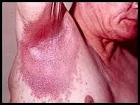
Flexural (inverse) psoriasis
|
|
|
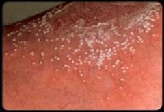
Pustular psoriasis
|
|
|
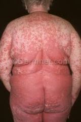
Erythrodermic psoriasis
|
|
|
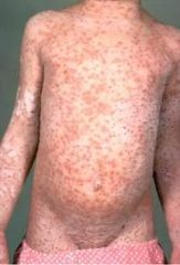
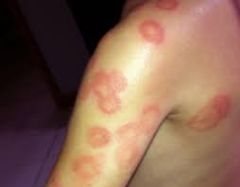
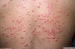
Guttate Psoriasis:
|
|
|
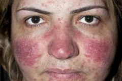
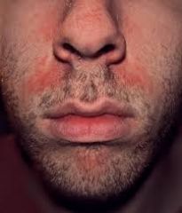
Rosacea
Rosacea primarily affects the convexities of the central face, including the cheeks, chin, nose, and central forehead. Some level of erythema is seen in virtually all patients early in the course of the disorder. All patients with the condition complain of some degree of redness. |
|
|
Telangiectases is common with which disease
|
Rosacea
|
|
|
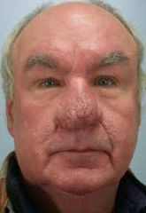
Rhinophyma
|
|
|
What are demodex?
|
Demodex:
Demodex are parasitic mites that live on human skin, primarily on the face. Dermodex infestation can trigger rosacea (especially in the immunosuppressed). Concurrent demodex infestation tends to induce ‘scaly’ papulo-pustular rosacea. Permethrin is used to manage dermodex. |
|
|
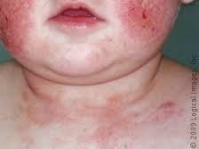
Atopic Dermatitis (EXCEMA):
|
|
|
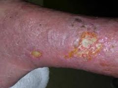
Venous Dermatitis
|
|
|
Seborrhoeic
Dermatits: |