Reading...
![]()
Play button
![]()
Play button
![]()
Use LEFT and RIGHT arrow keys to navigate between flashcards;
Use UP and DOWN arrow keys to flip the card;
H to show hint;
A reads text to speech;
79 Cards in this Set
- Front
- Back
|
Zygote
|
genome contains all info necessary to produce all structures and functions needed for survival
|
|
|
Commitment or DIFFERENTIATION of cells to specific functions have two phases
|
specification and determination
|
|
|
Specification
|
not permanent, can be reversed
cells know what they are to become but can be altered if left in current position will become what it was intended, if not will change in a neutral environment will still become what intended, however if placed elsewhere change |
|
|
Determination
|
later stage than specification
irreversible. will not change even if put in another part of the body |
|
|
Three different types of commitment:
|
autonomous
|
|
|
Autonomous Specification
|
particular blastomere removed early-removed blastomere will still make cells it would have if left in the embryo. Will lack cells that blastomere wouldve created.
gives rise to mosaic dev.: since embryo seems to be made of independent self differentiating parts mainly in invertebrates have morphogenic determinants (certain proteins and mrna) that are in different regions of egg and are apportioned to different cells as the embryo divides. |
|
|
Conditional specification
|
involves interactions with neighboring cells
originally each cell has abillity to become different cells however interactions with other cells limit so it is COND. fate of cell depends on conditions of embryos. If blastomere removed embryonic cells alter fate to take over roles of missing cells this ability of cells to change their fate is called REGULATION. isolated blastomere can give rise to variety of cell types |
|
|
Zygotes contains ______ in addition so signaling molecules.
|
cytoplasmic or morphogenic determinants (proteins and mRNA) during cleavage.
-these morphogenic determinants are not equally distributed -have same DNA but mor. factors can inhibit or activate cells |
|
|
zygote also has_____ in addition to Morphogenic determinants.
|
Signaling molecules: are produced by certain cells that cause change in another cell.
|
|
|
Cytoplasmic determinants and signaling molecules are the way cells undergo
|
specification and determination
|
|
|
Induction is
|
the process by which a cell or tissue influences the fate of another cell or tissue
|
|
|
Competence
|
ability of a cell to respond to an inductive signal.
|
|
|
What is a centrosome?
|
Microtubule organizing center: so i.e where microtubules are produced.
have centrioles with microtubules that grow into spindles which seperate chromosomes. |
|
|
Sperm function?
|
1. deliver centrosome to ovum
-mitotic spindle arises from south 2. activates ovum 3. restores diploid condition (egg is haploid so is sperm) |
|
|
Mammalian sperm structure
|
1.has nuclear region with hereditary material- this is where the acrosomal vesicle is.
2. Have mitochondral region which is the midpiece that produces ATP. 3. Flagellum for mobility |
|
|
the head of the sperm (mammalian)
|
-have acrosome th
|
|
|
Acrosomal reaction has two phases: in most marine invertebrates
|
1.fusion of acrosomal process with sperm cell membrane exocytosis of acrosomal vesicle (results in release of contents)
2. extension of acrosomal process |
|
|
IN sea urchins what triggers acr. rxn?
|
WHEN sperm contacts the egg jelly causes exocytosis and release of proteolytic enzymes that digest path through jelly to egg surface
|
|
|
Specificaly what happens?
|
interactions of sperm celll membrane with specific complex sugar in jelly, when these polysaccharides bind to specific receptors located on sperm cell membrane above acrosomal vesicle.
|
|
|
Once sperm reaches egg surface what happens?
|
acrosomal process adheres to the vitelline envelope
|
|
|
sometimes acrosomal rxn initiated by fucose sulfate, what happens when sulfate carb binds to receptor
|
when sulfated barbs bind to receptor on sperm, receptor activates three sperm membrane proteins:
activates three sperm membrane proteins 1.calcium transport channel> calcium enters sperm head 2. sodium hydrogen exchange pump that pumpsa Na+ into sperm and pumps H+ out 3. Phospholipase enzyme that makes IP3> which is able to release calcium from inside the sperm, prob from with in the acrosome itself. Elevated calcium level triggers fusion of acrosomal membrane with adjacent sperm cell membrane releasing enzymes that lyse path through egg jelly to vitelline envelope |
|
|
What will this calcium do?
|
fusion of acrosomal membrane and sperm plasm membrane and (#1 step of acrosomal rxn)exocytosis happens> then release enzymes that will digest through jelly
|
|
|
Second step of acrosomal rxn is
|
the extension of acrosomal process
|
|
|
the extension of acrosomal process
|
arises through polymerization of globular actin into actin filaments
will form a connection that widens the cytoplasmic bridge btw egg and sperm called the FERTILIZATION CONE |
|
|
What passes through the fertilization cone?
|
a little sperm tail (Centrioles) and nucleus
|
|
|
Bindin around acrosomal process is
|
a protein that recognizes whether the egg and sperm match
species specific |
|
|
sperm egg fusion causes
|
polymerization of actin to form fertilization cone
|
|
|
Sperm membrane fusion with egg plasma membrane is an active process that requires
|
fusogenic proteins.
|
|
|
The eggs membrane potential:
also look at A)potential difference and B)resting membrane potential |
-membrane is selective barrier>keeps ion conc. within and outside of egg different.
-difference mainly with Sodium and potassium -Sea water has high sodium ion conc. Na+: egg contains little sodium. -reverse with potassium i.e lots of potassium inside not a lot in sea water -maintained by membrane A)resting potential: constant difference in charge across cell membrane is generally -70 mV because inside is negative relative to outside |
|
|
two mechanisms for preventing polyspermy:
|
1. Fast reaction involving the egg plasma membrane
2.Slower prolonged rxn that involves the corticle granules of the egg |
|
|
Fast block prevention of polyspermy
|
reverses the membrane potential, shift to positive level of about +20 mV after first sperm enters
-caused by small influx of Na+ (sodium -prevents other sperm bc can only bind to vitelline envelope when -70 can't fuse to positively charged envelope -not permanent, only lasts maybe a minute |
|
|
Slow block prevention of polyspermy
(possibly initiated by:increase of Calcium concentration in cytoplasm> influx comes from within the egg itself |
bc fast not permanent need this
polyspermy could still occur if other sperm bound not removed. Sperm removal by cortical granule rxn. or slow block -Cortical granuels are directly beneath sea urchin egg cell membrane -when sperm fuses with egg cell membrane cortical granules fuse with egg cell membrane and release contents of proteases that cleave proteins linking vitelline envelope and cell membrane into space between membrane and fibrous mat of vitelline envelope proteins -components of cortical granules fuse with vitelline envelope to form fertilization envelope: which is elevated from cell membrane by mucopolysaccharides released by the cortical granules> sticky cmpds produce osmotic gradient that forces water to rush into the space between cell membrane and fertilization envelope causing it to expand and radially move away from egg. -peroxidase enzyme> released by corticle granules hardens fertilization env. by crosslinking tyroside residues on adjacent proteins. FInally fourth set of cortical granule proteins including hyalin forms a coating, microvilli extends and attach to hyaline layer>provides support to blastomeres during cleavage |
|
|
proteins released by cortical granule exocytosis
|
1.trypsin like protease CORTICAL GRANULE SERINE PROTEASE> this dissolves protein posts that connect vitelline envelope proteins to cell membrane and it clips off bindin receptors and any sperm attached.
|
|
|
Sperm entrance also triggers the Egg metabolism which:
|
-restores the mitotic cell cycle or causes cell to start undergoing mitosis again> also triggered by calcium release that initiates the slow block (occurs at fertilization sweeps across eggs in waves from intracellular storage)
-also reactivates egg protein synthesis -promotes DNA replication and movement in cytoplasm of morphogenic material ^(fusion causes intracellular pH to increase coupled with calcium elevation stimulate) |
|
|
Besides triggering egg metabolism, fusion of genetic material also occurring:
|
-when membranes of egg and sperm fuse sperm nucleus and centriole separate from mitochondria and flagellum-they disintegrate in the egg
-centrosome needed to produce mitotic spindle of subsequent division comes from the sperm centriole. -female already has pronucleus bc previously stopped after the second meiotic division - -male not case, decondenses in egg to be haploid pronucleus> once this happens DNA transciption and translation can occur -after enters cytoplasm male pronucleus rotates 180 degrees so that sperm centriole between egg and sperm pronucleus -centriole then acts a a microtubule organizing center, extending its microtubules and integrating them with the egg microtubules to form an aster. -microtubules extend throughout egg and contact the female pronuclues -pronuclei migrate toward each other -fusion forms nucleus (DNA synthesis can begin during pronuclear(migration stage) or after formation of zygote nucleus depending on Calsium levels released earlier in fertilization. |
|
|
Same release of Calcium that is responsible for cortical granule rxn, egg restoration to the cell cycle, and reactivation of protein synthesis also responsible for activation of what enzyme?
|
NAD+ kinase
>this converts NAD+ to NADP+ >imp. bc NADP+ (but not NAD+) may be imp. for lipid metabolism, bc can be used as a coenzyme for lipid biosynthesis. >therefore the conversion may be imp. in construction of new cell membranes required during cleavage. >NADP+ also used to make NAADP which seems to further boost calcium >Ca release also affects o2 rconsumprtion |
|
|
So to review sequence of events:
|
-Sperm egg binding -
Calcium levels increase -cortical/ slow block reaction occurs -fertilization envelope develops -elevated pH -increase protein synthesis -fusion of pronuclei -DNA synthesis-mRNA -THEN FIRST CLEAVAGE |
|
|
Cleavage is:
|
a period of rapid DNA replication and cell division
-series of mitotic division that divide egg cytoplasm into smaller nucleated cells early cleave no growth of cells also little gene expression of the zygote daughter cells result: called blastomeres timing and placement of production of blastomere is under control of maternal factor in egg initial rate of division and placement with respect to each other is under the control of proteins and Mrnas in oocyte. Only later do come under control of genome maternal factors determine orientation of mitotic spindle |
|
|
Cleavage:
transition from fertilization to cleavage caused by |
mitosis promoting factor (MPF) >regulates cell cycle of early blastomerees
|
|
|
Blastomeres generally progress through a biphasic cell cycle consisting of two steps:
|
M (mitosis) and S ( DNa synthesis)
|
|
|
MPF (mitosis promoting factor highest during?
|
M phase, undetectable during S
|
|
|
Shift between M and S phase in blastomeres driven solely by
|
gain and loss of MPF activity
|
|
|
What causes cyclic property of MPF? (Clue two subunits)
|
large subunit ( Cyclin B),>shows cyclic behavior key to mitotic regulation-accumulating during S (so i.e synthesis of Cyclin B allows to cell to enter mitosis) and being degraded after reach M (and going back to S)
-cyclin b often encoded by mRNA stored in oocyte cytoplasm and if translocation of message is inhibited won't undergo mitosis -Cyclin B regulates smaller cyclin dependent kinase> activates mistosis by phsphorylating several target proteins, including histones, the nuclear envelope lamin proteins, and the regulatory subunit of cytoplasmic myosin. >>>these bring chromatin condensation, nuclear envelope depolymerization, and organization of mitotic spindle. |
|
|
Controllers of MPF
|
-regulators often stored in cytoplasm
-therefore remains separate of the genome for some number of cell divisions -early divisions tend to be rapid and synchronous, however as cytoplasmic components used nucleus must then synthesize |
|
|
Cleavage is the result of two coordinated processes:
|
1. karyokinesis-takes place in central cytoplasm-> mitotic div. of cell nucleus- agent of karyokinesis is the mitotic spindle with microtubules made of tubulin (same type of protein that makes up sperm flagellum).
2. Cytokinesis-takes place in cortical cytoplasm> division of the cell> mechanical agent here is a contractile ring of microfilaments made of actin [found in cortex or outer cytoplasm of egg] (same type of protein that extends the egg microvilli and the sperm acrosomal process) |
|
|
First cleavage of a sea urchin egg
|
mitotic spindle and contractile ring perpendicular to each other, spindle being internal to the contractile ring
-contractile ring creates the cleavage furrow which eventually bisects the plane of mitosis, there by creating two genetically equivalent blastomeres -contractile ring of of microfilaments made of actin exist only during cleavage and extend into center of egg -ring resp. for exerting force that spits zygote into blastomeres; if the ring is disrupted, cytokinesis stops. -tightening of rings makes the clavage furrow -microtubules seen near furrow |
|
|
the pattern of embryonic cleavage of a particular species depends on two factors:
|
1. the amt. of yok and distribution of yolk proteins within the cytoplasm
2. factors in egg cytoplasm that influence the angle of the mitotic spindle and the timing of its formation. |
|
|
What pole of an egg is relatively yolk free?
And cell divisions here occur at a____ rate |
ANIMAL POLE
faster |
|
|
What pole of egg has a high yolk concentration?
And cell divisions here occur at a ____ rate |
VEGETAL POLE
slower |
|
|
In general yolk does what to cleavage
|
inhibits it
|
|
|
Hopoblastic cleavage
|
means that cleavage furrow extends through the entire egg
|
|
|
A)Isolecithal is what type of cleavage?
B)these eggs have what kind of yolk? C) What kinds of animals have these eggs? |
A)Hopoblastic, meaning cleavage extends through the entire egg
B)sparse equally spaced yolk C) sea urchins, mammals, and snails |
|
|
Since these isolecithal eggs have little yolk how do they obtain food?
|
most will generate a voracious larval form
mammals obtain from maternal placenta |
|
|
A)Mereoblastic cleavage
B)What types of animals have mereoblastic cleavage? C)how much yolk do they have? |
A) Only a portion of the cytoplasm is cleaved, cleavage furrow does not penetrate the yolky portion of the cytoplasm bc the yolk platelets impede membrane formation there
B)insects, fish, reptiles, and birds C) most of their cell bolume is yolk, because yolk must be sufficient to nourish these animals throughout embryonic development |
|
|
Different types of mereoblastic cleavage
|
-centrolecithal>yolk in center-insects
-telolecithal>only one small area of the egg free of yolk, therefore cell divisions occur only in small disc of cytoplasm giving rise to the discoidal pattern of cleavage |
|
|
Yolk is just one factor that influences cleavage what are the others
|
-inherited patterns of cell division superimposed upon constraints of the yolk
|
|
|
planes of hopoblastic cleavage
|
meridional (longitudinal: parallel to animal vegetal axis
equatorial>center perpendicular to AV axis Lattitudinal>perpendicular to AV but does not pass through equatorial plane |
|
|
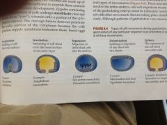
Types of cell movements during gastrulation.
|
invagination-infolding of cell sheet into embryo-sea urchins and endoderm
involution- inturning of cell sheet over the basal surface of an outer layer- amphibian mesoderm Ingression-migration of individual cells into the embryo; sea urchin mesoderm delamination: splitting or migration of one sheet into two sheet> mammalian and bird hypoblast formation -Epiboly: movement of epithelial sheets that spread as a unit (rather than individually) to enclose deepest layers of the embryo. Can occur by cell division or change of shape, or by several layers of cells intercalating |
|
|
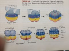
Sea Urchin cleavage have what type of cleavage
|
radial holoblastic cleavage
-first and second cleavages meridional and perpendicular ( or cleavage furrows pass through both animal and vegetal pole) -third is equatorial and separates vegetal and animal poles -fourth division different> four cells of the animal tier divide meridionally into eight blastomeres each with same volume: these are called mesomeres; >vegetal very different divides unequally equitorial cleavage to make four large macromeres and four smaller micromeres at vegetal pole. -As the 16 cell embryo cleaves, the eight animal mesomeres divide equatorially to produce two tiers an1 and an2 one staggered above the other. -Macromeres divide meridionally forming a tier of eight cells below an2k -micromeres also divide later producing small cluster between the largest tier. -At sixth division animal hemisphere divides meridionally, while vegetal divides equatorially then the reverse in the seventh division. AT THAT TIME NOW THE EMBRYO IS A 128 CELL BLASTULA |
|
|
worms.zoology.wisc.edu
|
look at dis
|
|
|
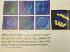
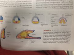
Blastula stage of sea urchin development
|
-begins at 128 cell stage
-cells now form a hollow sphere surrounding a central cavity or blastocoel (shown by f in pic) -By this time all cells same size, micromeres have slowed division. -every cell in contact with proteinaceous fluid of the blastocoesl on the inside and with the hyalin layer on the outside -tight junctions unite once loosely connected blastomeres into seamless epithelial sheet. -As cell continues to divide blastula remains one cell thick thinning as it expands> accomplished by adhesion of the blastomeres to the hyalin layer and by influx of water that expands blastocoel (blastula now called coeloblastula) -these rapid invariant cell divisions last through ninth or tenth division, by this time fates specificied and each cell gets ciliated on cell membrane farthest from the blastocoel. Ciliated blastula begins to rotate within fertilization envelop, soon after can see diff. in cells -cells at vegetal pole of the blastula begin to thicken to form vegetal plate. -cells of animal hemisphere synthesize and secrete a hatching enzyme that digests fert. envelope. -now a free swimming blastula |
|
|
Generally during gastrulation there is
|
- a large amt of coordinated movement
-portions of cells in blastula changed -cells destined to be endoderm brought inside cell -cells to be ectoderm spread over surface of blastula |
|
|
Overview of the whole process before go into details:
|

1. Invagination>infolding of cell sheet
2. Involution> in turn of cell sheet giving you mesoderm 3. Ingression> migration of indiv. cells into embryo 4.delamination-splitting or migration of one sheet into two. 5. epiboly expansion |
|
|
At end or late sea urchin blastulation you have...
|
single layer of about a thousand cells that form a hollow ball with a somewhat flattened vetetal end
-basal lamina and hyaline layer -cells bound tightly to each other and to hyaline layer, bound only minamally to basal lamina |
|
|
What happens in gastrulation after blastula hatches from fertilization envelope?
|
-egg hatches
-a grp of cells derived from micromeres undergo an epithelial to mesenchymal transformation -cells change cytoskeleton become bottle shaped and lose adhesions to lateral cells and break away from the apical layer to enter the blastocoel (8.15 diagram hours nine through ten) -These micromere derived cells called the primary mesenchyme> will form larval skeleton so often called skeletogenic mesenchyme. -skeletogenic mesenchyme begins extending and contracting long thin processes called filopodia -At first cells appear to move randomly along inner blastocoel surface, making and breaking filopodial connections to wall and blastocoel -Eventually become localized within the prospective ventrolateral region of blastocoel. Here they fuse into syncytial cables which will form axis of the calcium carbonate spicules of larval skeletal rods (8.14D-F) [see on previous card] |
|
|
Gastrulation continued: Why does the previous ingression of the previous card happen?
|
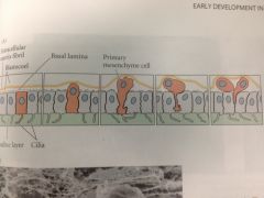
-Originally all the cells of the blastula connected on their outer surface to the hyaline layer, and on their inner surface to a basal lamina secreted by the cells. On lateral surface each cell has another cell for a neighbor
-before gastrulation micromeres bind tightly to one another and to hyaline layer but adhere only loosely to basal lamina. -Pattern changes after gastrulation other cells retain tight bond to hyaline layer and neighbors, primary mesenchyme (which is micromere derived) lose affinity for these structures while affinity for components of basal lamina and extra cellular matrix increases a hundred fold. -Change causes micromeres to release attachments to external hyaline layer and neighbors and is drawn in by basal lamina to migrate up into blastocoel (figure 8.16A) -this ingression that results bc primary mesenchyme cells lose affinity for neighbors and for the hyaline membrane and instead acquiring a strong affinity for a group of proteins that line the blastocoel. |
|
|
Gastrulation continued: Once inside the blastocoesl, what happens with the primary mesenchyme cells?
|
-primary mesenchyme cells have heavy concentration of extracellular material around the ingressing prim. mesenchyme cells.
-once inside, appear to migrate along extracellular matrix of blastocoel wall extending filopodia.>several proteins including fibronectin necessary to initiate and maintain migration -stop movement and form spicules near equator of blastocoel>arrange in a ring at specific position along animal vegetal axis |
|
|
Gastrulation continued: At two sites near the future ventral side of larva, now many of these primary mesenchyme cells ________
|
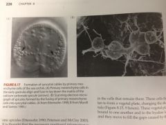
cluster together, fuse with one another, and initiate spicule formation.
|
|
|
Gastrulation continued: archenteron invagination
|
As primary mesenchyme cells leave vegetal region of the spheryical embryo imp. changes occurring.
-vegetal plate bends inward and invaginates one forth of the way into the blastocoel. Then suddenly ceases -invaginated region called the ARCHENTERON (primitive gut) and the opening on the archenteron at the vegetal pole is called the blastopore. |
|
|
Gastrulation continued: Archenteron formation and invagination most likely caused by
|
shape changes in vegetal plate cells and in extracellular matrix underlying them.
vegetal plate cells currounding the 2-8 cells at vegetal pole become bottle shaped, constricting apical ends, this change causes cells to pucker inward. -Also hyaline layer at vegetal plate buckles inward due to changes in composition. |
|
|
Gastrulation continued: changes in composition of the Hyaline layer-(a little about it background) has two layers
|
an outer lamina made primarily of hyaline protein and an inner lamina composed of fibropellin proteins.
-Fibropellins are stored in secretory granules within the oocyte and are secreted from those granules after cortical granules exocytosis releases the hyalin protein. -By blastula phase fibropellins have formed a mesh like network over embryo surface -@ time of invagination, vegetal plate cells and only those cells secrete a chondroitin sulfate proteoglycan into the inner lamina of the hyaline layer directly beneath them. THis water absorbing molecule swells the inner lamina but not outer and causes vegetal region of hyaline layer to buckle. -Slightly later, second force arising from the movements of epithelial cells adjacent to vegetal plate may facilitate invagination by drawing the buckled layer inward. |
|
|
At the stage where these skeletogenic/primary mesenchyme cells begin ingressing into the blastocoel, fates of vegetal plate cells have________
|
already been specified.
|
|
|
A)What is the first grp, of cells to invaginate and form the tip leading the way into the blastocoel?
B)What do these eventually form? |
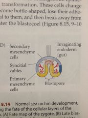
A)The Secondary mesenchyme, (remember primary mesenchyme has already migrated inside)
B)form pigment cells, musculature around gut, and coelomic puches/ |
|
|
Gastrulation of sea urchin continued: Second and third stages of archenteron invagination
|
Invagination of vegetal cells has discrete stages:
1.previously stated invagination AFTER brief pause then second phase. 2.-here archenteron extends dramatically, 3x previous length -goes from wide short gut to long thin tube -to accomplish extension, cells of archenteron rearrange by migrating over one another and by flattening (Convergent extension) 3. Third stage is final -initiated by tension provided by secondary mesenchyme cells which form tip and remain there. -Sec. mes. have filopodia that extend through blastocoel fluid to contact inner surface of cell wall -filopodia attach to specific site that differes from other regions of the animal hemisphere. -extend, touch random spots then retract. When touch particular region however remain attached there and flatten against region and pull archenteron toward it. -there appears to be target region on what is to become ventral side of larva that is recognized by secondary mesenchyme cells and which positions the archenteron in the region where the mouth will form. |
|
|
Sea Urchin gastrulation continued: 3rd phase of archenteron formation: As the top of the archenteron meets the blastocoel wall in target region...
|
-secondary mesenchyme cells disperse into blastocoel where they proliferate to form mesodermal organs (8.15 13.5 hrs)
-Where the archenteron contacts the wall, a mouth is eventually formed. The mouth fuses with the archenteron to create a continuous digestive tube. Blastopore marks position of the anus. |
|
|
What happens then as the pluteus larva we now have elongates?
|
coelomic cavities form from secondary mesenchyme.
Under influence of nodal proteins right coelomic sac remains rudimentary However left coelomic sac undergoes extensive development to form many of the structures of adult sea urchin -left sac splits into three sacs (smaller) -an invagination from the ectoderm fuses with the middle sac to form the imaginal rudement (8.22) -This rudiment develops fivefold symmetry and skeletogenic mesenchyme cells enter the rudiment to synthesize the first skeletal plates of the shell. -Left side of pluteus becomes in effect the future oral surface of the adult sea urchin. -During metamorphosis, imaginal rudiment separates from the larva which then degenerates, while the imaginal rudiment is reforming its digestive tract and settling on ocean floor it depends on nutrition it recieved from the jettisoned larva. PROVIDES PATTERN FOR DEUTEROSOMES DEVELOPMENT-first opening anus then mouth |
|
|
What do nearby cells around secondary mesenchyme become?
|
Endodermal cells adjacent to micromere derived mesenchyme become foregut, migrating farthest into blastocoel, next layer of endodermal cells become midgut and circumferential row to invaginate forms the hindgut and anus
|
|
|
Gastrulation will give you:
|
three tissue layers, echindoderms have three
-ectoderm: outer layer-gives rise to nervous system, skin, and its derivatives -mesoderm-middle layer-gives you coelomic lining, muscles, skeletal systtem -endoderm-inner layer- main portion of gut and structures like organs.. |
|
|
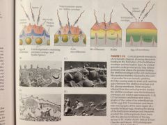
different morphogenic movements
|
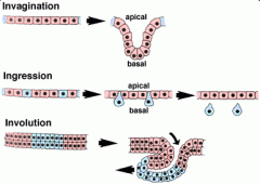
Invagination
During invagination, an epithelial sheet bends inward to form an inpocketing. One way to think of this in three dimensions is to imagine that you are poking a partially deflated beach ball inward with your finger. The resulting bulge or tube is an invagination. If the apical side of the epithelium forms the lumen (central empty space) of the tube, then the movement is termed invagination. If the lumen is formed by basal surfaces, then the movement is termed an evagination. Ingression During ingression, cells leave an epithellial sheet by transforming from well-behaved epithellial cells into freely migrating mesenchyme cells. To do so, they must presumably alter their cellular architecture, alter their program of motility, and alter their adhesive relationship(s) to the surrounding cells. Primary mesenchyme cells are an example of a mesenchymal cell type that emigrates out of an epithelium (do you know which one?). Involution During involution, a tissue sheet rolls inward to form an underlying layer via bulk movement of tissue. One helpful image here is of a tank tread or conveyor belt. As material moves in from the edges of the sheet, material originally at the sites of inward rolling (shown in blue here) is free to move further up underneath the exterior tissue. Epiboly During epiboly, a sheet of cells spreads by thinning. i.e., the sheet thins, while its overall surface area increases in the other two directions. Epiboly can involve a monolayer (i.e. a sheet of cells one cell layer thick), in which case the individual cells must undergo a change in shape. In other cases, however, a sheet that has several cell layer can thin by changes in position of its cells. In this case, epiboly occurs via intercalation, one of the other movements described on this |