![]()
![]()
![]()
Use LEFT and RIGHT arrow keys to navigate between flashcards;
Use UP and DOWN arrow keys to flip the card;
H to show hint;
A reads text to speech;
34 Cards in this Set
- Front
- Back
|
When to order an inspiratory & expiratory film? |
Foreign body |
|
|
When to order an expiratory film? |
Small pneumothorax |
|
|
When to order a decubitus film? |
Pleural Effusions |
|
|
The quality of the film is dependent on which three factors? |
Inspiration Penetration Rotation |
|
|
How to know if the patient had an adequate degree of inspiration? |
Count 6 ribs on the right anterior hemidiaphragm, 10-11 ribs on the posterior. The anterior 7th rib should meet at the midclavicular line. |
|
|
If the patient didn't inspire enough, what can the film mimic? |
CHF, pneumonia |
|
|
Area of increased density in the lung means one of four possibilities: |
Consolidation Interstitial Nodule/Mass Atelactasis |
|
|
Any pathologic process that fills the alveoli with fluid, pus, blood, cells (including tumor cells) or other substances resulting in lobar, diffuse or multifocal ill-defined opacities. When it reaches a fissure the spread stops there. |
Consolidation |
|
|
A Pneumothorax is diagnosed using which modality? |
Lateral decubitus CXR Patient lies on opposite site of the suspected pneumothorax to accentuate the view. |
|
|
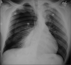
Pneumothorax |
|
|
|
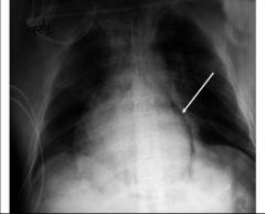
Pneumothorax |
|
|
|
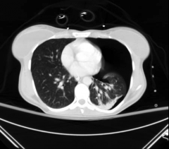
Pneumomediastinum |
|
|
|
Unilateral Pleural thickening is likely due to what? |
Empyema |
|
|
Bilateral Pleural thickening is likely due to what? |
Asbestos Poisoning |
|
|
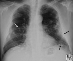
Pleural thickening |
|
|
What is fundamental in diagnosing interstitial lung disease? |
High resolution CT |
|
|
Three appearances of interstitial lung disease? |
Reticular Micronodular Reticulonodular |
|
|
What is ground glass opacity on an HRCT a sign for? |
Active interstitial pulmonary disease |
|
|
What helps differentiate between Lung fibrosis and ground glass opacity? |
HRCT |
|
|
Ground glass opacity responds well with what type of treatment? |
Steroids |
|
|
Honeycombing is a sign for? |
Advanced lung fibrosis |
|
|
What is the most common cause of lobar/segmental consolidation? |
Pneumonia |
|
|
Air bronchogram sign is seen on? |
CT |
|
|
Diagnostic study of choice for a bronchiectasis? |
HRCT |
|
|
Diagnostic study of choice for COPD? |
HRCT |
|
|
Most common cause of a benign pulmonary tumor? |
Granuloma |
|
|
Dilatation of pulmonary vessels proximal to embolism along with collapse of distal vessels, often with a sharp cut off. |
Westermark sign - Pulmonary embolism |
|
|
Diagnostic study for Pulmonary Embolism? |
Pulmonary Angiography |
|
|
Diagnostic study for proximal DVT? |
Venous Ultrasonography
When there is clinical suspicion - use contrast venography |
|
|
Creatinine with >1.8 |
Not able to do Contrast CT |
|
|
Diagnostic study for Pulmonary hypertension? |
Gold standard: Right sided Cardiac Catheterization (ECHO w/ doppler is useful) |
|
|
4 reliable signs of CHF |
Stage 2 Kerley B lines Fluid in the fissure Stage 2 Peribronchial cuffing Stage 4 Bilateral Pleural effusion |
|
|
Bat wings appearance |
Stage 3 CHF |
|
|
Double lumen in a CT |
Aortic Dissection |