Reading...
![]()
Play button
![]()
Play button
![]()
Use LEFT and RIGHT arrow keys to navigate between flashcards;
Use UP and DOWN arrow keys to flip the card;
H to show hint;
A reads text to speech;
47 Cards in this Set
- Front
- Back
|
What holds down the biceps brachii tendon instead of the humeral retinaculum in the horse?
|
The tendons of insertion of supraspinatus m. inserting on both greater and lesser tubercle.
|
|
|
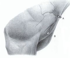
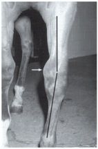
Where do you inject into a horse shoulder joint?
|
(A)
Insert needle at cranial margin of infraspinatus tendon approximately 2 cm proximal to caudal part of greater tubercle Direct needle ventromedially, approximately 4-5 cm deep |
|
|
Do intraarticular anesthetic blocks block structures distal to the joint?
|
No, they just anesthetize locally.
|
|
|
What is most important in performing a joint injection?
What reasons would you perform a joint injection? |
Sterile everything
Anesthetics, antiobiotic delivery, steroid delivery, draining infection. |
|
|
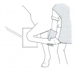
What is your approach to a joint injection of the bicipital bursa?
|

Bicipital (intertubercular) bursa (B)
Insert needle between biceps and humerus, slightly above the level of the deltoid tuberosity Direct needle proximally |
|
|
Which tendons act as the lateral collateral ligaments of the equine elbow?
|
Laterally - infraspinatus m.
Medially - subscapularis m. |
|
Name this stuff - especially #6
|

how was it?
|
|
|
What carpal bones are most commonly fractured in racing horses? What type of fractures?
|
Radial carpal bone (chip fractures) and 3rd carpal bone (slab fractures - across two joints)
|
|
|
Why is the radial carpal bone one of the most commonly fracture in horses?
|
Most of the weight is carried medially.
|
|
|
What are the ligaments of the carpus in the horse?
|
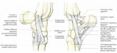
Collateral ligaments
Ligaments of the accessory carpal bone Short ligaments - running transversely |
|
|
How many medial and lateral collateral ligaments do equine carpal joints have?
|

3 medially, 2 laterally
|
|
|
Which aspect of the equine carpal bones have the most movement - proximal or distal?
|
Most movement in the proximal joint, distal – minimal movement.
|
|
|
What shape should the carpal bones be used?
|
Carpal bones should be cuboidal in shape
Rounded margins indicate incomplete ossification |
|
|
When are the equine carpal bones fully developed?
|
18 months
|
|
|
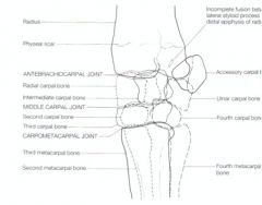
What are the ossification centers of the distal radius in the equine? When do they close?
|
Lateral styloid process
Fuses in first year of life Distal radial physis Closes at approximately 20 months of age |
|
|
What is the clinical relevance of the distal radial physis of the equine carpus?
|
Distal radial physis will tell you the level of maturtity and whether the animal is ready for heavy work
|
|
|
What is the epiphysis of the metacarpal III in the horse? When does it fuse?
|
Metacarpal III (cannon bone)
has a proximal epiphysis which is fused at birth |
|
|
What are the three distinct articulations of the equine carpus?
|
Radiocarpal (antebrachiocarpal) joint
Middle carpal (midcarpal) joint Carpometacarpal joint KNOW THIS |
|
|
The equine carpus has a fibrous joint capsule, how many of the articulations in the carpus does it articulate with? What ligament is a part of the capsule?
|
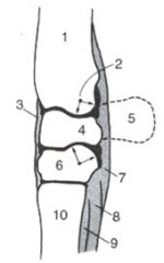
Fibrous joint capsule (3) is common to all three articulations
Palmar carpal ligament (7) is part of the fibrous joint capsule |
|
|
Do any of the articulations of the equine carpus communicate?
|

Synovial membranes enclose the individual articulations
Except for small communication between middle and distal levels (THIS IS IMPORTANT TO KNOW!) |
|
|
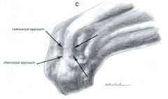
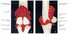
What are the needle placements of the equine carpus?
|
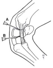
Needle placement in radiocarpal joint (A) and the intercarpal joint (B) of the right forelimb
|
|

What type of deformity is this? How do you treat it?
|
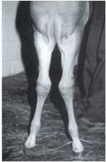
carpal valgus
Valgus deformity Lateral deviation of a part of the limb distal to a joint Periosteal stripping |
|

What type of deformity is this?
What is the treatment? |
carpal varus -
Varus deformity Medial deviation of a part of the limb distal to a joint Periosteal stripping |
|
|
How would you define the radiocarpal joint of the equine carpus? What is its degree of flexion?
|
Between radius and proximal row of carpal bones
90-100 degrees flexion |
|
|
What is the degree of flexion of the middle carpal joint of the equine carpus?
|
45 degrees of flexion
|
|
|
What is the degree of flexion of the carpometacarpal joint of the equine carpus?
|
No significant movement
|
|
|
How and where do you inject into the radiocarpal joint of the equine carpus?
|

Flex carpus
Palpate between radius and proximal row of carpal bones ****Medial or lateral to extensor carpi radialis tendon (IMPORTANT) |
|
|
How and where do you inject into the middle carpal joint of the equine carpus?
|
Flex carpus
Palpate between the two rows of carpal bones ****Medial or lateral to extensor carpi radialis (IMPORTANT!) |
|
|
Where do you inject into the carpometacarpal joint of the horse?
|

Communicates with middle carpal joint - so you need to follow that injection site:
Flex carpus Palpate between the two rows of carpal bones ****Medial or lateral to extensor carpi radialis |
|
|
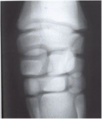
What are the standard radiographic views of the equine carpus?
|
Lateromedial (lateral)
Dorsopalmar (“AP”) Dorsal 45° lateral-palmaromedial oblique (medial oblique) Dorsal 45° medial-palmarolateral oblique (lateral oblique) Flexed lateromedial (flexed lateral) |
|

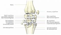
What would 6 be?
|

Carpal bone 1
|
|
|
Which radiograph view will give you the best view of the 4th carpal bone, ulnar carpal bone and metacarpal 4?
|
Dorsal 45 degree lateral-palmaromedial oblique
|
|
|
Which radiograph view is best to view the 2nd carpal bone, radial carpal bone and 2nd metacarpal bone of horses?
|
Dorsal 45° medial-palmarolateral oblique
|
|

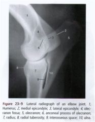
What radiographic view is this? What does it let you see?
|

Dorsal 45° medial-palmarolateral oblique
Best to see: 2 c, radial carpal and 2nd metacarpal. |
|
What radiographic view of the equine carpus is this? What does it let you see best?
|
Dorsal 45° lateral-palmaromedial oblique
To see: Mc 4, 4th carpal, and ulnar carpal bone |
|
|
What are the landmarks for the proximal palmar pouch of the the fetlock of the horse for the fetlock joint injection?
|

Proximal to proximal sesamoids and collateral sesamoidean ligament
Distal to the distal end of the splint bone Palmar to MC III Dorsal to the suspensory ligament (interosseus) (D) in the picture |
|
|
What are the injection site boundaries for the pastern joint in the horse?
|
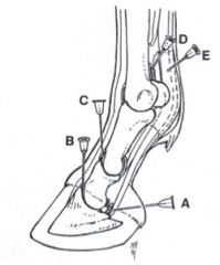
(C) in the picture
Medial or lateral to midline near the palpable epicondyles of P II Insert needle vertically for approximately 2.5 cm Minimal movement in this joint |
|
|
Are pastern of coffin joint injections down frequently? Why or why not?
|
No, because perineural blocks are safer and easier.
|
|
|
What are the boundaries of injection into the coffin joint?
|

Insert needle 1.5 cm proximal to the coronet approximately 2 cm lateral to the vertical center of the pastern
Direct needle obliquely ventral to the common digital extensor tendon toward the extensor process (B) in the picture |
|
|
How can blocking the coffin joint effect the navicular bursa?
|
Though the joints don't communicate, anesthetic can diffuse from one joint to the other.
|
|
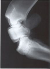
What radiographic view is this?
|

Flexed lateromedial view
|
|

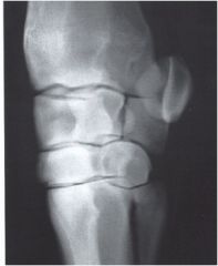
What radiographic view is this of the equine carpus?
|
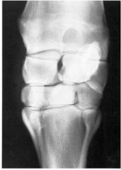
Dorsopalmar view
|
|

What radiographic view is this?
|
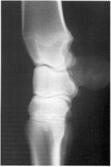
This is the lateromedial view.
|
|
|
What is the best radiographic technique for diagnosis of slab fractures and degenerative joint disease?
|

Dorsoproximal-dorsodistal oblique (“skyline”)
Distal radius - 85° to cassette Proximal row of carpal bones - 55° to cassette Distal row of carpal bones - 35° to cassette |
|
|
T/F in the ox, there is a carpal bone I
|
False - there may or may not be one in the horse, but it is absent in the ox.
|
|
|
T/F in the ox, the carpal bones II and III are fused.
|
True.
|
|
|
Is the fetlock very extensive?
|
Yes
|