Reading...
![]()
Play button
![]()
Play button
![]()
Use LEFT and RIGHT arrow keys to navigate between flashcards;
Use UP and DOWN arrow keys to flip the card;
H to show hint;
A reads text to speech;
107 Cards in this Set
- Front
- Back
|
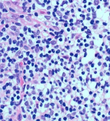
Lymphocytes. Appear as naked nuclei because of limited amounts of cytoplasm
|
|
|
|
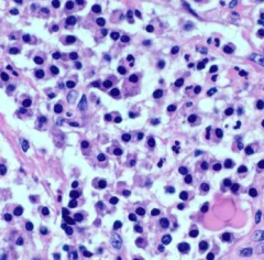
B cell derived plasma cells with eccentrically placed nuclei with prominent cytoplasm
|
|
|
|
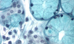
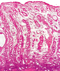
Plasma cells in loose connective tissue between mucus-secreting glands
|
|
|
|
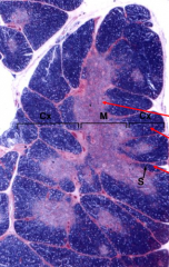
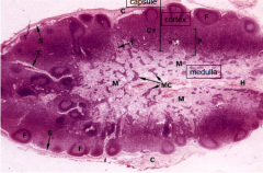
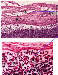
Infant thymus. Lobulated organ surrounded by loose collagenous capsule, short interlobular septa with blood vessels. Divided into inner medulla and outer cortex
|
|
|
|
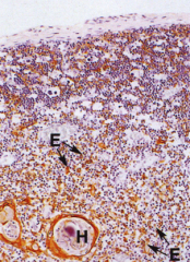
Thymus. Epithelial framework more finely branced in cortex (reticular) than medulla. Cells have desmosomes and contain keratin in intermediate filaments. H - Hassals corpuscles: degenerating keratinized epithelial cells
|
|
|
|
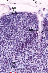
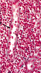
Thymic cortex: Contains immature and mature T-Cells. Macrophages phagocytose apoptotic T cells are cortico0medullary junction
|
|
|
|
Lymph node. Contains outer capsule, macrophages, and lymphocytes
|
|
|
|
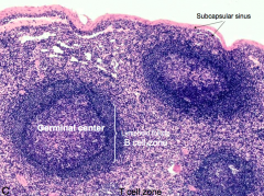
Lymph node. Capsule, cortex, and medulla
|
|
|
|
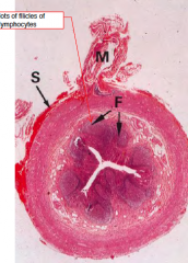
Capsule and Subcapsular Sinus: Capsule is pierced by afferent lymphatic vessels with valves to ensure one-way flow. Underlying subcapsular sinus S contains endothelial cells and Dendritic cells that capture Ags
|
|
|
|
Lymph node with high endothelial venule
|
|
|
|
Lymph node. Subcapsular and medullary sinus filled with reactive macrophages
|
|
|
|
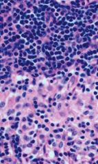
Lymph node: lymphocytes on top and macrophages on bottom
|
|
|
|
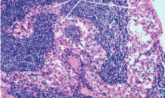
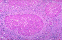
Lymph node: B cells in lymphoid follicle and germinal center. Resting B cells in mantle zone (outer circle) and B cells proliferating in germinal center
|
|
|
|
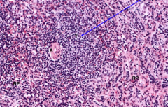
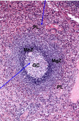
Splenic lymphoid tissue: White pulp (area containing lymphocytes suspended on reticular fibers and involved in immune function), T cell zone
|
|
|
|
Spleen. Perilymphoid cells contains lymphocytes migrating towards white pulp
|
|
|
|
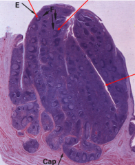
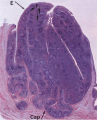
Palatine tonsil. Leukocyte surveillance of antigens
in oral cavity, gastointestinal tract & at other mucosal surfaces. Ag enters in crypts phacocytosed by epithelial cells of crypt lining, passed to follicles. |
|
|
|
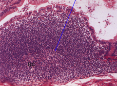
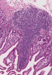
Peyer’s Patch in gut with germinal center.
Contains M cells-specialized to take up antigen from lumen of gut & transport to PP |
|
|
|
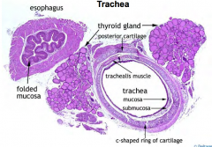
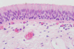
Trachea. Cartilage rings and epithelium
|
|
|
|
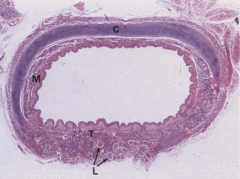
Trachea
|
|
|
|
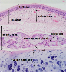
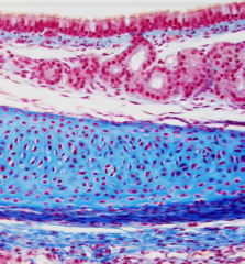
Trachea. Mucosa (epithelium and lamina propria), submucosa (submucous gland - serious and mucous part), and Hyaline Cartilage
|
|
|
|
Trachea
|
|
|
|
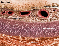
Trachea - epithelium, basement membrane, lamina propria, hyaline cartilage, and adventitia
|
|
|
|
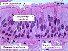
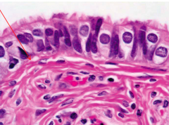
Trachea - respiratory epithelia, ciliated pseudostratified with goblet cells
|
|
|
|
Trachea
|
|
|
|
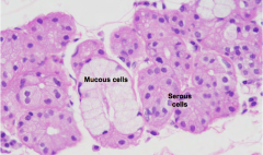
Trachea - submucosal gland, mucous and serous secretory cells (secrets lysozyme)
|
|
|
|
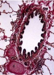
Bronchus - Cartilage patches (not rings), fewer seromucous glands, smooth muscle, lymphoid tissue in submucosa
|
|
|
|
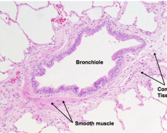
Bronchiole - no longer contains cartilage
|
|
|
|
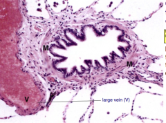
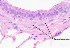
Bronchiole - smooth muscle and connective tissue
|
|
|
|
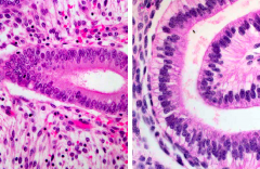
Bronchiole - simple, columnar ciliated epithelium
|
|
|
|
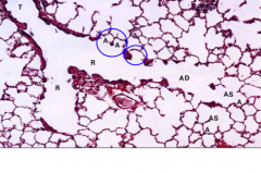
Respiratory portion: Terminal bronchiole, respiratory bronchiole, alveolar duct, alveolar sac, alveoli
|
|
|
|
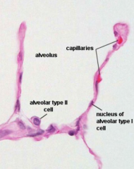
Alveoli - Type 1 cell for efficient gas exchange, type 2 cell secrets surfactant
|
|
|
|
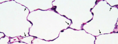
Alveoli with elastic fibers stained
|
|
|
|
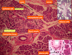
Salivary glands
|
|
|
|
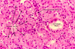
Acini enter into intercalated ducts then striated ducts
|
|
|
|
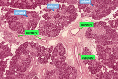
Striated ducts empty into excretory ducts
|
|
|
|
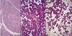
Parotid gland: Neonate, mid age, and elderly
|
|
|
|
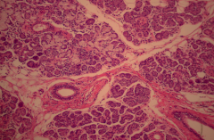
Parotid gland, Serous acinar cells and lots of adipose
|
|
|
|
Submandibular gland with mixed mucous and serous cells and less adipose tissue
|
|
|
|
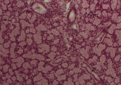
Sublingual gland with only mucous acini
|
|
|
|
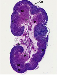
Kidney
|
|
|
|
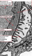
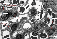
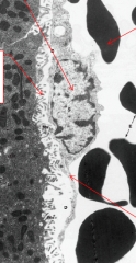
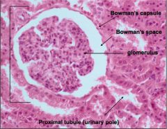
Glomerular filter
|
|
|
|
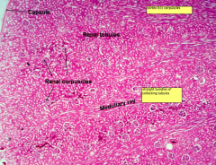
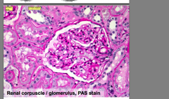
Renal corpuscle, glomerulus
|
|
|
|
Kidney
|
|
|
|
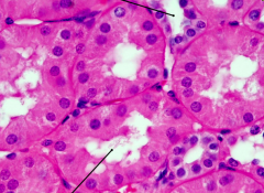
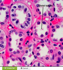
Proximal and Distal Convoluted Tubule
|
|
|
|
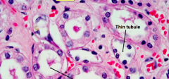
Distal and Thin tubule
|
|
|
|
collecting duct and distal tubule
|
|
|
|
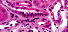
Distal tubule, afferent arteriole, and juxtaglomerular cells with renin granules
|
|
|
|
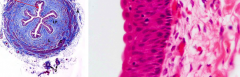
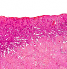
Transitional epithelium
|
|
|
|
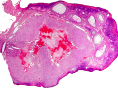
Ovary with follicles
|
|
|
|
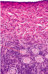
Germinal epithelium (continuous with viceral peritoneum) tunica albugenia, and stroma with follicles
|
|
|
|
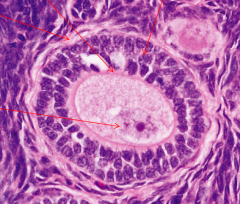
Primary oocytes. ZP, granulosa, and theca cells
|
|
|
|
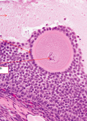
Graafian follicle. Antrum, granulosa, theca, ZP
|
|
|
|
Fallopian tube
|
|
|
|
Corpus luteum, foamy cytoplasm contains progesterone
|
|
|
|
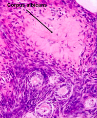
Corpus albicans
|
|
|
|
Proliferative endometrium (uterus) Stratum compactum, stratum spongiosum, stratum basalis, myometrium
|
|
|
|
Secretory endometrium. Stratum functionalis (= compactum+ spongiosum)at the onset of menstruation spiral arterioles constrict in the absence of progesteron
|
|
|
|
Proliferative (many mitotic nuclei) vs. secretory (basal glycogen vacuoles)
|
|
|
|
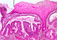
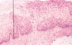
Cervix - squamocolumnar jxn
|
|
|
|
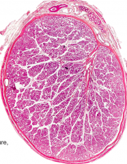
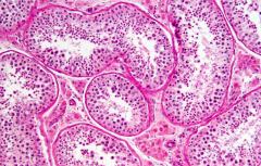
Testis
|
|
|
|
Seminiferous tubules
|
|
|
|
Spermatazoa
|

|
|
|
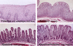
GI Tract epithelium
|
|
|
|
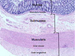
Layers of epithelium in GI tract
|
|
|
|
E: epithelium
LP: lamina propria SM: Submucosa MM: muscularis mucosae |
|
|
|
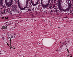
Submucosa: LP: Lamina propria
MM: muscularis mucosa SM: submucosa PG: parasympathetic ganglia |
|
|
|
Two Layers of Muscularis Propria
C: circular muscle layer L: longitudinal muscle layer AP: Auerbach’s plexus |

|
|
|
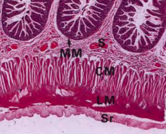
Adventitia/Serosa
S: Submucosa MM: muscularis mucosa CM: Circular muscle LM: Longitudinal muscle Sr: Serosa/Adventitia |
|
|
|
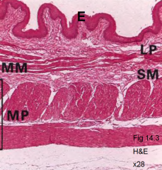
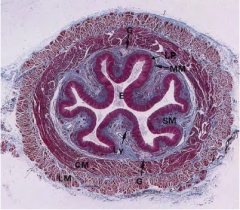
Esophagus
E: stratified squamous epithelium (protective mucosa) LP: lamina propria Ly: lymphoid aggregates MM: muscularis mucosa SM: submucosa G: seromucous glands CM: circular muscle LM: longitudinal muscle |
|
|
|
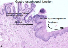
Gastro-esophageal junction
|
|
|
|
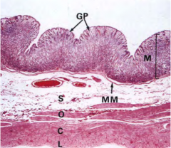
Stomach
GP: gastric pits M: mucosa MM: muscularis mucosa S: submucosa O: oblique muscle layer C: circular muscle layer L: longitudinal muscle layer |
|
|
|
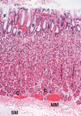
Gastric Mucosa
F: Gastric pits C: Chief (peptic) cells MM: muscularis mucosa SM: submucosa |
|
|
|
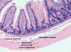
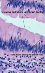
Small intestine
|
|
|
|
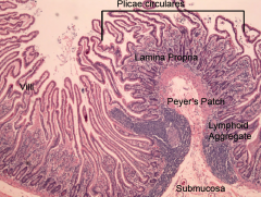
Peyer's patch
|
|
|
|
Small intestine
|
|
|
|
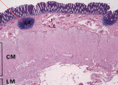
Large intestine
|
|
|
|
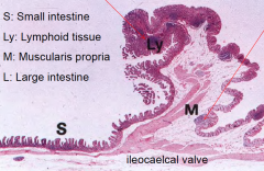
Illeoceacal junction
|
|
|
|
Rectoanal junction
|
|
|
|
Appendix
|
|
|
|
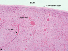
Liver
|
|
|
|
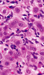
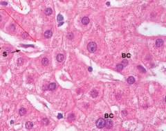
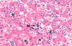
Hepatocytes with sinusoids and binucleated cells
|
|
|
|
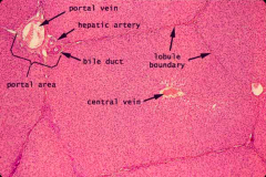
Hepatic lobule
|
|
|
|
Hepatocytes space of Disse
|
|
|
|
Liver cirrhosis
|
|
|
|
Hepatic hematopoiesis
|
|
|
|
Bile canaliculi
|
|
|
|
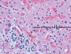
Bile canaliculi merge to form canals of Hering
|
|
|
|
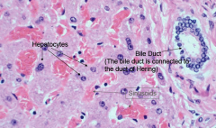
Bile ducts
|
|
|
|
Gall bladder
|
|
|
|
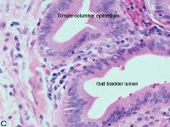
Gall bladder wall
|
|
|
|
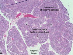
Exocrine and endocrine pancreas
|
|
|
|
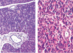
Exocrine pancreas: acini and duct system
A glandular acini C centroacinar cells D intercalated ducts I intralobular ducts S support tissues |
|
|
|
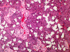
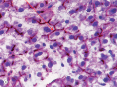
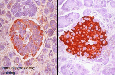
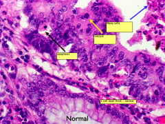
Endocrine pancreas: islets of Langerhans
Antibody to insulin identify the β cells. Insulin induces increased glycogen synthesis in liver (and muscle) Antibody to glucagon identifies the α cells. Glucagon causes the liver to release glucose - stored in the form of glycogen |
|
|
|
Cervix - normal and dysplasia
|
|
|
|
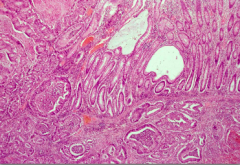
Invasive Colonic Adenocarcinoma
Invading, glands go into different wall of colon. abmoral glands in submucosa |
|
|
|
Invasive Colonic Adenocarcinoma
|
|
|
|
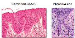
Carcinoma in situ vs microinvasion
|
|
|
|
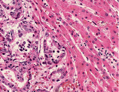
Metastatic Colonic Adenocarcinoma
|
|
|
|
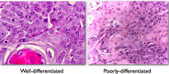
Squamous cell carcinoma
|
|
|
|
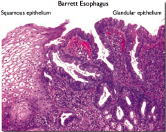
Barret's esophagus
|
|
|
|
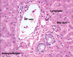
Portal tract
|
|
|
|
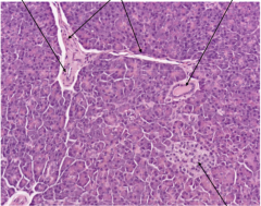
Pancreas
|
|
|
|
Kidney
|
|
|
|
Renal Corpuscle
|
|
|
|
Tonsil
|
|
|
|
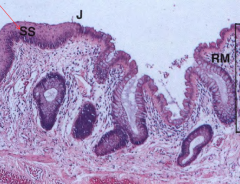
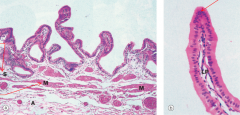
Duodenum
|
|
|
|
Peyers Patch
|

|