Reading...
![]()
Play button
![]()
Play button
![]()
Use LEFT and RIGHT arrow keys to navigate between flashcards;
Use UP and DOWN arrow keys to flip the card;
H to show hint;
A reads text to speech;
99 Cards in this Set
- Front
- Back
|
How is lymphoid tissue organised in the body
|
1) Diffuse: MALT (mucosa associated lymphoid tissue) eg GALT, NALT, BALT, SALT
2) Aggregated: thymus, lymph nodes, spleen 3) connective tissue |
|
|
What type of tissue is bone marrow, describe its functions
|
1 deg lymphoid: immature B&T produced from stem cells. B cells mature in BM. T cells migrate & mature in thymus.
|
|
|
Where do APCs present antigen? What is the response called?
|
2 degree lymphoid organs (lymph nodes, spleen, MALT, connective tissue). It is an adaptive immune response.
|
|
|
How are lymphatic capillaries different to blood capillaries
|
Blind ended, valves, larger diameter, thinner walls, low pressure
|
|
|
What are the lymphatic capillaries inthe small intestine
|
lacteals
|
|
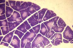
Name the features
|
Thymus: lobules, cortex & medulla, septa, thymocytes, reticular cells (visible only in medulla)
|
|
|
What types of cells attach to the thymic reticular cell processes
|
T lymphocytes and macrophages
|
|
|
Describe the process of maturation of lymphocytes in the thymus
|
1) Immature T cells produced in bone marrow
2) Enter cortex & divide 3) Migrate & differentiate. Macrophages destroy self-recognising T-Cells 4) Mature T-Cells in medulla leave to 2 deg lymphoid organs |
|
|
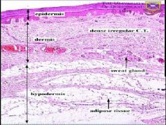
Describe the layers of the skin
|
1) Epi: epithelial layer
2) Dermis: ieergular connective tiss, blood vessels, nerves, pilosebaceous units 3) Hypodermis: temp regulation (sweat glands, lots of fat), attachment of upper layers |
|
|
Describe histologically the stratum basale
|
1) Basal/germinative: thin layer of basophilic columnar bound to basement membrane via hemidesmosomes (desmosomes bind each other)
|
|
|
Describe histologically the stratum spinosum
|
Thick layer of basophilic cells, slightly flattened and adjacent to stratum basale
|
|
|
Describe histologically the stratum granulosum
|
Thin layer of intensely basophilic cells (keratohyalin -> keratin) above stratum spinosum
|
|
|
Describe histologically the stratum lucidum
|
Thin, pale eosinophilic (no nuclei) above stratum granulosum, only in glabrous epithelium
|
|
|
Compare glabrous and thin skin
|
In thin skin
1) stratum corneum is much thinner 2) stratum lucidum absent 3) stratum spinosum reduced |
|
|
Describe histologically the stratum corneum
|
Eosinophillic (no nuclei), flattened, dead squamous cells
|
|
|
How is the skin waterproofed
|
1) Stratum granulosum produces hydrophillic lipid
2) Stratum corneum is tightly packed, dead squamous epithelium. |
|
|
What factors determine the colour of skin
|
1) Blood flow
2) Epidermal thickness (allowing capillaries to show through) 3) Pigmentation: B-carotine, melanin |
|
|
How and where is melanin produced
|
Melanocytes (round cells in stratum basale) contain melanosomes, passed to epidermal cells by dendritic processes. Activity controlled by hormones & sunlight.
|
|
|
What cells are involved in an immune function in the epidermis
|
Langerhans cells: APCs
|
|
|
Describe the structure(s) of the dermis
|
1) Papillary layer: fibroblasts, blood vessels, collagen,
|
|
|
What happens during G1, S and G2 phases of the cell cycle
|
G1: cell growth
S: DNA synthesis G2: Doubling of each DNA strand into pairs |
|
|
What is the M phase of the cell cycle
|
Mitosis: separation of duplicated strands of DNA and recombination
|
|

What stage of cell cycle
|
Prophase
|
|

What stage of cell cycle
|
anaphase
|
|

What stage of cell cycle
|
metaphase
|
|

What stage of cell cycle
|
cytokinesis
|
|

What stage of cell cycle
|
cytokinesis
|
|

What stage of cell cycle
|
Telophase
|
|
|
List the roles of simple squamous epithelia
|
1) endothelia
2) alveoli 3) lining of body cavities |
|
|
List the roles of simple cuboidal cells
|
1) secretory (glandular)
2) terminal bronchii |
|
|
List the roles of simple columnar
|
1) secretion & absorption, enhanced by microvilli
|
|
|
Describe pseudostratified cells
|
Irregularly placed nuclei, appearance of stratification, ciliated & secrete mucous
|
|
|
Where is stratified epitheliam and describe it's main features
|
1) Skin, pharynx, oesophegus, oral cavity
2) Flattened, non nucleated, dessicated, keratinised |
|

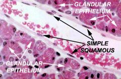
Describe the cells present
|
Simple squamous epithelium in endothelium. Columnar cells beneath.
|
|
Describe the cells present
|
Simple squamous epithelium in endothelium. Columnar cells beneath.
|
|

Why are the central cells lighter in colour
|
Genuine duct (not artifact) surrounded by cuboidal epithelium.
|
|
Why are the central cells lighter in colour
|
Genuine duct (not artifact) surrounded by cuboidal epithelium.
|
|
Describe the main epithelial features
|
Simple columnar cells lining white duct, attached to pink basement membrane
|
|
|
List 7 causes of cell injury
|
mechanical, chemical, oxygen deprevation, nutritional, genetic, infection, inflammation
|
|
|
How is necrotic heart tissue identified
|
pale
|
|
|
List the critical factors leading to cell death
|
1) mitochondrial injury
2) ATP depletion 3) increased intracellular calcium 4) oxygen free radicals 5) changes in membrane permeability |
|
|
In hypoxia or mitochondrial, impairment, list the biochemical consequences
|
ATP depletion causes: Na+/K+ 1) pump stops -> cellular swelling
2) Ca+ pump stops -> necrosis 3) glycolysis increases -> lactic acid buildup |
|
|
What are the effects on mitochondria during hypoxia
|
1) mitochondrial membrane potential drops (mitochondrial membrane transition
2) leakage of cytochrome-C (happens in apoptosis) |
|
|
What are the intracellular effects of increased intracellular calcium
|
Induction of phospho lipases, proteases -> cell death
|
|
|
What are the intracellular consequences of oxygen free radicals present
|
Oxidation/modification of DNA, enzymes, membranes.
|
|
|
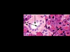
Why is the liver pale
|
Fatty liver due to alcohol. Reversible form of cellular damage.
|
|
|
List the morphological changes defining necrosis (as opposed to just cell death)
|
1) autolysis and/or lysosomal breakdown of RNA & proteins -> eosinophillic (red)
2) glassy (no glycogen), moth eaten cytoplasm (digested contents) 3) karyolysis (breakdown of DNA), karyohexis (disappearance of DNA), |
|
|
List the 5 types of necrosis
|
1) Liquefactive (2) Gangrenous (3) Coagulative (4) Caseous (5) Fat
|
|
|
Describe coagulative necrosis
|
Cell structural proteins preserved, enzymes denatured
|
|
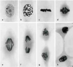
Identify A - F
|
A Interphase
B Prophase C Metaphase D-F Anaphase G Telophase H Interphase |
|
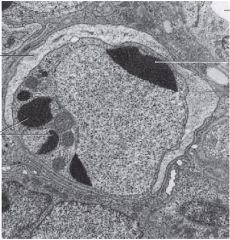
What is 1 in the diagram & how will these be removed from teh cell
|
Chromatin bodies - apoptosis - will form blebs and be engulfed by macrophages
|
|
List the features of the lymph node
|
1) Capsule
2) Marginal sinus 3) Medullary sinus 4) Cortex showing numerous B cells |
|
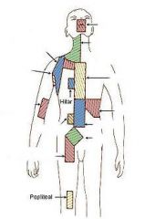
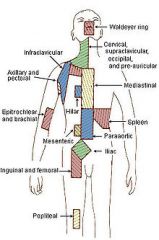
Name the lymph node regions
|
|
|
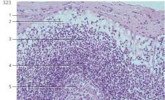
Describe 2, 3 & 5 of a lymph node
|
2) Marginal sinus showing reticulum cells
3 Cortex 5 Germinal center |
|
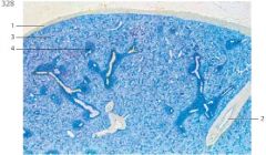
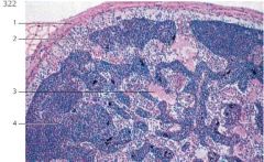
List the features of the spleen
|
1 Fibrous capsule
2 Spleen trabeculae with vein 3 Red pulp 4 Splenic nodule with central artery |
|
|
What parts of the GI does the mucosa change most abruptly
|
1) Gastro-oesophageal jn
2) Gastro-duodenal junction 3) Ileo-coecal jn 4) Recto-anal jn |
|
|
Where is the submucosa of the GI tract & what does it do
|
1) in between the muscularis mucosa and themuscularis externa
2) loose connective tissue, lymphatics, blood vessels, nerves |
|
|
Describe the adventitia
|
1) visceral peritoneum
2) contains major nerves & blood vessels 3) derived from mesoderm |
|
|
List the features if the lip & how these can orientate the inner & outer surfaces
|
Outer: hair follicles, sweat glands, sebacious glands, epithelium is keratinised
Inner: labial salivary glands in the lamina propria of the mucosa, thick stratified non-keratinised squamous epithelium |
|
|
How is the organisation of glands different in the stomach compared to the duodenum and oesophegus
|
Glands located in the mucosa in stomach. Outer epithelium is all columnar mucous cells, beneath in mucosa are parietal cells and chief cells.
|
|
|
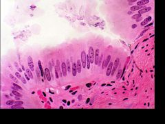
How can you histologically differentiate between parietal & chief cells
|
Parietal cells: dark nucleus surrounded by clear zone,
Chief cells: stain dark red |
|
|
How can we histologically differentiate between jejunum and duodenum
|
Duodenum: numerous large Brunner's glands in sub-mucosa
Jejunum: no significant glands in sub-mucosa |
|
|
How can we differentiate between ilium & jejunum
|
1) submucosa full of lymphocytes - lymphoid aggregates
|
|
|
How can we histologically differentiate between colon & ilium
|
1) circular Peyers patches
2) Peyers patches lined with M-cells, antigen presenting cells with pale white cytoplasm 3) folding less prominent |
|
|
What are the main differences between identifying oesophageal and stomach tissue
|
Oesophagus: stratified squamous, secretory glands in sub-mucosa
Stomach: columnar mucosal cells, deep formation of crypts, distinct chief & parietal cells in mucosa |
|
|
Describe each layer of the skin
|
1) basale: cuboidal/columnar (dividing) sits on basale
2) spinosum: flattening, forming keratin 3) granulosum: keratin producing, intensely basophilic, nucleus degenerating 4) lucidum (thick skin), pale, nucleus degenerated 5) corneum: flattened, dead, anuclear, keratinised cells |
|
|
Describe the features of the dermis
|
Papillary dermis: papilla project into epidermis, contain fibroblasts, connective tissue, Langerhan's cells (APCs), mast cells, vascular, innervated
Reticular epidermis: dense irregular connective tissue, sweat, sebaceous, pilosebaceous units. |
|
|
What kind of gland is a sweat gland, how is it inervated
|
1) Exocrine gland (merocrine)
2) sympathetic cholinergic fibres, post ganglionic, muscarinic receptors |
|
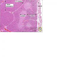
Describe the features
|
Thymus lobule:medulla is paler following apoptosis of self recognising T-cells, epitheliocytes form framework and degenerate to Hassal's corpuscles, septa continuous with capsule to enclose lobule
|
|
|
How does the spleen differ from the thymys
|
1) spleen 2°, thymys 1°
2) thymus has medulla (sparse Ts) & cortex (dense Ts), spleen has white (Tcells surround arteriole, B cells close by) and red pulp (open capillaries drain into sinuses containing slitted venule walls allowing passage of competent RBCs. Stiff RBCs are degraded by macrophages) |
|
|
How do the main 2 roles of lymph nodes and the spleen differ
|
Lymph nodes: filter lymph, Ag presentation
spleen: filters blood (RBCs), Ag presentation |
|
|
What are the features of the cortex & medulla of a lymph node
|
1) cortex: 1° follicles of B cells prior to Ag
2) Ag from aferent lymph -> B cell proliferation in cortical germinal centres. Can present antigen to T-cells in paracortex 3) Paracortex: T cells encounter APCs from high endothelial venules & B-cells, proliferation of Th (effector & memory), Tc 4) Medulla: proliferation of mature plasma cells producing ABs |
|
|
List the parts of the upper body having pseudostratified columnar epithelium
|
1) nasal cavity including nasopharynx
2) conduction airways ( trachea, bronchi (not oropharynx & vocal chords) |
|
|
Describe pseudocolumnar tissue
|
1) each cell irrecular shaped columnar but all join basement membrane
2) goblet cells in between 3) cilia on free surface |
|
|
List the areas stratified squamous epithelium is found
|
1) skin
2) oral cavity 3) oropharynx 4) vocal chords 5) oesophagus |
|
|
What is the primary role of columnar cells
|
Absorption via microvilli but also some secretion
|
|
|
What is the primary role of cuboidal cells
|
secretory ie in glands.
|
|
|
What type of cells are endothelium
|
simple squamous
|
|
|
What are the 4 main roles of simple squamous epithelium
|
1) mechanical barrier (eg endothelium)
2) filtration (kidneys) 3) diffusion (alveoli) 4) secretion (serous fluid into cavity spaces eg peritoneal, pleural) |
|
|
What is the difference between G1 & G2 of the cell cycle
|
G1 = normal growth of cytoplasm
G2 = growth of cell preparing for mitosis |
|
|
What is the difference between S & M of the cell cycle
|
S = DNA synthesis
M = mitosis (separation of identical strands of DNA) |
|
|
Describe in one word each phase of mitosis
|
Prophase - cluster
Metaphase - line Anaphase - circle Telophase - pair |
|
|
Define metaplasia and give 2 examples
|
Reversible replacement of one differentiated cell type with another
1) smoking - airways pseudostratified replaced with squamous 2) Barrett's Oesophagus: stratified squamous replaced with columnar |
|
|
What is the replacement of normal cells with abnormal immature cells
|
dysplasia
|
|
|
List 3 type of cell replacement processes leading to abnormal tissue
|
Metaplasia (dislike with dislike)
Dysplasia (like with like but immature) Neoplasia (abnormal proliferation) |
|
|
What are Langerhans Cells
|
Dendritic
|
|
|
What are teh germ layer origins of skin
|
Ectoderm - epidermis
Mesoderm - dermis |
|
|
Describe the stratum spinosum
|
1) Proliferating layer between basal and granulosum
2) Contains Merkel & Langerhans |
|
|
Describe the stratum basale
|
1) Keratinocytes & stem cells
2) Adjacent to basement membrane |
|
|
What germ layers are skin appendages derived from and which are developed at birth
|
1) Mesoderm, ectoderm
2) Hair, nails, sweat, sebaceous glands |
|
|
Describe the keratinocyte journey from stratum basale to the surface
|
1) division: one stem cell remains, other (TAC - transient amplifying cell) multiplies
2) 2 weeks through nucleated layers (basale, spinosum, terminal differentiation through granulosum) 3) apoptosis - 2 weeks through non-nucleated layers (corneum) |
|
|
How is colour produced in the skin
|
Immediate: melanosomes released & dispersed from melanocytes
Delayed: production of melanosomes Uptake: keratinocytes uptake melanin from melanosomes |
|
|
What are 3 proliferation disorders of melanocytes
|
1) Benign: increased melanosomes - freckles
2) Benign: increased melanocytes - melanocytic naevi 3) Neoplastic: melanoma |
|
|
What are the 3 components of the dermis
|
1) Collagen (strength)
2) Elastin 3) Ground substance (filler) |
|
|
What are the 3 major cell types of the dermis
|
1) fibroblasts (produce connective tissue - collagen, elastin, GS)
2) Immune: Dendritic, macrophages, mast 3) Adipose |
|
|
Describe the 3 layers of the tympanic membrane
|
External: stratified squamous
Middle: fibrous collagen Internal: cuboidal mucous membrane continuous with middle ear |
|
|
What accessory skin appendages are present in the auditory canal
|
1) Ceruminous glands
2) Sebaceous glands |
|
|
List the properties of the 5 types of epithelium
|
1) Squamous: simple (exchange), stratified (mechanical barrier)
2) cuboidal (glands, linings of ducts, tubules) 3) columnar (stomach, intestine, modification: microvilli) 4) pseudostratified (columnar with cilia: respiratory tract including non-terminal bronchioles) 5) transitional: renal calices to urethra) |
|
|
What kind of connective tissue is found in the auricle, pubic symphysis, trachea
|
1) auricle: elastic
2) pubic symphysis: fibrous 3) trachea: hyaline |