Reading...
![]()
Play button
![]()
Play button
![]()
Use LEFT and RIGHT arrow keys to navigate between flashcards;
Use UP and DOWN arrow keys to flip the card;
H to show hint;
A reads text to speech;
32 Cards in this Set
- Front
- Back
- 3rd side (hint)
|
Developmental stages of tooth
|
1. primary epithelium band, 5wks: initiation
2. bud stage, 6-8wks: ingrowth 3. cap, 8-12wks: morphogenesis 4. bell, 12-16wks: differentiation 5. crown, 18wks: mineral forms |
|
|
|
Bud stage of tooth development
|
1. epithelial cells move into the underlying ectomesenchyme
2. ectomesenchyme cells pack closer together, around the bud |
|
|
|
tooth germ
|
dental organ, dental papilla, dental follicle
|
|
|
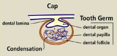
Cap stage of tooth development
|
1. the bud splits into a cap-like structure due to proliferation
2. the epithelium forms the enamel organ 3. ectomesenchymal cells aggregate beneath the enamel organ in a process called condensation, to form the dental papilla 4. the dental follicle (sac) forms |
|
|
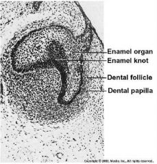
Describe enamel knot
|
The enamel knot is a group of closely oriented cells within the enamel organ. This structure signals the formation of cusps. After providing the signal, enamel knot cells die by apoptosis.
|
|
|
|
Fgf-4
|
gene expressed at the enamel know, induce cusp development
|
|
|
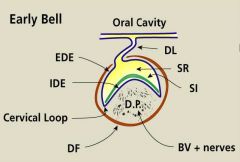
Describe bell stage tooth development
|
1. during bell stage, the undersurface of the enamel organ deepens, and cellular histo-differentiation begins. The inner dental epithelium folds according to tooth type
2. dental lamina disintegrates into small islands 3. epithelial cells assume different appearance in preparation for formation of the hard tissue (enamel). stellate reticulum is reduced in thickness at location of mineral formation. |
|
|
|
4 epithelial cell types observed during bell stage are
|
outer dental epithelium: cuboidal cells
stellate reticulum: star-shaped cells stratum intermedium: several flattened layers inner dental epithelium: short columnar |
|
|
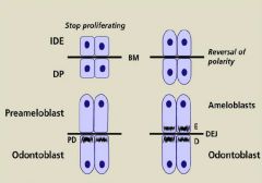
Describe the interaction between the IDE and DP cells
|
1. cells of IDE and DP communicate with each other though the BM.
2. IDE and DP stops proliferating. Reversal of polarity: nuclei move opposite direction from the BM and become taller. 3. preameloblast send signals below and the cells of DP become odontoblasts. Predentin forms when mineral is secreted to the ECM. The undifferentiated pre-odontoblast sit below the the odontoblasts. 4. Presence of dentin signals the preameloblast to make enamel and becomes ameloblasts Ameoblasts make enamal and odontoblast make dentin, DEJ = dental enamal junction |
|
|
|
permanent teeth
|
permanent molar tooth germs (w/o precursors) begin to develop as the dental lamina burrows back from the second premolar.
|
|
|
|
reciprocal interaction
|
important in determining the location at which teeth will develop
|
|
|
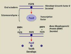
BMP4 + FGF8 ->
FGF8 -> Pax9 -> |
BMP4 + FGF8 -> no tooth development
FGF8 -> Pax9 -> tooth development |
some of the epithelium cells begin to secret Fgf8 and the protein go down to ectomesenchyme and induce expression of Pax9.
Below. When BMP4 and Fgf8 are expressed, teeth do not develop |
|
|
3 mechanisms of signaling
|
1. single signal
2. combined signals resulting in antagonism 3. combined signals resulting in synergism |
antagonism (FGF and BMP in tooth undevelopment)
synergism (FGF and BMP work together in heart development) |
|
|
Late bell, early crown stage
|
1. Mineralized dentin and enamel have begun to form
2. First odontoblasts differentiate at the tip of the cusp or incisal edge 3. Subsequently, ameloblasts begin to differentiate at the same location. 4. Both cell types progressively differentiate down the slopes of the cusp, with odontoblasts always earlier. |
|
|
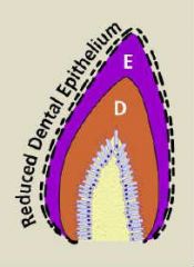
reduced dental epithelium
|
reduction in thickness of the enamel organ where dentin and enamel have been secreted.
|
|
|
|
Is inner dental epithelial cells involved in root dentin formation?
|
No, inner dental epithelial cells are not involved in root dentin formation, they are only found in the crown
|
|
|
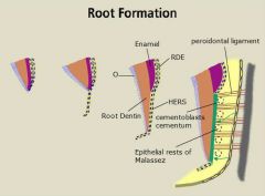
Root Formation
|
1. Internal and external dental epithelial cell layer grows downward from the cervical loop to form HERS
2. The inner layer of cells secrete some enamel related proteins, but don't become ameloblasts 3. HERS cells induce dental papilla cells to differentiate into odontoblasts to produce root dentin. 4. HERS then breaks away to form epithelial rests of Malassez 5. Dental follicle cells migrate inward (green arrow) and become cementoblasts and starts to secrete cementum |
HERS: Hertwig's epithelial root sheath
|
|
|
cementoblasts
|
-precursors are in the dental follicle.
-cementocytes are found w/i mineralized cementum, and have cytoplasmic processes in canaliculi, oriented toward the vascularized PDL. -cementum forms continually and thickens with age. |
PDL: periodontal ligament
|
|
|
epithelial cell rests of Malassez
|
found in the periodontal ligament, residue of HERS that didn't completely disappear
|
|
|
|
Dental follicle produces 3 cell types
|
cementoblasts, periodontal ligament fibroblasts and alveolar bone osteoblasts
|
|
|
|
percent of minerals in these tissues:
enamel dentin cementum bone |
95% mineral
70% 50-61% 45-47% |
|
|
|
Events during tooth eruption
|
1. bone remodeling: create a pathway thru bone
2. root growth 3. mucosal penetration: penetrate the oral mucosa 4. preocclusal eruption: until the occlusal plane is reached 5. PDL remodeling: to permit movement |
|
|
|
odontoclasts
|
bone resorption
|
|
|
|
2 required structures in regulation of tooth eruption
|
1. dental follicle 2. reduced dental epithelium
|
|
|
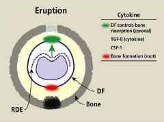
mechanisms of tooth eruption
|
RDE = reduced dental epithelium generate TGF-beta, which is received by the dental follicle and secrete CSF-1, which brings in osteoclast, teeth erupt. another signal produced at the root promote bone formation
|
|
|
|
reciprocal interactions: epithelial-mesenchyme relationships
|
mesenchyme determines the fate of the epithelium
|
|
|
|
Mandibular central incisor erupts first
|
Ture
|
|
|
|
diphyodont dentition
monophyodont dentition |
diphyodont: 2 sets of teeth
monophyodont: 1 set of teeth |
|
|
|
Andontia
|
congenitally missing teeth, complete or partial. 3rd molar, maxillary lateral incisors and MD second premolars are most commonly affected.
(oligodontia, hypodontia) |
|
|
|
Oligodontia
|
congenital absence of many but not all teeth
|
|
|
|
Hypodontia
|
only a few teeth are missing
|
|
|
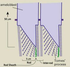
Rod Sheath
|
Rod sheath is a space created by crystal orientation at right angels. Small amounts of protein remains here (sheathlin). The rod sheath separates enamel rods/prisms.
|
|