![]()
![]()
![]()
Use LEFT and RIGHT arrow keys to navigate between flashcards;
Use UP and DOWN arrow keys to flip the card;
H to show hint;
A reads text to speech;
29 Cards in this Set
- Front
- Back
|
Hyoid bone |

Assoicated with skull but not directly in contact with any other bone Lies inferior to the mandible in anterior neck The only bone with no direct articulation with any other bone Acts as a moveable base for the tongue |
|
|
Vertebral column |
Formed from 26 bones in the adult Transmit weight of trunk to the lower limbs Surronds and protects the spinal cord Serves as attachment sites for muscles of neck and back |
|
|
Five major regions of Vetebral column |
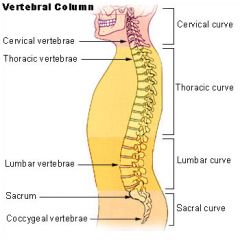
7 cervical vertebrae of the neck region 12 Thoracic vertebrae 5 lumbar vertebrae 1 sacrum (5 fused bone= 1 bone ) 1 coccynx inferior to saccum |
|
|
Normal curvatures of vertebral column |
Cervial and lumbar curvatures concave posteriorly Thoracic and sacral curvatures : convex posteriorly |
|
|
Ligaments which stabilize the vertebral column |
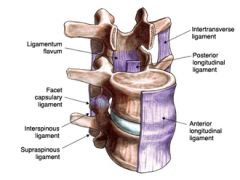
Anterior longitudinal ligaments: wide and attach strongly to both boney vertebrae and intervertebral discs and prevent hypertension of back ( prevent leaning back ) Posterior longitudinal ligament : narrow , relatively weak, and attaches only to the vetebral disc and prevents hyperflexion of back Ligamentum flavum : contains elastic connective tissue and connects lamina of adjacent vertebrae |
|
|
Intervertebral disc : Cushion-like pads between vertebrae |
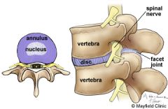
Nucleus pulposus Gelatinous inner sphere Absorbs compressive shock Anulus Fibrous Outer rings formed of ligament Innter ring formed of fibrocartilage These rings function to contain the nuclues pulposus |
|
|
Common structures of vertebrae |
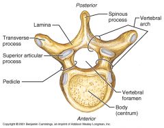
Body Vertebral arch Vertebral foramen Spinous process Transverse process Superior and interior articular processes Intervertebral foramena- between every pair of vertebrae are two apertures (openings) which allow for the passage of the spinal nerve root , dorsal root ganglion |
|
|
Vertebral region characteristic |
Specific regions of the spine perform specific functions Types of movement that may occur between vertebrae Flexion and extension Lateral flexion Rotation in the long axis |
|
|
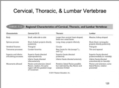
Cervical Vertebrae |
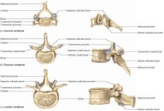
seven cervical vertebrae C1-C7 are the lightest vertebrae in the spine Body: small and wide ( laterally) Spinuous process: short and bifid (except C7) and project posteriorally Vertebral foramen : triangular and large Tranverse processes : Contains foramina Superior facets directed superposteriorly Inferior facets directed inferoanteriorly Spine region with the greatest range of motion with the folowing movment allowed : flexion, extension , lateral flexion, rotation |
|
|
The Atlas C1 |
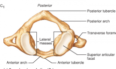
lacks a body and spinous process supports the skull - superior articular facets receive the occipital condyle - Allows flextion and extension of neck , nodding the head yes |
|
|
Axis C2 |
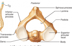
Has a body and spinous process |
|
|
Den of C2 |
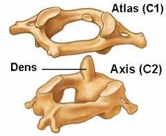
Dens - is a knoblike structure which projects superiorly from the body of axis and is cradlled in the anterior arch of the atlas - acts as a pivot for rotation of the atlas and skull , ( rotating the head from side to side ) |
|
|
Comparison ( IMPORTANT ) |
|
|
|
Thoracic Vertebrae |
All articulate with ribs Body: larger than cervical bodies and heart shaped from superior view Spinous processes are long and point inferiorly Vertebral foramen are circular |
|
|
Costal Facets of Thoracic Verebrae which interface with ribs |
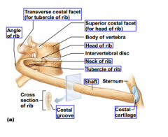
Inferior costal facet for head of rib Superior costal facet for head of rib Transverse costal facet for tubercle of rib except T11 and T 12 Each of these above three facets are presented on both sides of vetebrae , so each vertebrae has a total of 6 facets that interface with ribs The head of a rib is attached to the bodies of two vertebrae , the inferior costal facet of the superior vertebrae and the superior costal facet of the inferior vertebrae |
|
|
Connections between Thoraic Vertebral Bodies |
Laterally each side of the vertebral body bears two facets( demifacets) , one at the superior edge and one at the inferior edge These demifacets interface with vertebral bodies above and below Superior articular facets point posteriorly Inferior articular processes point anteriorly Allows rotation and limits flexion and extension |
|
|
The thoracic cage |
Forms the framework of the chest Components Thoracic vertebrae - posteriorly Ribs - laterally Sternum and costal cartilage - anteriorly Protects throacic organs Supports shoulder gridle and upper limbs Provides attachment sites for muscle |
|
|
Sternum has 3 sections |
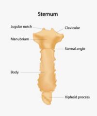
Manubrium - superior section: articulates with medial end of clavicles and rib 1 Body - Bulk of sternum: sides are notched at articulation for costal cartilage of ribs 2-7 Xiphoid process - inferior end of sternum ossifies around age 40 |
|
|
Sternum : anatomical marks |
Jugular notch : central indentation at superior border of the manubrium Sternal angle : Horizontal ridge where manubrium joins the body of the sternum Xiphisternal joint - where sternal body and xiphoid process fuse at the level of the 9th thoracic vertebra |
|
|
Ribs attach to vertebral column posteriorly |
Ribs pair 1-7- superior 7 pairs of ribs which attach to sternum by coastal cartilage Rib pair 8-10 - pairs if rubs which attach to the sternum indirectly Rib pair 11-12 ( floating ribs) - are not attached to the sternum Ribs 8-12 are sometimes called false ribs because they attach to the sternum indirectly or not at all |
|
|
Lumbar vertebrae |
Bodies - are thick and robust Transverse process are thin and tapered and nearly perpendicular to spinous process Spinuous processes are thick, blunt and point posteriorly Vertebral foramina - are triangular Superior articular facets face posteromedially or medially Inferior articular facets face anterolatterally or laterally Allows flexion and extension Rotation is prevented |
|
|
Sacrum (S1-S5) |
Shaped the posterior wall of pelvis Formed from 5 fused vertebrae Superior surface articulates with L5 Inferiorly articulates with coccyx |
|
|
Sacrum ( anterior view ) |
Sacral promontory - where the anterosuperior margin of the first sacral vertebrae bulges into pelvic cavity Human body's center of gravity is 1 cm posterior to sacral vertebrae Four transverse ridges - cross the anterior surface of the sacrum, marking the lines of fusion of sacral vertebrae Sacral spinal nerves pass through the sacral formaina |
|
|
Sacrum ( posterior view ) |
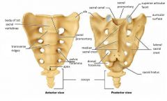
Facets of superior articular processes On the posterior surface in the midline is the bumpy median sacral crest which represents the fused spinuous processes of the sacral vertebrae Lateral to the medial sacral crest are the sacral foramina through which sacral spinal nerves pass Ala - wing are in superior lateral part of sacrum The alae articulate with the hip bones and form the sacroilac joints which are sites where the axial skeleton bone (sacrum) interfaces with an appendicular skeleton bone ( ileum of coxal)
|
|
|
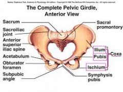
Bones of the Pelvic Gridle |
|
|
|
Coccynx |
Is the tail bone Formed from 3-5 fused vertebrae Offers only slight support to pelvic organs Long filament of connective tissue (filum terminale) attaches the coccynx which helps anchor spinal cord in place |
|
|
Fontanelles |
Fontanelles - are unossified remnants of membranes present at birth Anterior , posterior , mastoid and sphenoidal fontanelles Allows skulls to be safely compressed and molded as infant passes through narrow birth canal A visible arterial pulse may be seen in the fontanelles and can look like "fountain" Fontanelles tend to be replaced by bone by the end of the 1st year however anterior fontanelle may take 1.5 to 2 years to ossify and close |
|
|
Skull and face growth |
9 months of age : skull 1/2 adult size 2 years of age : skull 3/4 adult size 8-9 years - cranium almost adult size 6-13 years - accelerated growth of jaws cheekbones, large permanent teeth, nose and paranasal sinuses |
|
|
Axial skeleton throughout life |
water content of the intervetebral discs decrease with age by age 55 loss of a few cm in height is common thorax becomes more rigid as costal cartilage gradually ossifies Bone lose mass with age |