Reading...
![]()
Play button
![]()
Play button
![]()
Use LEFT and RIGHT arrow keys to navigate between flashcards;
Use UP and DOWN arrow keys to flip the card;
H to show hint;
A reads text to speech;
55 Cards in this Set
- Front
- Back
|
Epimysium
Structure of Skeltal Muscle |

...is a layer of connective tissue which ensheaths the entire muscle.
|
|
|
Perimysium
Structure of skeletal muscle |

...is a sheath of connective tissue which groups individual muscle fibers (anywhere between 10 to 100 or more) into bundles or fascicles.
|
|
|
Endomysium
Structure of skeletal muscles |

...literally meaning within the muscle, is a layer of connective tissue that ensheaths a muscle fiber and is composed mostly from reticular fibers. It also contains capillaries, nerves and lymphatics.
|
|
|
Fascicle
Structure of skeletal muscle |
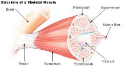
...a bundle of skeletal muscle fibers surrounded by connective tissue.
|
|
|
Fundamental contractile units made up of two protein filaments (actin and myosin).
|
myofibrils
|
|
|
The cell membrane of muscle cells.
|
sarcolemma
|
|
|
The cytoplasm of muscle cells.
|
sarcoplasm
|
|
|
Ability to shorten when stimulated.
|
contractility
|
|
|
Ability to be stimulated with a nervous impulse.
|
excitability
|
|
|
Ability to propagate an electrical stimulus.
|
conductivity
|
|
|
Ability to mechanically relax.
|
extensibility
|
|
|
Ability to be stretched beyond resting length.
|
elasticity
|
|
|
The addition of many single twitches.
A sustained contraction (tetanic contraction). |
summation
|
|
|
Progressively increasing the involvement of additional motor units.
Tend to recruit slow twitch fibers first. |
recruitment
|
|
|
Force is generated and muscle shortens.
|
concentric contraction
|
|
|
Force is generated and muscle is lengthened.
Associated with DOMS. |
eccentric contraction
|
|
|
Force is generated with NO MOVEMENT in the JOINT.
|
isometric contraction
|
|
|
A sustained contraction.
|
tetanic contraction
|
|
|
Elbow and knee.
Flexion and extension movement. |
hinge joint
synovial |
|
|
Bones of the wrist and ankle.
Sliding movement. Joints NOT fit together like other synovial joints, though they move in many positions. |
gliding joint
synovial |
|
|
Proximal end of the radius and ulna.
Rotation around central axis. |
pivot joint
synovial |
|
|
Between thumb and carpal.
Various movements. many movements that other primates can't do. |
saddle joint
synovial |
|
|
Knuckle between metacarpals and phalanges.
Various movements. |
condyloid joint
synovial |
|
|
Shoulder and hip.
Movement in all planes. |
ball and socket joint.
synovial |
|
|
Bone forming cells.
Create the spongey bone of intramembranous bone: flat bones of the skull. Deposit thin layers of bone during the "ossification" of the endrochondrial bones. |
osteoblasts
|
|
|
Specialized bone cells that "break down" the calcified cartilage allowing osteoblasts to invade the space and create new osteocytes.
|
osteoclasts
|
|
|
endosteum
|
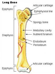
...thin layer that lines the medullary cavity.
|
|
|
periosteum
|

...outer covering of the bone.
Continuous with the fibers of tendons and ligaments. |
|
|
diaphysis
|

...shaft of the long bone.
|
|
|
epiphysis
|

...the expanded end that forms a joint.
|
|
|
The function of yellow marrow is...
|
fat storage.
|
|
|
The function of red marrow is...
|
production of red blood cells.
|
|
|
The joint created by the two pubic bones.
|
symphisis pubis
|
|
|
Bone is made up of...
|
33% collagen.
66% calcium. |
|
|
A single α-motor neuron and all of the corresponding muscle fibers it innervates.
|
motor unit
|
|
|
The progressive increase number of action potentials sent down motor neurons, per given unit of time, to stimulate a muscle to increase in strength of contraction.
|
summation
|
|
|
A muscle fiber contracts, or it doesn't.
Contracts completely. |
all or none response
|
|
|
Number of lumbar vertebae.
|
5
|
|
|
Number of true ribs.
|
7
|
|
|
What causes rigor mortis?
|
ATP reserves are quickly exhausted from the muscle contraction and other cellular processes. This means that the actin and myosin fibers will remain linked until the muscles themselves start to decompose.
|
|
|
Can training completely convert fiber types?
|
No. You can convert up to 10% of fast twitch subgroup muscle fibers. Detraining will reverse conversions.
|
|
|
Small motor units.
|
eyes, fingers
|
|
|
Large motor units.
|
legs
|
|
|
1. An action potential travels down a MOTOR NERVE to a...
|
motor end plate.
|
|
|
2. ACH (acetylcholine) is released from...
|
presynaptic vesicles.
|
|
|
3. ACH diffuses across the synaptic cleft and...
|
depolarizes the sarcolemma (muscle membrane).
|
|
|
4. Wave of depolarization (excitation)...
|
spreads across the sarcolemma and down the t-tubules.
|
|
|
5. T-tubules interact with the sarcoplasmic reticulum and...
|
the wave of excitation causes Ca++ (calcium) to be released from it.
|
|
|
6. Ca++ binds with...
|
troponin.
|
|
|
7. Troponin moves tropomyosin...
|
out of the way of the actin active site.
|
|
|
8. Myosin Head with ATP attached...
|
cleaves the ATP to ADP+Pi
|
|
|
9. The Myosin Head (ADP+Pi) binds to the...
|
exposed active site on actin.
|
|
|
10. Pi (inorganic phosphate) is released from the Myosin Head and the head...
|
changes shape and pivots to pull.
|
|
|
11. ADP is then discarded and...
|
a new ATP binds with Myosin causing it to release from actin.
|
|
|
12. Ca++ is resequestered by the...
|
sarcoplasmic reticulum (SR) and is ready for the next wave of depolarization.
|