![]()
![]()
![]()
Use LEFT and RIGHT arrow keys to navigate between flashcards;
Use UP and DOWN arrow keys to flip the card;
H to show hint;
A reads text to speech;
61 Cards in this Set
- Front
- Back
- 3rd side (hint)
|
What are the 2 compartments of the arm?
|
Anterior/Flexor compartment and Posterior/Extensor compartment.
|
To do with action or position.
|
|
|
What does the anterior compartment contain? |
1. Biceps Brachii 2. Brachialis 3. Coracobrachialis |
BBC |
|
|
What is the function of Biceps Brachii? |
Suppination of the Forearm and flexes arm at the elbow and shoulder. |
Opening a bottle with a corkscrew |
|
|
What is the function of the Brachialis?
|
1. Upper arm muscle that flexes the elbow joint. 2. Assists Biceps Brachii to flex at elbow. 3. Partially makes up floor of Cubital Fossa |
Cubitus=Elbow (Latin), BB help, movement |
|
|
What is the function of the Coracobrachialis?
|
1. Flexes and adducts the arm at the Glenohumural joint. 2. Resists deviation of the arm during abduction (from the frontal plane) |
It both draws the humerus forward, (causing shoulder flexion) also draws the humerus toward the torso, causing shoulder adduction
|
|
|
What are these muscles innervated by?
|
The Musculocutaneous Nerve
|
It's Cute
|
|
|
Where does the nerve arise from? Blood Supply? |
5th and 6th Cervical Spinal Nerves; Brachial Artery |
Latin for 'of the hand'? |
|
|
What does the posterior compartment contain?
|
Triceps Brachii
|
Wussy :-(
|
|
|
What is the function of the Triceps Brachii |
The Triceps Brachii performs extension of the arm at the elbow. |
|
|
|
What is this muscle innervated by? Blood Supply?
|
The Radial Nerve (C6, C7, C8, mainly C7); The Profunda Brachii/Deep Brachial Artery
|
Radical Dude
|
|
|
What are the bones of the upper limb?
|
Clavicle, Scapula, Humerus, Ulna, Radius, Carpal Bones, Metacarpal Bones, Phalanges
|
Carpus=Wrist (Latin) Phalanx: Line of Soldiers |
|
|
Name the Carpal Bones (Lateral to Medial)
|
Proximal: scaphoid, lunate, triquetrum, pisiform Distal: trapezium, trapezoid, capitate, hamate |
Scaphoid: Bone-Shaped Lunate: Moon shaped (longitu.) Pisiform: Pea-shaped Trapezium: "the thumb swings on the trapezium" Capitate: largest carpal bone She Looks Too Pretty, Try Catch Her |
|
|
What are the Base, Body and Head of the Metacarpals and Phalanges?
|
Base: Proximal End (articulates with carpal/metacarpals) Body/Diaphysis: Slender shaft Head: Distal End, Usually articulates with . |
|
|
|
What muscles make up the Rotatory Cuff? |
Supraspinatus, Infraspinatus, Teres Minor, Subscapularis |
SITS |
|
|
What is the function of Rotatory Cuff muscles? |
1. They all stabilise the Glenohumeral Joint 2. Individually, they function as rotors of the humerus 3. Exception: Supraspinatus initiates abduction of the arm |
Stabilise... |
|
|
Define Brachial Plexus. |
A complex network of nerves which arises from the neck and extends into the Axilla where it mostly branches out. |
Supplies the Upper Limb the most |
|
|
Roots of the Brachial Plexus are formed from the... |
Anterior Rami of Spinal nerves: C5, C6, C7, C8 and T1 |
Cervical and Thoracic nerves |
|
|
What is the Brachial Plexus divided into? |
Roots, Trunks, Divisions, Cords and Branches |
Reach To Drink Cold Beer; Draw 3 Ys (Middle Reflected), X between C5 &C6 |
|
|
What nerves are found on the Superior Trunk? Innervations? |
Subclavius Nerve (Subclavius Muscle); Suprascapular Nerve (Supraspinatus and Infraspinatus muscles) |
S-nerves |
|
|
Which nerve is found near the root of C5 and what does it innervate? |
Dorsal Scapular Nerve (Rhomboid Major and Rhomboid Minor) |
DSN |
|
|
What nerve arises from the roots of C5, C6 and C7? Innervation? |
Thoracic Nerve (of Bell); Serratus Anterior Muscle |
C5, 6, 7 Bells of Heaven, Pointing to heaven needs this muscle to contract |
|
|
What are the five terminal nerves? |
Musculocutaneous, Axillary, Radial, Median and Ulnar nerves. |
MARMU |
|
|
What are the nerves found on the Medial Cord? Innervations? |
1. Medial (Antebrachial) Cutaneous Nerve of the Forearm. (Skin) 2. Medial (Brachial) Cutaneous Nerve of the Arm. (Skin more medial) 3. Medial Pectoral Nerve (Pectoralis Major and Minor Muscles) |
M-Nerves Note: Lateral Pectoral Nerve innervates Pec M. From Lateral Cord! Cutaneous: relating to or affecting the skin. |
|
|
What are the nerves of the Posterior Division? Innervations? |
Upper (Subscapularis muscle) and Lower (Subscapularis and Teres Major) Subscapular nerves, Thoracodorsal (Latissimus Dorsi Muscle), Radial (a lot) and Axillary (Deltoid and Teres Minor) |
ULTRA |
|
|
What are the 3 categories of the anterior compartment of the forearm? |
Superficial, Intermediate and Deep |
SID, muscles mostly perform flexion and pronation |
|
|
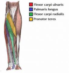
What are the superficial muscles? |
1. Flexor Carpi Ulnaris 2. Palmaris Longus 3. Flexor Carpi Radialis 4. Pronator Teres |
Place thumb on their Origin (Medial Epicondyle) and 4 fingers on forearm. From Medial (pinky) to Lateral (index): FPFP |
|
|
What is the 1. Function, 2. Origin, 3. Attachment and 4. Innervation of the Flexor Carpi Ulnaris? |
1. Flexion and Adduction of the wrist. 2. Medial Epicondyle and Ulna 3. Pisiform of Carpus 4. Ulnar Nerve |
Pinky |
|
|
What is the 1. Function, 2. Origin, 3. Attachment and 4. Innervation of the Palmaris Longus? |
1. Flexion at the wrist 2. Medial Epicondyle 3. Flexor Retinaculum of wrist 4. Median Nerve |
Ring, Just distal to the wrist, reflecting back PL will find you the median nerve underneath it. |
|
|
What is the 1. Function, 2. Origin, 3. Attachment and 4. Innervation of the Flexor Carpi Radialis? |
1. Flexion and Abduction of the wrist 2. Medial Epicondyle 3, Base of Metacarpals II and III 4. Median |
Middle |
|
|
What is the 1. Function, 2. Origin, 3. Attachment and 4. Innervation of the Pronator Teres? |
1. Pronation of the Forearm 2. Medial Epicondyle and Coronoid Process (Ulna) 3. Mid-shaft of Radius 4. Median |
Index, its lateral border forms the medial border of the Cubital Fossa |
|
|
What is the Cubital Fossa? |
An area of transition between anatomical arm and forearm |
Located as a depression on anterior surface of elbow joint, 'anatomical triangle' appearance |
|
|
The Cubital Fossa is triangular in shape, what are its borders? |
1. Lateral Border (Medial border of Brachioradialis Muscle) 2. Medial Border (Lateral border of Pronator Teres Muscle) 3. Superior Border (imaginary line between the Humerus' Epicondyles) |
Floor: Prox. by Brachialis and Dis. by Supinator Muscle Roof: Skin and Fascia Bicipital Aponeurosis reinforces it. Note: Within the Roof is the Median Cubital Vein (Venipuncture site) |
|
|
What are the contents of the Cubital Fossa? (4) |
1. Radial Nerve (near it) 2. Biceps Tendon (Attaches to Radial Tuberosity) 3. Brachial Artery (bifurcates into Radial and Ulnar at apex of CF) 4. Median Nerve (Supplies flexor muscles in forearm) |
Really Need Beer To Be At My Nicest |
|
|
List the clinical relevance of the Cubital Fossa |
1. Brachial Pulse: palpating medial to biceps tendon to feel 2. Blood Pressure: Stethoscope placed here for Korotkoff sounds 3. Venipuncture: Median Cubital Vein is superficial and accessible |
Median Cubital Vein connects Basilic and Cephalic veins |
|
|
Which muscle is in the Intermediate Compartment? |
The Flexor Digitorum Superficialis |
Sometimes classed as Superficial, but often deeper in Cadavers Note: Anatomical Landmark; Median and Ulnar Nerve pass between 2 heads and travel posteriorly. |
|
|
What is the 1. Function, 2. Origin, 3. Attachment and 4. Innervation of the Flexor Digitorum Superficialis? |
1. Flexes Metacarpophalangeal, prox. interphalangeal joints (at the 4 fingers) and wrist 2. 2 heads; Medial Epicondyle and Radius 3. Attaches to middle phalanges of 4 fingers 4. Median Nerve |
The muscle splits into four tendons at wrist which travel down carpal tunnel.
|
|
|
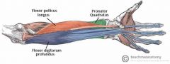
What are the muscles of the deep compartment? |
1. Flexor Digitorum Profundus 2. Flexor Pollicis Longus 3. Pronator Quadratus |
Profundus: Deep Pollicis: from Pollex (Thumb) Quadratus: roughly Square or rectangular |
|
|
What is the 1. Function, 2. Origin, 3. Attachment and 4. Innervation of the Flexor Digitorum Profundus? |
1. The only muscle that can flex the distal interphalangeal joints (fingers). Also flexes at metacarpophalangeal joints and at the wrist. 2. Ulna and associated interosseous membrane 3. Distal phalanges of the four fingers 4. A) Medial half: Ulnar nerve B) Lateral half: Median nerve |
Anterior interosseous branch of the median nerve |
|
|
What is the 1. Function, 2. Origin, 3. Attachment and 4. Innervation of the Flexor Digitorum Profundus? |
1. The only muscle that can flex the distal interphalangeal joints (fingers). Also flexes at metacarpophalangeal joints and at the wrist. 2. Ulna and associated interosseous membrane 3. Distal phalanges of the four fingers 4. A) Medial half: Ulnar nerve B) Lateral half: Median nerve |
anterior interosseous branch of the median nerve |
|
|
What is the 1. Function, 2. Origin, 3. Attachment and 4. Innervation of the Flexor Pollicis Longus?
|
1. Flexes the interphalangeal joint and metacarpophalangeal joint of the thumb 2. Anterior surface of the radius and surrounding interosseous membrane 3. Base of the distal phalanx of the thumb 4. Median nerve |
Median nerve (anterior interosseous branch) |
|
|
What is the 1. Function, 2. Origin, 3. Attachment and 4. Innervation of the Pronator Quadratus
|
1. Pronates the forearm 2. Anterior surface of the Ulna 3. Anterior surface of the Radius 4. Median nerve |
A square shaped muscle, found deep to the tendons of the FDP and FPL.
Median nerve (anterior interosseous branch) |
|
Superficial Compartment |
Intermediate Compartment |
Deep Compartment |
|
|
Do the Reflex Dance!!
|
S1, S2: Ankle Reflex L3, L4: Knee Reflex C5, C6:BicepsReflex C7, C8: Triceps Reflex |
Ankle Knee Biceps Triceps |
|
|
What does the Axial Skeleton/Appendicular Skeleton consist of? |
Axial: Bones of the Head (Skull/Cranium), Neck (Cervical Vertebrae) and Trunk (Ribs, Sternum, Vertebrae and Sacrum)
|
|
|
|
What does the Appendicular Skeleton consist of?
|
Appendicular: Bones of the Limbs (including those forming the Pectoral and Pevic Girdles)
|
Pectoral Girdle: Clavicle and Scapular
|
|
|
Where is the Clavicle (collarbone) located?
|
Extends between the Manubrium of the sternum and the Acromion of the Scapula
|
Classified as a Long Bone, S-Shaped
|
|
|
What is the Clavicle's Function?
|
1. Attaches the upper limb to the trunk. 2. Protects the underlying neurovascular structures supplying the upper limb. 3. Transmits shocks (traumatic impacts) from the upper limb to the axial skeleton |
Attaches Axial Skeleton to Appendicular Skeleton.
|
|
|
What is the common fracture site of the Clavicle?
|
The Junction of the medial 2/3 and lateral 1/3. After fracture, the lateral end of the clavicle is displaced inferiorly by the weight of the arm, and medially, by the Pectoralis Major. The medial end is pulled superiorly, by the sternocleidomastoid muscle.
|
Most commonly fractured bone; from fall onto shoulder or outstretched hand
|
|
|
What are the consequences of the Clavicular Fracture?
|
Damage results in unopposed medial rotation of the upper limb – the ‘waiters tip’ position/Erb's Palsy
|
The Suprascapular nerves (medial, intermedial and lateral) may be damaged by the upwards movement of the medial part of the fracture. They innervate the lateral rotators of the upper limb at the shoulder.
|
|
|
What are the functions of the Scapular?
|
1. It articulates with the Humerus at the Glenohumeral joint, 2. It articulates with the clavicle at the acromioclavicular joint. (In doing so, the scapula connects the upper limb to the Trunk) 3. Attachment site of 17 muscles |
The Shoulder joint is dislocated anteriorly, everything else is posteriorly dislocated.
|
|
|
What does the Costal (Anterior) Surface of the Scapular have?
|
1. Subscapular Fossa 2. Superolateral surface has Corocoid Process (Attachment site of Short head of BB, Pec.) |
Anterior surface of the scapula is termed ‘costal’; as it's the side facing the ribcage.
|
|
|
What does the Lateral Surface of the Scapular have?
|
1. Glenoid Fossa 2. Supraglenoid tubercle 3. Infraglenoid tubercle |
1. Articulates with Humerus to form Glenohumeral joint. 2. Attachment site of long head of BB 3. Attachment site of long head of TB |
|
|
What does the Posterior Surface of the Scapular have?
|
1. Spine 2. Infraspinous Fossa 3. Supraspinous Fossa |
1. It runs transversely across the scapula, dividing the surface into two. 2. Area below spine 3. Area above spine |
|
|
What does the Subclavian artery go through to become the Axillary artery? |
The lateral border of the 1st rib
|
Not Clavicle |
|
|
Where does the Axillary nerve run? Innervates?
|
1. Behind the Surgical Neck of the Humerus 2. Deltoids ad Teres Minor |
Shoulder, Surgical Neck Fracture: No abduction of arm (15 to 90 degrees prevented)
|
|
|
The Median Nerve Runs Anterior to... Innervates... |
1. The Elbow 2. Thenar Eminence, Lateral 3 and a 1/2 finger tips. |
THenaR: Thumb via Recurrent Branch. Thenar Atrophy
|
|
|
The Median Nerve's injured to function may lead to Carpal Tunnel Syndrome. Suspected by...
|
1. Myxedema 2. Edema 3. Diabetes 4. Idiopathic 5. Acromegaly 6. Neoplasm 7. Trauma 8. Rhumatoid Arthritis 9. Amyloidosis 10. Pregnancy |
MEDIAN TRAP
|
|
|
Where is the Radial Nerve located? Innervations? Injured by?
|
1. Posterior part of the Arm, Radial Groove. 2. Triceps, Brachioradialis and Extensors of the Wrist. Medial 3 and a 1/2 below finger tips. |
Injured by the fracture of the body of the Humerus, Saturday Night Palsy (Compression of Axilla i.e. Crutches): Causes wrist drop.
|
|
|
Ulnar Nerve runs... Injured by... |
1. Posterior to the Medial Epicondyle. 2. Medial Epicondyle and Hook of Hamate Fracture. |
Results in the inability to adduct or abduct fingers. In Ulnar Claw Hand U can't extend the 4th & 5th digits open from fist. |