![]()
![]()
![]()
Use LEFT and RIGHT arrow keys to navigate between flashcards;
Use UP and DOWN arrow keys to flip the card;
H to show hint;
A reads text to speech;
86 Cards in this Set
- Front
- Back
|
What happens during the 4th-5th week of development of liver? |
-liver, gallbladder and bile ducts develop |
|
|
What happens during the 6th week of development of liver? |
-right lobe becomes larger, quadrate and caudate lobes develop from right lobe, left lobe only grows slightly (same size up till this point) |
|
|
What does the liver tissue differentiate into? |
-hepatic cells -kupffer cells (for metabolism) -hemopoetic cells (formation of blood cells) -hemopoiesis (peaks at 12-14 weeks) |
|
|
What happens during the 10th week of development of liver? |
-lymphocyte formation occurs in the liver (ceases at birth) |
|
|
What happens during the 10th-12th week in development of liver? |
-coagulation factors are manufactured in the liver |
|
|
What happens during the 13th-16th week in development of liver? |
-bile is produced but fetal liver does not function in digestion until after birth |
|
|
What does the umbilical cord consist of? |
-2 arteries -1 vein: brings oxygenated blood and nutrients to the fetus |
|
|
The umbilical vein divides into 2 branches and goes where? |
Left branch- joins portal vein and enters liver Right branch- called ductus venosus goes directly to IVC bypassing the liver |
|
|
What happens to the umbilical vein branches after birth? |
-both turn into fibrous cords Left branch- becomes ligament trees or round ligament Right branch- ductus venosus becomes ligament venosum |
|
|
Both ligaments from the umbilical vein can do what? |
-recanalize with certain liver disease processes (mostly ligament teres) |
|
|
Location of the liver? |
-both RUQ and LUQ -both right and left hypochondrium and part of epigastrium -within peritoneal cavity, with exception of gallbladder fossa, portal hepatic and bare area |
|
|
Where is the bare area located? |
-posterior aspect of the right lobe of liver -attached to right diaphragm by the coronary ligament, right and left triangular ligament |
|
|
What is the Glisson's capsule? |
-fibrous capsule that encloses the liver -contains small blood vessels, lymphatic vessels and nerves |
|
|
What is the approximate diameter of the liver? |
Transverse: 20cm- 22cm AP right lobe (at mid-clavicular line): 10cm- 12cmRight lobe craniocaudad length (at mid-clavicular line): 15cm- 17cm |
|
|
What is the weight of the liver in an adult? |
1200g - 1600g |
|
|
What's the largest lobe of the liver? |
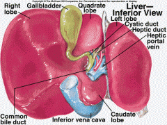
-Right lobe (6 x's larger than left) |
|
|
What separates the right from left lobe of the liver? |
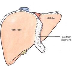
-falciform ligament on diaphragmatic surface |
|
|
What are the 3 fossae (grooves)? |
On posterior and visceral (inferior) surfaces -porta hepatis -GB fossa -IVC fossa |
|
|
Left lobe of the liver |

-smaller and flatter than right lobe -left of falciform ligament -left of ascending portion of prox left portal vein |
|
|
Where is the Caudate lobe of the liver located? |

-posterior to the right lobe and portal hepatis -anterior and medial to IVC -posterior to ligamentum venosum -superior to main portal vein |
|
|
Where is the quadrate lobe of the liver located? |

-between gallbladder fossa and falciform ligament -visceral surface -anterior to porta hepatis |
|
|
Ligaments of the liver |
-liver is attached to anterior abdominal wall, diaphragm, the retroperitoneum and the stomach to seven ligaments |
|
|
Coronary ligaments |
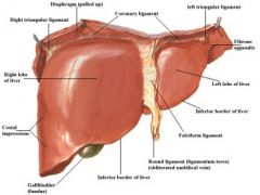
-connect posterior liver to the diaphragm -continuous with triangular ligament on each side, and with the falciform ligament anteriorly -forms ant and post borders of bare area |
|
|
Falciform ligament |

-connects anterior and superior surface of liver to anterior abdominal wall between the umbilicus and diaphragm -contains ligament teres |
|
|
What does the falciform ligament look like on ultrasound? |
-oblique echogenic line (in transverse) |
|
|
Ligament Teres |
AKA round ligament -from inferior margin of falciform ligament to umbilicus -remnant of left branch of fetal umbilical vein |
|
|
What does the ligament teres look like on ultrasound? |
-slightly irregular, rounded echogenicity just right of midline
|
|
|
Ligament Venosum |
-seperates left lobe from caudate lobe -remnant of right branch of fetal umbilical vein |
|
|
What does the ligament venosum look like on ultrasound? |
-echogenic line extending transversely from portal hepatis (in transverse and longitudinal) |
|
|
Triangular ligaments (right and left) |
-both are continuation of coronary ligament -attach liver to diaphragm |
|
|
Gastrohepatic ligament |
AKA lesser omentum -on visceral surface of the liver, continuous with the ligament venosum, lesser curvature of stomach and first portion of duodenum |
|
|
Hepatoduodenal ligament |
-surrounds portal triad just porx to portal hepatis -on right free edge of gastrohepatic ligament |
|
|
Main lobar fissure |
-division separating right lobe from left lobe Sonographically: seen as hyperechoic lone extending from the right portal vein to neck of gallbladder |
|
|
Right intersegmental fissure |
-right hepatic vein lies in this fissure -landmark for division of anterior segment from posterior segment of right lobe |
|
|
Left intersegmental fissure |
-left hepatic vein lies in this fissure -landmark for division of medial and lateral segments of left lobe |
|
|
Porta hepatis |
-portal vein and hepatic artery enter the liver and the bile duct exits the liver -on posteromedial aspect of liver |
|
|
Functional segmental division divides the liver into what? |
-3 lobes (right, left and caudate) -4 segments --Right lobe into anterior and posterior --Left lobe into medial and lateral |
|
|
Segmental divisional landmarks |
Hepatic veins (right, middle, left) -courses between lobes (interlobar) and between segments (intersegmental) Major Portal Vein branches -course with segments (intrasegmental) Fissures & ligaments |
|
|
What two things divide the right lobe from the left lobe? |
-Main lobar fissure -Middle hepatic vein |
|
|
The right hepatic vein subdivides what? |
-the right lobe into anterior and posterior segments |
|
|
The left hepatic vein subdivides what? |
-the left lobe into medial (quadrate lobe) and lateral segments |
|
|
The right portal vein branches into what? |
-anterior branch courses within anterior segment of right lobe -posterior branch courses within posterior segment of right lobe |
|
|
The left portal vein branches into what? |
-medial branch courses within medial segment of left lobe -lateral branch courses within lateral segment of left lobe |
|
|
Where is the caudate lobe located and what divides it from the left lobe? |
Located: posterior to porta hepatis, btw ligament venosum and IVC Divided by ligament venosum |
|
|
How is the caudate lobe functionally distinct? |
-receives blood from both right and left hepatic arteries and right and left portal branches -has its own bile duct -venous blood directly to IVC |
|
|
Why is it important for us to determine segmental locations? |
-incase of neoplasms or other pathologies for preparation of possible surgical resections |
|
|
Couinaud's anatomy |
-detailed method of dividing the liver into segments for hepatic surgeries |
|
|
Segments of the liver in Couinaud's |
-based on portal and hepatic venous supply -each has own blood supply (portal and hepatic), lymphatics and biliary ducts -each has at least one branch of portal vein in center bounded by hepatic vein |
|
|
What are the divisions in Couinaud's |
Vertically- by the three planes of hepatic veins (right, middle, left) Horizontally- through right and left portal veins |
|
|
Caudate lobe |
(segment 1) -supplied by branches of both right and left portal veins (can be divided into two sections- Brisbane 2000 segmentation) |
|
|
Lateral Segment of Left Lobe (superior) |
(segment 2) -supplied by ascending segment of left portal vein |
|
|
Lateral Segment of Left Lobe (inferior) |
(segment 3) -supplied by descending segment of left portal vein |
|
|
Medial Segment of Left Lobe (quadrate lobe) |
(segment 4) -supplied by the horizontal segment of portal vein |
|
|
Anterior Segment of Right Lobe (inferior) |
(segment 5) -supplied by the anterior branch of the right portal vein |
|
|
Posterior Segment of Right Lobe (inferior) |
(segment 6) -supplied by the posterior branch of the right portal vein |
|
|
Posterior Segment of Right Lobe (superior) |
(segment 7) -supplied by the posterior branch of the right portal vein |
|
|
Anterior Segment of Right Lobe (superior) |
(segment 8) -supplied by the anterior branch of the right portal vein |
|
|
The Hepatic Artery supplies the liver with what percent of blood? |
20-25% |
|
|
What do the hepatic arteries look like on ultrasound? |
RHA- circular structure lying between the Right Portal Vein and Common Bile Duct (mickey) LHA- typically not seen |
|
|
Is the Hepatic Artery high or low resistive? |
LOW |
|
|
The Portal Veins supply the liver with what percent of blood supply? |
75-80% |
|
|
What do the Portal Veins look like sonographically? |
-walls more echogenic than surrounding vessels -interlobar and follow a horizontal course with largest caliber at portal hepatis |
|
|
Are the portal veins pulsatile or phasic? Hepatopetal or hepatofugal? |
-mild phasicity -hepatopetal |
|
|
How many hepatic veins are there and what is their course? |
Three -drain blood from liver to IVC -intersegmental and interlobar |
|
|
What do the Hepatic Veins look like sonographically? |
-caliber increases as it gets closer to IVC -best visualized in transverse plane |
|
|
Are the Hepatic Veins pulsatile or phasic? Hepatofugal or hepatopetal? |
-pulsatile (bc of proximity of IVC to heart) -Hepatofugal |
|
|
Reidel's lobe (anatomic variant) |
-tongue like projection of inferior right lobe
-common in females -can give hepatomegaly impression |
|
|
Situs Inversus (anatomic variant) |
-mirror image of the liver: right lobe on left side of body |
|
|
Polysplenia/asplenia complex (anatomic variant) |
-affects location& size of liver -liver may be symmetric in size of right and left -may be located in midline |
|
|
Right hepatic artery / common hepatic duct reversal (anatomic variant) |
-Right hepatic artery crossing above the common bile duct -use color to differentiate (artery will have color) |
|
|
Liver Lobule |
(microscopic level) -basic functional unit of the liver 50,000-100,00 in liver parenchyma |
|
|
Bile production and secretion |
-bile produced in the liver and secreted to the gallbladder 800ml- 1000ml produced per 24 hrs -produced to breakdown fats |
|
|
Activity of reticuloendothelial tissue |
-starts before birth with production of blood cells (hemopoiesis) -produce plasma and antibodies -destroy old RBC's and destroy bacteria thru Kupffer cells |
|
|
Metabolism |
-physical and chemical synthesis of food products to produce energy |
|
|
What are the three things that get broken down for metabolism and into what? |
Carbohydrates- break down into simple sugars, such as glucose Fats (lipids)- broken down into fatty acids Proteins- broken down into amino acids |
|
|
Storage Depot |
For various compounds it metabolizes: -Glycogen/Glucagon -Amino Acids -Fats -Vitamins A, D, and B complex -Iron and copper |
|
|
Blood reservoir |
-reserves blood in case of hemorrhage or other blood loss |
|
|
Heat production |
-energy as result of metabolism |
|
|
Detoxification |
-converts waste products, drugs and poisons to compounds that are non toxic (medications, hormones, alcohols, bilirubin) |
|
|
How long must a patient fast for a liver exam? |
NPO for at least 6 hours |
|
|
What type of transducer should we us for liver protocol? |
Adults: 3-5 MHz Children: 5-7 MHz Obese: 2 MHz Curved liner: wider field of view Sector: better access btw ribs |
|
|
Image Optimization of the liver |
-homogenous from ant to post liver -should have same echogenicity throughout -medium gray -focal zone placed at posterior aspect -subcostal or intercostal can be used -use color to document blood flow |
|
|
Normal sonographic appearance of liver in grayscale |
-homogenous medium gray echogenicity (slightly hyper to renal cortex and hypo to pancreas) Left lobe- wedge shaped Right lobe- rounded contour Hepatic veins- sonolucent with thin walls Portal veins-sonolucent & hyperechoic |
|
|
What is the doppler signal of portal veins |
-hepatopetal flow -phasic |
|
|
What is the doppler signal of hepatic veins |
-hepatofugal flow -pulsitile |
|
|
What is the doppler signal of hepatic artery |
-low resistive |