![]()
![]()
![]()
Use LEFT and RIGHT arrow keys to navigate between flashcards;
Use UP and DOWN arrow keys to flip the card;
H to show hint;
A reads text to speech;
41 Cards in this Set
- Front
- Back
|
What is the protein hierarchy? |
Primary Secondary Tertiary Quaternary |
|
|
Primary Structure. Just an unfolded amino acid sequence |
|
|
Secondary Structure. Secondary structure refers to regularly repeating elements within a protein, in which hydrogen bonds form between polar atoms in the backbone chain.Local structure (alpha helix & beta pleated) held together by Hydrogen bonds between the peptide backbone. The portion of a protein that has neither α helices nor β conformation is composed of loops and turns that allow secondary structural elements to reverse direction back and forth to form a folded, globular protein. |
|
|
Tertiary Structure. 3D structure held together by weak interactions like disulfide bonds. This is the proteins lowest energy state. |
|
|
Quaternary Structure: Multiple subunits in large complex. Not held together by peptide bonds. Are held together by - Hydrogen bonds between polar R-groups, Hydrophobic effect between Non-polar R-groups in presence of polar groups, - Ionic bonds between charged R-groups - Van der waals between non-polar R-groups - Covalent: Disulfide bonds |
|
|
What are some important chemical bonds? |
Van Der Waals interaction between non-polar molecules. Pi Stacking between aromatic molecules (adjacent base pairs in DNA). Hydrogen bonds in proteins and nucleic acids (polar molecules). Hydrophobic effect: Tendency of non-polar molecules to exclude water |
|
|
How do proteins enable themselves to perform different functions? |
They form homo/hetero dimers. |
|
|
What is a dimer? |
A protein consisting of two polypeptide subunits. This is the quaternary structure. |
|
|
Homodimer |
Two copies of the same protein to perform a function. |
|
|
Heterodimer |
Two different proteins coming together to perform a function. |
|
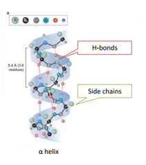
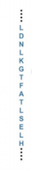
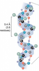
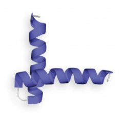
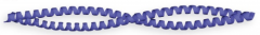
Secondary Structure: Alpha Helix |
Has Hydrogen bonding just between the peptide backbone (unlike DNA double helix which has H bonds in side chains). R groups stick out and are available to interact with other secondary structures. The secondary structure is held together by the hydrogen bonds. The hydrogen in the amide nitrogen forms a hydrogen bond with the carbonyl oxygen, this makers one helical turn. |
|
|
How do the Hydrogen bonds of an alpha helix point? |
All the hydrogen bonds of an α helix point in the same direction, and this sets up an electric dipole that gives a partial positive charge to the N-terminus and a partial negative charge to the C-terminus of the helix. Because the last four residues at either end of an α helix are not fully hydrogen-bonded, the dipole charges are spread out on these residues. |
|
|
What are some guidelines we can use to predict where an alpha helix will form? |
- Consecutive stretches of amino acid residues with long or bulky R groups cannot approach one another closely enough to form the tightly packed α helix. - Also, polar side chains can hydrogen-bond to the peptide backbone, thereby destabilizing the helix. - Consecutive like-charged R groups repel one another in the close confines of the α helix. - Proline is infrequent in α helices because its cyclic structure lacks an amide hydrogen-bond donor and restricts N–Cα bond rotation. |
|
|
What kind of R groups can stabilize the alpha helix? |
Side chains spaced four residues apart are stacked upon one another in the helix. Oppositely charged side chains that are close together can form an ion pair, which stabilizes the helix. Likewise, aromatic side chains with this four-residue spacing can form hydrophobic effects that stabilize the helix. Amino acids with a charge opposite to the partial charge of the helix dipole are sometimes located at the ends of a helix, which adds stability. |
|
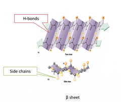
Secondary Structure: Beta Pleated Sheet |
Have R groups facing opposite sides. This alternating geometry prevents the interaction of R groups of adjacent residues. Also because of this, one side can be polar or non-polar & provide a shield. |
|
|
How is a beta pleated sheet formed? |
The β sheet, like the α helix, is formed by hydrogen bonds between backbone amide and carbonyl groups, but unlike the α helix, the β sheet structure cannot form from one β strand. Instead, all hydrogen bonds are formed between the backbones of two different β strands. |
|
|
How can you tell which is parallel and anti-parallel and which is more stable? |
They are parallel when all sheets have the hydrogen pointing in the same direction. They are anti-parallel if the hydrogen's point in the same direction but shift and alternate. Anti-parallel sheets are more stable because they form nearly straight hydrogen bonds. |
|
|
What type of interaction is directly responsible for the formation of secondary structure? |
Hydrogen bonds between sections of the polypeptide backbone |
|
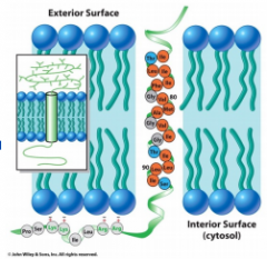
Describe the alpha helix of a transmembrane protein |
Lipids are extremely hydrophobic & water is excluded from the lipid bilayer. Polar amino acids wont go through this span. We need a lot of hydrophobic amino acids when spanning through the barrier. |
|
|
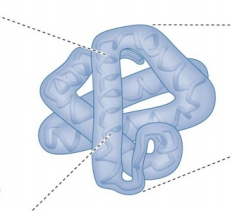
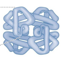
What is a domain? |
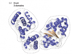
For a protein with 150 to 200 amino acid residues, the polypeptide chain usually folds into two folding units known as domains. Different parts of domains can have different functions. |
|
|
What is a motif? |
They are particularly stable and common arrangements of multiple secondary structural elements. They are linked together to form sections of domains, or even whole domains. Motifs are made of alpha helix's or beta-pleated sheets. |
|
|
What are some examples of helix motifs? |
Helix-turn-helix Motif Four-Helix Bundle Motif Coiled-Coil Motif |
|
What motif is this? |
Helix-Turn-Helix motif. Proline is found in the turn, not in the main structure. Sometimes found in proteins that bind specific DNA sequences |
|
What motif is this? |
Four-Helix bundle motif |
|
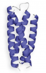
What motif is this? |
Coiled-Coil motif. Used to bind/grab DNA. Common in DNA & binding proteins. Two α helices pack against each other and gently twist around one another in a left-handed supercoil. The two α helices interact through hydrophobic contacts along the sides of each helix that form the coiled-coil interface. |
|
|
What allows secondary structures to fold? |
The size of α helices and β strands is limited by the diameter of a globular protein, and these structures must repeatedly reverse direction back and forth to form a properly folded protein. These are called reverse turns. |
|
|
How do alpha helices interact with each other? |
The interaction between α helices in a protein occurs through hydrophobic surfaces that face one another. |
|
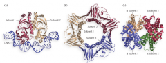
Identify these |
A) Homodimer B) Homotrimer C) Homooligomer (multiple non-identical subunits) |
|
|
Why do cells assemble such large oligomers from multiple subunits, rather than producing a large protein as a single, multidomain polypeptide chain? |
The folding of the many different domains in a single, very large polypeptide chain may be problematic. In addition, if one domain did not fold properly, the entire protein would lack function, so the investment of energy in making it would be wasted. A multi=subunit composition avoids these problems. If a protein subunit misfolds, it will not be included in the oligomer, but at least only the investment of cellular resources to make this one domain will be wasted.If one subunit subsequently denatures (becomes unfolded) or becomes inactive for any reason, it can be replaced by another subunit. The accuracy of translation is also an issue for very large proteins. During protein synthesis, approximately one mistake occurs in every 100,000 peptide-bond joining events. Therefore, a large protein of Mr exceeding 106 may accumulate mistakes, perhaps becoming inactive; smaller, individual subunits that do not fold properly can simply be discarded. Finally, the architecture of many large oligomers includes multiple copies of some subunits. More DNA would be required to encode a single large protein than is needed to encode multiple |
|
|
Many signaling and receptor molecules at the cell's surface are embedded in the cell membrane. Which amino acids would be most suitable for insertion into the lipid bilayer? |
Neutral non-polar |
|
|
At which position in the protein would a change from phenylalanine (non-polar) to glutamine (polar) make the biggest difference? |
On this inside of the protein. |
|
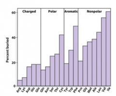
By order which of the following amino acids would be found buried in the protein? |
Nonpolar, aromatic, polar, charged. Nonpolar are the best. |
|
|
Examine |
|
|
If you know the primary structure of a protein, can you determine if there is a transmembrane region? |
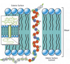
Yes. Look at the amino acid sequence from the N-Terminus to the C-Terminus and measure he hydrophibicity. You can detect the region of hydrophobic nature. |
|
|
Alpha-Keratin and your hair |
Your hair is a coil of coils. Amino Acids are linked together in hair by disulfide bonds between keratin chains and this gives it strength and structure. |
|
|
When you go to the beauty parlor to get a "perm" one of the first steps usually includes treating the hair with a sulfhydryl reducing agent. What is this likely to do to the hair? |
It breaks disulfide bonds between alpha helices so hair shape can change |
|
|
What is anti-venom? |
Anti-Venom are just antibodies to the venom. |
|
|
What are antibodies? |
Antibodies (immunoglobulin) are proteins produced by the immune system. Antibodies bind antigens very specifically. In some cases, antibodies can distinguish between proteins that differ by a single amino acid. |
|
|
What is an antigen? |
A foreign substance that elicits production of an antibody. |
|
|
How do antibodies work? |
B cells in the body become plasma cells when they are secreting antibodies. These antibodies are specific for a certain pathogen thats in the body. Scavenging cells in the body look around for things that shouldn't be there. When they find it, they kill it and present pieces of foreign agent on the surface. B-cells will make antibodies specific for this foreign agent to target it for destruction. |
|
|
Describe the structure of the antibody. |
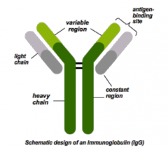
These are 4 individual proteins brought together to function as an antibody. The heavy chain is one region, it is constant. The bottom light chain is also constant. There are two variable regions. |