![]()
![]()
![]()
Use LEFT and RIGHT arrow keys to navigate between flashcards;
Use UP and DOWN arrow keys to flip the card;
H to show hint;
A reads text to speech;
20 Cards in this Set
- Front
- Back
- What is the GENUS name? -What stage? -This is very rare in the US today but how can humans become infected? |
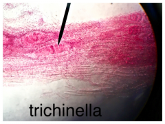
- Genus name: Trichinella - Stage: worm's larval stage - Humans can become infected by: consuming undercooked pork or sometime bear meat |
|
- Identify this as? - What do you see? - The gravid proglottids would be shed in? |
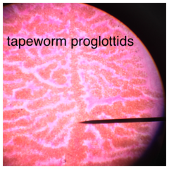
- Identify this as: Mature tapeworm proglottids - What do you see: the presence of fertilized eggs in the gravid ones -The gravid proglottids would be shed in: In the definitive host's feces |
|
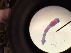
- What is the GENUS name? - Do you know where the Scolex is? - Who is the definitive hosts? - Who are the intermediate hosts? - Humans occasionally ingest Echinococcus eggs, becoming? - * A life threatening disease may result due to tissue damage by the developing larvae |
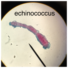
- Genus name: Echinococcus
- Location of Scolex: YES! the tip mister ;) - Definitive hosts: Canids are usual the definitive hosts - Intermediate hosts: Deers and other grazing animals Becoming: ACCIDENTAL intermediate hosts |
|
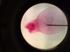
- Identify this as? - This larva stage will be found where? |
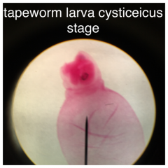
Identify as: A tapeworm cysticercus larva - Larva stage will be found: in the muscle and/or viscera of the worm's intermediate host. |
|
- Identify this as? - These are shed in ? |
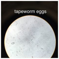
Identify as: tapeworm eggs Shed in: the feces of the tapeworm's definitive host, sometimes encased in proglottids |
|
- What is the GENUS name? - Identify male and female individual - How will the Flukes be found? - What is the estimated # of people infected? |
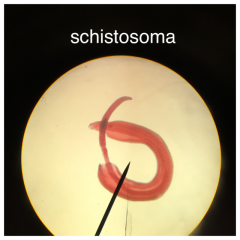
Genus name: Schistosoma Male and female individual: Thick one= MALE; Thin one= WOMEN Flukes will be found: coupled like this when in the intestinal blood vessel, liver tissue, and intestinal wall of the human(definitive) host - # of people infected: 240 million people worldwide |
|
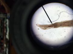
- What is the GENUS name & it's other name? - Adults like this live? - How do Humans get infected? |
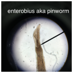
Genus name & other name: Enterobius & "Pinworm" Adults live: in the human colon, emerging at night onto the perianal skin to mate and lay eggs. - Humans get infected by: ingesting these eggs, and that symptoms are mostly confined to perianal itching and discomfort |
|
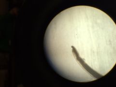
- What is the GENUS name & it's other name? - This worm was historically common in? - Adults like this live? - A heavy hookworm can cause? |
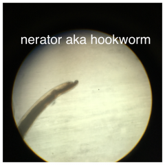
Genus name & other name: Necator and "hookworm" Common in: American South; other hookworms are found in the tropics and subtropics. - Adults live: attached by their mouthparts to the human intestinal wall heavy hookworm can cause: anemia |
|
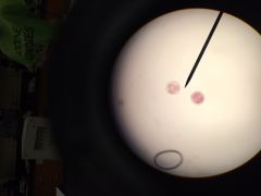
- Identify these as? - Where during this stage are these worms found? - They will remain in this stage until? - |
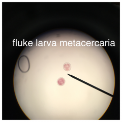
- Identify these as: Metacercaria (larval) stage of the fluke life cycle Found: on aquatic plants (like watercress, cattails, etc.) Until: they die or until a suitable definitive host (sheep, occasionally humans) eats the plants. |
|
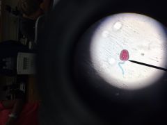
- Identify these as? - Where during this stage are these worms found? |
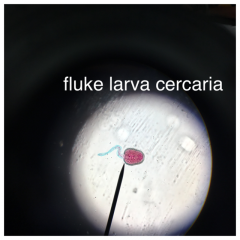
- Identify these as: The cercaria (larval) stage of the fluke life cycle Found: swimming in water, after leaving the snail *stage doesn't last that long |
|
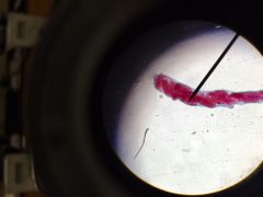
- Identify these as? - Where during this stage are these worms found?- |
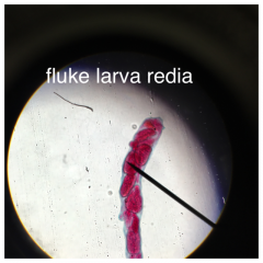
- Identify these as: redia (larval) stage of the fluke life cycle - found: in the body of the appropriate host snail (intermediate host) ** developing cercaria are inside the redia |
|
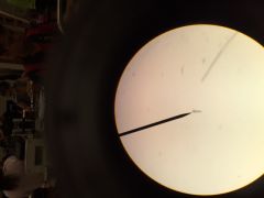
- Identify these as? - What do they do? - Where during this stage are these worms found? - They are stained what color? - What is the shape? |
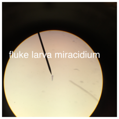
- Identify these as: larval flukes (miracidium stage) - Do: these are what hatch out of the eggs - Found: swimming in water until they either die about 8 hours after hatching or encounter the appropriate host snail. Stained: red, blue-green, or purple Shape: bullet-shaped and are about the same width as the microscope's pointer when viewed at 100x (10x objective) |
|
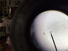
- Identify these as? - Where during this stage are these worms found? |
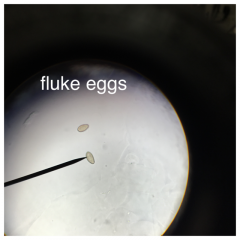
- Identify these as: fluke eggs - found: in water, get there by being expelled in the feces of an infected definitive host |
|
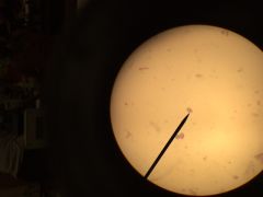
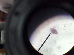
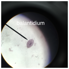
- What are there shape? - Stained what color? - What objective do you view them with? - Identify this as? - Trophozoite are mostly abundant where? - These protists move by? -They are not infective but once established in a host intestine.... -Cyst stage of this organism is infective when? |
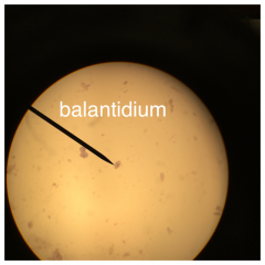
Shape: large, football-shaped
Stained what color: Red and are easy to find on the slide Objective: 40x Identify as: Balantidium trophozoite Mostly abundant: in the host's small intestine Move by: Cilia Once in hosts intestine: Trophozoite do cause the symptoms of balantidiasis When: Ingested |
|
same thing |
same thing |
|
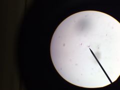
- Viewed at? - Stained with? - What is the GENUS name? - How do they move? - A human could acquire a _______ infection by? |
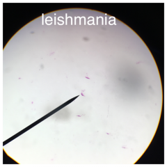
- Viewed at: 40x objective - Stained with: a purple dye - Genus name: Leishmania - Move: means of flagella Get a Leishmania infection: getting bit by a sand fly |
|
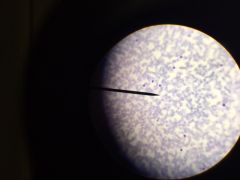
- Viewed at? - Stained with? - What is the GENUS name? - How do they move? - A human could acquire a _______ infection by? |
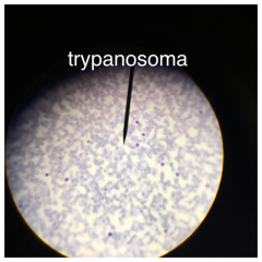
- Viewed at: 40x objective - Stained with: a purple dye - Genus name: Trypanosoma - Move: means of flagella Get a Trypanosoma infection: by a kissing bug |
|
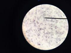
- How do they look? - A median body, a dark diagonal line in the cyst, may be seen - Identify it as ? -Cysts are shed in? - While cysts do not cause Giardiasis symptoms, they are infective when ingested |
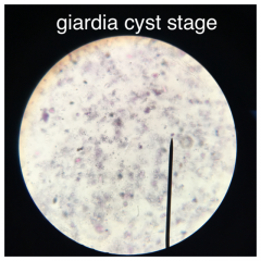
- Look: like a small oval object, gray and tan, and slightly longer than the width of the microscope's pointer when the 40x objective is in use. -Identify as: Giardia cyst -Shed in: Host's feces |
|
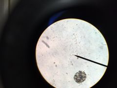
- How do they look like? - May be able to see 2 faint nuclei - Identify it as? - Trophozoites are most abundant in? - How do they move? - while they are infective, once established in a host's intestine....? |
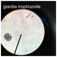
look like: a small teardrop-shaped object, gray or tan, and slightly wider than the width of the microscope's pointer when the 10x objective is in use - Identify it as: Giardia trophozoite - Trophozoites are most abundant in: host's small intestine - move: by means of flagella - Once in hosts intestine: trophozoites do cause the symptoms of GIARDIASIS |
|
View at? This _______ cyst will look like a small round gray object, its diameter about twice the width of the microscope's pointer when the 40x is in use. You may be able to see granules in its cytoplasm, and possibly one or two nuclei - Identify as? - Shed in? - How do they move? - They don't cause ________ symptoms but they are infective when? |
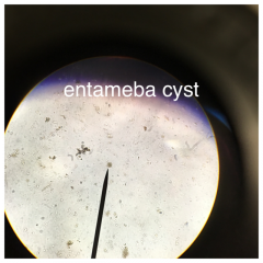
View at: 40x objective bright light, and closed Iris This______ cyst: Entameba Identify as: Entameba cyst - Shed in: the host's feces - Move: by means of pseudopods - they don't cause: Entamebiasis; they are infective when ingested |