Reading...
![]()
Play button
![]()
Play button
![]()
Use LEFT and RIGHT arrow keys to navigate between flashcards;
Use UP and DOWN arrow keys to flip the card;
H to show hint;
A reads text to speech;
155 Cards in this Set
- Front
- Back
|
What happens during binary fission?
|
1. DNA is attached to cytoplasmic membrane
2. Cell enlarges & DNA Duplicates 3. Cross wall forms, cell divides into 2 cells & cells separate into daughter cells |
|
|
What is a Cationic dye?
|
a dye that has a + charge within it that allows it to adhere to negatively charged areas of the sample (e.g. Crystal violet, Safranin & Methylene blue) Cationic stains bacteria
|
|
|
What is an Anionic dye?
|
A dye that has a negative charge within it that causes the dye to be forced away from negatively charged areas of the specimen (e.g. Nigrosin) Anionic stains background
|
|
|
What is Simple Staining?
|
Uses 1 dye either cationic or anionic. Reveals shape and arrangement of cells
|
|
|
What is Differential Staining?
|
Uses 2 or more dyes (usually both are cationic) reveals shape, arrangement & differences in chemical makeup
|
|
|
What are the 3 types of differential staining?
|
1. Gram Stain
2. Acidfast Stain 3. Endospore stain |
|
|
What is the 1st step in Gram Staining & what is the state of the Bacteria?
|
Add the primary stain crystal violet for 1 minute & rinse. Bacteria cell with stain purple.
|
|
|
What is the 2nd step in Gram Staining & what is the state of the Bacteria?
|
Add the mordant Iodine for 1 minute and rinse. Bacteria cell remains purple.
|
|
|
What is the 3rd step in Gram Staining & what is the state of the Bacteria?
|
Add 3 drops of Alcohol and quickly rinse with distilled water. Gram positive cells remain purple; Gram negative cells become colorless.
|
|
|
What is the 4th step in Gram Staining & what is the state of the Bacteria?
|
Add the counterstain Safranin for 30 sec. Gram positive cells remain purple; Gram negative cells appear pink or red.
|
|
|
Define Acidfast Stain
|
1. Used to identify Mycobacterium & Nocardia
2. Uses Carbol fuchsin as primary stain 3. Acid alcohol as decolorizer 4. Uses Methylene blue as secondary stain 5. Red areas are Acidfast |
|
|
What are the principles of Microscopy?
|
1. Total magnification
2. Resolution/Resolving Power 3. Refraction 4. Contrast |
|
|
What is Total Magnification?
|
1. Microscope has multiple lenses that magnify
2. Total Mag = mag objective x mag ocular 3. Maximum magnification = 1000x |
|
|
How does a lens magnify?
|
1. Curved regions of lens refracts/bends light
2. Light rays in the center of the lens are spread out 3. This yields a larger image that is also inverted |
|
|
What is the formula for Resolving power?
|
RP = wavelength/ 2NA
wavelength of light use .55um, thiss is the average of white light wavelengths NA = Numerical aperture (light trapping ability of objective) or widest cone of light that can enter objective |
|
|
What does RP mean?
|
The closest distance two points can be seen distinctly. If your distance is < or smaller than the RP you will see a blob, If your distance is > or = the RP then you will see 2 points
|
|
|
Define Refraction
|
Bending of light when it passes through different densities. Makes a pencil in water look as if it's broken.
|
|
|
How do you prevent refraction?
|
Use of immersion oil
Between 100x objective & sample Oil is same refractive index (density) as glass |
|
|
How do you get maximum contrast?
|
Decrease light reaching the sample by:
lowering rheostat closing iris diaphragm lowering condenser or stain the microorganism |
|
|
You are trying to observe 2 points within your specimen that are .15 um apart. You are observing the specimen using a 45X objective with a NA of .85. Will you see two points or a blob?
|
Answer:
|
|
|
What is Brightfield Microscope?
|
A Light Microscope
|
|
|
What is the pathway of light?
|
Light source
Condenser Iris Diaphragm Through Stage Sample Objective Body tube with mirror Ocular |
|
|
How do you perform a Smear Preparation?
|
1. Spread thin film of specimen over slide
2. Allow to air dry 3. Pass slide through flame to fix specimen 4. Flood with stain, rinse & dry 5. Examine with microscope |
|
|
Why is oxygen so important for aerobes, facultative aerobes & microaerophiles?
|
It acts as a final electron acceptor for production of ATP using Glycolysis, Krebs cycle & electron transport chain
|
|
|
Define Glycolysis
|
C6H1206 - broken into 2 Pyruvates
2 ATP are produced + NADH Each pyruvate is converted to 1 Acetyl CoA & 1 NADH |
|
|
Define Krebs Cycle
|
AKA Tricarboxylic Cycle
Acetyl CoA enters cycle For each Acetyl CoA (1 ATP, 3 NADH & 1 FADH2) Total for cycle = 2ATP, 6 NADH & 2FADH2 |
|
|
Define Electron Transport Chain
|
Where majority of ATP are formed
Occurs within Plasma membrane of bacterial cell as no mitochondria Using NADH & FADH2 to make ATP Total ATP from this reaction is 34 |
|
|
Why can't Obligate anaerobes handle O2?
|
No production of Superoxide dismutase or Catalase therefore O2- & H2O2 build up
|
|
|
Why can't Aerotolerant anaerobes handle O2?
|
Production of superoxide dismutase, no production of catalase & H2O2 builds up
|
|
|
Organisms are sensitive to changes in pH because:
|
H+ & OH- interfere with hydrogen bonding within proteins & nucleic acids
|
|
|
How well can cell wall protect cell?
|
Intracellulary must remain neutral!
|
|
|
How is Bacterial growth in nature?
|
Never grow in isolation, but grow in communities called Biofilms
|
|
|
Where are Biofilms found?
|
covering rocks in stream beds
coating kitchen drains accumulating in toilet bowls In form of dental plaque |
|
|
What is Agar?
|
Non-nutritive Derivative of sea weed
Melting point 100 C & solid at 40 C |
|
|
How do we streak a plate?
|
1. Loop is sterilized 2. Loop is inoculated 3. 1st set of streaks made 4. Loop is sterilized 5. 2nd set of streaks made 6. Loop is sterilized 7. Final set of streaks made 8. Isolated colonies develop after incubation
|
|
|
What are the four main types of media?
|
1. Broth
2. Slants 3. Deeps 4. Petri dish |
|
|
What is the condensor of a microscope?
|
device found above the light source that converges the light beams into a point so that they can enter the specimen.
|
|
|
What is the light source?
|
on/off switch and intensity knob.
|
|
|
What is the Rheostat knob?
|
controls the intensity of the light coming from the bulb
|
|
|
What is the knob?
|
raises and lowers the condensor, so that the point of light entering the specimen can be varied. For most smears the condensor should be all the way in the up position,
|
|
|
What is the Iris Diaphragm Lever/Control?
|
works like the iris of your eye. It controls the amount of light entering the specimen. It is found within the condenser and a lever is provided so the light can be varied. For oil magnification this lever should be positioned to allow maximum light to the specimen.
|
|
|
What is the stage?
|
where the slide is placed and has a central opening to allow light from the condensor to pass through the specimen.
|
|
|
What is the slide clip?
|
Pulling the slide clip towards you allows for the placement of the slide, flat on the stage
|
|
|
What are the stage adjustment knobs?
|
a double knob found below the stage that allows for movement of the slide.
|
|
|
What does the lower knob of the stage adjustment knob do?
|
moves slide clip left to right
|
|
|
What does the lower knob of the stage adjustment knob do?
|
moves slide clip forward and back
|
|
|
What are the objectives?
|
after the light passes through the specimen it then is picked up by the objectives that are housed within the revolving nosepiece. It magnifies the image that is received.
|
|
|
What is the revolving nosepiece?
|
allows for the movement of different power objectives into position over the specimen
|
|
|
How many objectives are housed on the revolving nosepiece and what are their magnifications?
|
There are 4.
4x, 10x, 40x, 100x |
|
|
What is the body tube?
|
light from the objective after being magnified travels through the body tube and is directed to the ocular by being reflected off a mirror.
|
|
|
What is the ocular?
|
there are two eyepieces or oculars therefor this microscope is called a binocular scope. The oculars have the ability to magnify the image by 10x
|
|
|
What is the Intrapupillary Distance Adjustment?
|
Adjust the oculars by sliding them either closer together or further apart until you see one field of view when using both eyes.
|
|
|
If you are unable to see 1 image what should you ?
|
Adjust the intrapupillary distance.
|
|
|
What are the fine adjustment knobs?
|
gives exact focus while looking through the oculars
|
|
|
Define parfocal
|
when you switch to a different objective the image should remain in perfect focus
|
|
|
What happens to the field of view as you increase the power of the objective?
|
the field of view decreases
|
|
|
What is the formula for total magnification?
|
ocular power (always 10) times objective power.
|
|
|
Define resolving power.
|
a numerical measure of the resolution of the lens. The smallest distance between two objects that the microscope is able to distinguish as two distant points.
|
|
|
What is the formula for RP
|
= wavelength of light/ 2NA
|
|
|
Define wavelength of light
|
the distance between 2 troughs of the light wave, we will be using the average wavelength of visible light or .55 um. the wavelength of light limits resolution to approx .2 um
|
|
|
What is NA?
|
Numerical Aperature. The light concentrating power of the objective. This is listed on the side of the objective after the magnification power.
|
|
|
Calculate the resolving power for each objective:
RP of 4x objective RP of 10x objective RP of 40x objective RP of 100x objective |
.10
.25 .65 1.25 |
|
|
Define field of view
|
the area that is visible
|
|
|
Define binocular
|
adapted to the use of both eyes
|
|
|
Define Brightfield
|
A type of microscope that produces a dark image and is the simplest of all microscopy techniques
|
|
|
Define microorganism
|
a microscopic form of life that can only be studied under a microscope, including bacteria, viruses, fungi, algae, protozoa and some multicellular parasites.
|
|
|
What are Eukaryotes?
|
complex organism whose cells contain organelles as well as a nucleus with multiple chromosomes and a nuclear membrane, reproduction of the organism is by mitosis.
|
|
|
What groups fall under Eukaryotes?
|
Protozoans
Algae Fungi |
|
|
What are Prokaryotes?
|
a simple microorganism composed of single cells and having a single chromosome and few intracellular organells, reproduction does not involve mitosis
|
|
|
What groups fall under Prokaryotes?
|
Cyanobacteria
Eubacteria |
|
|
What are the 2 domains
|
Bacteria
Archaea Eukarya |
|
|
Define Bacteria
|
unicellular microorganisms that are mostly cylindrical (rod, sphere or spiral) rigid cell wals of peptidoglycan, multiply by binary fission, has appendages called flagella
|
|
|
When observed under a microscope, what does an Amoeba proteus look like and how does it move?
|
animal like single cell microorganism w/ pseudopodia (false feet) and moves by cytoplasmic streaming
|
|
|
On the brightfield microscope, what happens to the field of view as you increase the power of the objective?
|
decreases field of view
|
|
|
What is the resolving power for each objective:
RP of 4x RP of 10x RP of 40x RP of 100x |
.10
.25 .65 1.25 |
|
|
Define Protozoans
|
single celled animal like microorganisms found within aquatic environments, capable of motility, food is obtained by engulfing, some are pathogenic, others are good aquatic indicators and on the lowest rung of food chain
|
|
|
Define Algae
|
can be single celled or multi-celled microorganisms, capable of photosynthesis, therefore are called producers, also good aquatic indicators and on the lowest rung of food chain
|
|
|
Define Cyanobacteria
|
Formerly called blue green algae, possesses a prokaryotic cell structrue, possess chlorophyll, arranged as single cells, within a gelatinous matrix or filamentous colonies, Heterocyst if present stores nitrogen from the atmosphere, found in fresh water, damp soils and anaerobic environments
|
|
|
Define heterocyst
|
are specialized nitrogen-fixing cells
|
|
|
Define Eubacteria
|
also known as true bacteria, have cell wall of peptidoglycan and lipids, vast majority of bacteria fall in this group
|
|
|
What is the method of locomotion for Amoeba proteus?
|
moves by cytoplasmic streaming, animal like single cell w/ psudopodia (false feet)
|
|
|
Who first observed Giardia lamblia?
|
Leeuwenhook in 1681
|
|
|
What area does Giardia lambia infect in humans and how does it attach itself?
|
the small intestines and it has adhesive disks that attach to the bowel wall.
|
|
|
How would one contract this microorganism?
|
Contaminated water
|
|
|
What symptoms does the microorganism produce in humans?
|
diarrhea, indigestion, severe vomiting, flatulence
|
|
|
Why must back packers both chlorinate & filter their water?
|
It kills Giardia Lamblia
|
|
|
What are the spiral shaped organelles within the spirogyra called?
|
chloroplast
|
|
|
What are diatoms?
|
yellow brown algae
|
|
|
What is the light trapping pigment within Diatoms called?
|
carotenoids and phycocyanin
|
|
|
After Diatoms die it still has 2 important uses, name them:
|
silver polish and filtration for pools
|
|
|
List the 2 characteristics which indicate that Euglena could be classified as either a protozoan or algae:
|
Some Euglena are considered to have both plant and animal features.
It has animal like characteristics like protozoans because it eats food by engulfing and has algae characteristics because uses chloroplast to produce sugar by photosynthesis |
|
|
What is Anabaena?
|
a symbiotic bacterium often found in association with Azolla a water fern. It is found within the fores of the fern and falls under Cyanobacterium.
|
|
|
How can you differentiate the heterocyst from the other cell of Anabaena?
|
it's clear
|
|
|
What is the function of the heterocyst?
|
perform nitrogen fixation
|
|
|
Where is Gleocapsa typically found?
|
on wet rocks and other damp areas where it forms masses of jelly. The jelly contains masses of single cells.
|
|
|
What is the function of the capsule?
|
Holds the cell together
|
|
|
Why is Gleocapsa important?
|
It's a producer and performs nitrogen fixation.
|
|
|
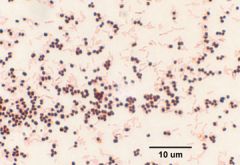
What is the typical coccus?
|
cells appear as spherical, in clusters that often are referred to as grapes
|
|
|
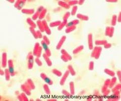
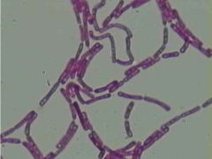
What is the typical bacillus?
|
cells appear long and narrow, they may contain stacked or be arranged in chains.
|
|
|
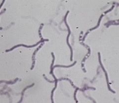
What is the typical spirillum?
|
cells are wavy in appearance, but have no particular arrangement.
|
|
|
What is true bacteria?
|
can be characterized as to shape biochemical reactions or metabolism except for bacteria in the Archae Domain.
|
|
|
What is trypanosoma brucei gambiense?
|
a pathogenic protozoan parasite that causes the disease call "African Sleeping Sickness"
|
|
|
What is the vector for African Sleeping Sickness?
|
daytime biting tseste fly.
|
|
|
What is the late symptom for the African Sleeping Sickness?
|
loss of activity, indifference to food, coma & ultimately death
|
|
|
What is Paramecium?
|
a slipper shaped microorganism that is motile by the presence of rows of cilia, this allows for smooth and rapid movement through water.
|
|
|
How does paramecium take in its food?
|
oral cilia within their oral groove
|
|
|
Why is paramecium an important microorganism?
|
good environmental indicator
|
|
|
What are the ingredients found within Trypticase soy agar?
|
Soybean
Casein Sodium Agar |
|
|
What are the benefits of agar?
|
it is used for the sole purpose of producing a solid surface on which the bacteria can grow. It remains in a solid state even at high temperatures and does not liquify until it reaches 100 degrees celcius. This allows the growth of thermophiles within the lab. Agar solidifies at 40 degrees celcius. It remains stable for bacteria.
|
|
|
Why are buffers put in the media?
|
to keep the pH of the medium from changing drastically. Most bacteria prefer a pH around neutral. As the bacteria grow and reproduce ont he medium, acidic and basic by products of metabolism are produced. If buffers were not included the bacteria would be quickly killed off by their own waste.
|
|
|
When making agar, if you only wish to make 600 ml of agar assuming the directions call for 40 grams of agar for a liter of media how many grams of agar will you need?
|
40 grams of agar/ 1000 ml of water =
x grams of agar/ 600 ml of water Cross multiply: 1000x = 40 x 600 1000x = 24000 x = 24g of agar |
|
|
At what temperature does agar solidify?
|
40 degrees Celsius
|
|
|
At what temperature does agar liquefy?
|
100 degrees Celsius
|
|
|
What is the function of the agar that is within a media?
|
allows the media to stay solid through various temperatures
|
|
|
Why are buffers added to mediums?
|
to keep the pH from changing too drastically
|
|
|
Are there any buffers in Trypticase soy agar?
|
no
|
|
|
At what temperature, pressure and time is media autoclaved?
|
121 degrees, 15 psi above atmospheric pressure, 20 minutes
|
|
|
If a media requires 19.5 grams of media in order to make a liter of the media, how much powdered media should be weighed out if you are only going to make 375 ml of the media?
|
(19.5/1)(x/.375) x = 7.37
|
|
|
Why are plates incubated in the inverted position?
|
to prevent condensation
|
|
|
'What is the purpose of using a control plate?
|
to test the sterility of the media
|
|
|
Why is the petri dish from your skin sample incubated at 37 degrees Celcius?
|
it is considered the average body temperature
|
|
|
What is bacteria morphology?
|
Cocci
Bacilli Spirillum |
|
|
What is the arrangement for
cocci bacilli spirillum |
cocci: single,diplo, tetrads, strepto or staphylo
bacilli: stacked, chained or irregular spirillum: single or irregular |
|
|
Which stain is considered the primary stain?
|
Crystal violet
|
|
|
Which stain is considered the secondary stain?
|
Safronin
|
|
|
Why is Gram's iodine called a mordant?
|
it helps the sample stick to the slide
|
|
|
What happens if you over decolorize in the Gram stain procedure?
|
If you put too much alcohol on sample you can remove the crystal violet pigment.
|
|
|
Why do some bacteria turn purple when you use the Gram staining procedure?
|
a gram + cell wall will accept the crystal violet into its pores. The iodine will act as a mordant to hold the dye into the pores. The alcohol will shrink the pores, blocking the entry of Safronin. Therefore the cell wall stays purple in color.
|
|
|
Why do some bacteria turn red when you use the Gram staining procedure?
|
A gram - cell wall will accept the crystal violet into its LPS layer. Once the alcohol is applied the crystal violet is removed. Alcohol is a solvent and dissolves the LPS layer. The saffronin is now able to be absorbed into the pores of the peptidoglycan layer turning the layer red.
|
|
|
How are colonies formed?
|
by a single bacterium setting onto the agar and starting to utilize the nutrients within the media. The bacterium uses the media as a nutritional foundation for replication.
|
|
|
How long will it take for a colony to appear on the agar that is microscopically observable?
|
within 24 hours
|
|
|
What does each individual colony represent?
|
a single bacterium that has replicated at this location and has approximately several hundred bacterial cells within it.
|
|
|
A pure culture may be obtained by 2 different methods, what are they?
|
pour plate and the streak plate method.
|
|
|
What does it means to have a sterile media/agar plate?
|
means that the media is devoid of any live organisms, such as bacteria.
|
|
|
Describe the pigment of the growth in each of the slant tubes and whether they are motile or non-motile: Bacillus subtilis, Escherichia coli, Micrococcus luteus, Pseudomonas aeruginosa, Serratia marcescens, Staphylococcus epidermidis
|
Bs: cream/ motile
Ec: cream/ motile Ml: yellow/ non-motile Pa: green / motile Sm: red/pink/ motile Se: cream/ non-motile |
|
|
Explain the technique of streak plate isolation.
|
the purpose of streaking for isolation is to produce isolated colonies of an organism on an agar plate. This is useful when you need to separate organisms in a mixed culture or when you need to study the colony morphology of an organism.
|
|
|
What part of the flame is the best part to use to sterilize an inoculating loop or needle?
|
In the lighter blue section of the flame just above the dark blue area.
|
|
|
Explain how a motile microorganism looks when it grows in a Motility Test medium culture.
|
Answer:
|
|
|
Why was the Serratia marcescens incubated at room temperature and not at 37 degrees Celcius?
|
Answer:
|
|
|
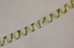
Spirogyra
|
|

|
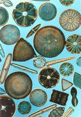
Diatoms
|
|

|
Spirogyra
|
|
|
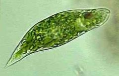
Euglena
|
|
|
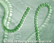
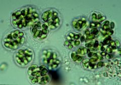
Anabaena
|
|
|
Gleocapsa
|
|
|
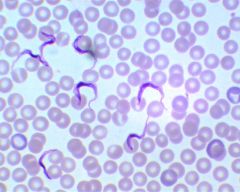
Trypanosoma brucei gambiense
|
|
|
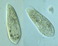
paramecium
|
|
|
diplococcus
|
|
|
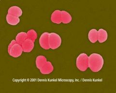
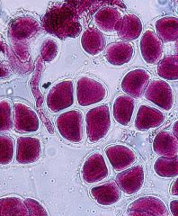
Tetrad
|
|
|
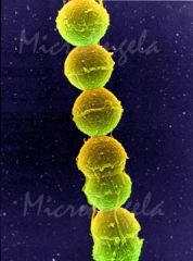
Streptococcus
|
|
|
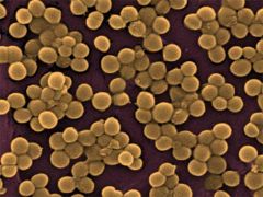
staphylococcus
|
|
|
Stacked Bacilli
|
|
|
Chained Bacilli
|
|
|
Irregular Bacilli
|
|
|
Irregular spirillum
|