Reading...
![]()
Play button
![]()
Play button
![]()
Use LEFT and RIGHT arrow keys to navigate between flashcards;
Use UP and DOWN arrow keys to flip the card;
H to show hint;
A reads text to speech;
64 Cards in this Set
- Front
- Back
|
What is the germ theory of disease? What was a competing theory?
|
Theory that microorganisms cause disease as apposed to the theory of spontaneous generous.
|
|
|
Who is credited with the compound microscope?
|
Robert Hooke
|
|
|
Famous lens maker who coined the phrase 'animalcules'.
|
Anton van Leeuwenhoek
|
|
|
Who is credited for our current naming system and what is that system called?
|
Carl Linnaeus.
Binomial Nomenclature |
|
|
Franceso Redi
|
Gauze and cork experiment
|
|
|
Florence Nightingale
|
Promoted cleanliness as a way to prevent disease during Crimean War.
|
|
|
Semmelweis
|
sanitary practice (hand washing)
|
|
|
Louis Pasteur
|
Swan necked flasks, wine pasteurization, rabies vaccine
|
|
|
Koch's Postulates
|

1.agent must be present in every disease.
2. agent must be isolated and grown in pure culture. 3. agent must cause disease in inoculated host 4. same agent must be reisolated from diseased host. |
|
|
What is the implication of Koch's postulate.
|
1 organism causes 1 disease
|
|
|
Reproduction in Prokaryotes vs Eukaryotes
|
Prokaryotes: usually asexual through binary fission or by budding
Eukaryotes: sexually through meiosis |
|
|
How does budding differ from binary fission?
|
In binary fission, the parent cell disappears with the formation of two equal-sized offspring; in contrast, a bud is often much smaller that its parent, and the parent pre mains to produce more buds.
|
|
|
What are the 7 morphologies of prokaryotes?
|
1. coccus (round)
2. Coccobacillus (oval) 3. Bacillus (rod) 4. Vibrio (bent rod) 5. Spirillum (stiff corkscrew) 6. Spirochete (flexible corkscrew) 7. Pleomorphic (variable shape) |
|
|
What are the 5 arrangements of cocci?
|
1. diplococci (2 cocci)
2. streptococci (chain) 3. tetrad (4) 4. sarcinae (cube) 5. staphylococci (grape-like cluster) |
|
|
What are the 4 arrangements of bacilli?
|
1. single
2. diplobacilli (2) 3. Streptobacilli (chain) 4. palisade (side by side) 5. v-shape |
|
|
What is a glycocalyx? What is it's purpose?
|
A sticky gelatinous substance made of polysaccharides that surround the outside of the cell. It prevent desiccation, prevents the cell from being devoured, and helps attach cell to surroundings.
|
|
|
What is a capsule? Give an example of a microorganism that has one.
|
A secreted glycocalyx that acts a protective layer.
1. Streptococcus pneumoniae |
|
|
What is a slime layer? Example of microorganism that has one.
|
A thiner glycocalyx that protects agains desiccation and temperature.
1. Streptococcus mutans (found in oral cavity-dental plaque) |
|
|
What is an endospore? Give a microorganism example.
|
Highly resistance resting stage. 1. Bacillus subtilis
2. Clostridium botulinum |
|
|
What is the spore coat made of?
|
dipicolinic acid and Ca++
|
|
|
Parts of flagella?
|
filament, hook, basal body
|
|
|
one flagella
|
monotrichous (polar)
|
|
|
flagella at each end
|
amphitrichous
|
|
|
group of flagella at one end
|
lophotrichous (tuft)
|
|
|
flagella spread out around organism
|
peritrichous
|
|
|
no flagella
|
atrichous (cocci usually have no flagella)
|
|
|
Rotation of flagella for a 'run'
|
counterclockwise
|
|
|
Rotation of flagella for a 'tumble'
|
clockwise
|
|
|
movement towards chemical/light
|
chemotaxis/phototaxis
|
|
|
Number of protein rings and lipid bilayers in G+ versus G-
|
G+ have 1 protein ring and 1 lipid bilayer.
G- have 2 protein rings and 2 lipid bilayers |
|
|
What is an axial filament and give a microorganism example.
|
Internal flagella found in spirochetes.
1. Treponema pallidium (syphilis) |
|
|
1. tiny hollow projection
2. short sticky projections 3. grappling hook |
1. Pili (sex pilis)
2. fimbria 3. hamus |
|
|
What are the layers of the cell wall of G+ and G-
|
G+:
1. peptidoglycan 2. periplasm 3. lipid bilayer G-: 1. Lipid bilayer 2. periplasm 3. peptidoglycan 4. periplasm 5. lipid bilayer |
|
|
Layer of acid fast organism. Give example of organism that has this.
|
1. mycolic acid
2. peptidoglycan layer 3. periplasm 4. lipid bilayer 1. Mycobacterium leprae (Leprosy) 2. Mycobacterium tuberculosis (TB) |
|
|
Antibiotic resistant bacteria that lacks cell wall.
|
Mycoplasm pneumoniae
|
|
|
What is a bacterial pili?
|
Tiny hollow projection used to transfer DNA from one cell to another.
|
|
|
Describe the various ways substances are moved across the membrane.
|
1. diffusion: no energy required, molecule moves with concentration gradient
2. facilitated diffusion: no energy requires, molecule moves with concentration gradient but through channels or pores. 3. active transport: requires energy, against gradient, (uniport, antiport, symport) 4. Osmosis: diffusion of water |
|
|
1. cell regular size in solution
2. cell shrunk in solution 3. cell enlarged or lysed in solution |
1. isotonic
2. hypertonic 3. hypotonic |
|
|
Phase contrast microscope
|
- based on principle that cells differ in refractive index from their surroundings
-phase ring in objective lens amplifies this contrast - no stains |
|
|
dark field microscope
|
Bright specimen agains dark background.
|
|
|
fluorescent microscope
|
fluorescent structures agains dark background. UV light fluoresce chemical or dyes
|
|
|
Differential interference contrast (Normarski)
|
Image appears three-dimensional.
|
|
|
Confocal
|
single plane stained with fluorescent dye
|
|
|
Two types of electron microscopes.
|
1. transmission: 2D of ultrastructure
2. scanning: 3D of surface |
|
|
2 types of probe microscopes
|
1. sanning tunneling: atomic level
2. atomic force: atomic level |
|
|
What are special stains used for? Give organism examples
|
1. Negative stain for capsule (Klebsiella pneumoniae)
2. Flagellar stain for flagella (Proteus Vulgaris) |
|
|
What are the three differential stains we used?
|
1. Acid fast (Mycobacterium Tuberculosis)
2. Gram stain (G+ Bacillus cereus and G- Escherichia coli) 3. endospore stain (Clostridium botulinum) |
|
|
Describe typical microbial growth curve.
|

1. Lag phase
2. Log (exponential phase) 3. Stationary Phase 4. Death (decline) phase |
|
|
Define generation time
|
time for population to double
|
|
|
How long does Escherichia coli take to double.
|
about 20 minutes
|
|
|
Define synchronous growth
|
Doubling at exact intervals
|
|
|
Define non-synchronous growth
|
doubling at irregular intervals (nature)
|
|
|
How many cells are need before they are visible to naked eye.
|
10^8
|
|
|
How can we directly measure bacterial growth?
|
1. Serial Dilutions (# CFU)
2. Most Probable Number 3. Filtration Plate Count (CFU) 4. Microscope Counts (Hemocytometer) 5. Flow Cytometry |
|
|
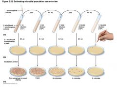
Describe serial dilutions to estimate microbial growth.
|

The number of colonies is multiplied by 10 because .1 ml was added and then multiplied by the reciprocal of the dilution. For example:
65*10*1000=650,000 bacterial/ml |
|
|
Describe Membrane Filtration to estimate microbial growth.
|

Bacteria is trapped by filter and then the filter is transferred to media to be incubated. The CFU are counted and divided by volume of media.
colonies/L |
|
|
Describe the most probably number method for estimating microbial growth.
|

5 tubes are used for each of the three dilution. After incubation the results are compared to a MPN table.
|
|
|
Describe the Microscopic Count method for estimating microbial growth.
|
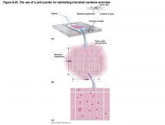
Bacteria suspended in solution placed next to cover slip slide under through capillary action. The slide is marked with grids and is looked at under the microscope. The bacteria are counted. Calculations are made to estimate cm^3 which is ml.
|
|
|
What are the indirect methods for estimating bacterial numbers.
|
1. pH test
2. dry weight 3. turbidity |
|
|
What is the pH range for acidophiles. Give an example of an organism.
|
<5.4
1. Thiobacillus ferrooxidons |
|
|
What is the pH range for alkalinophiles? Give an example of an organism.
|
>7
1. Bacillus cereus |
|
|
What is the pH range range for nuetrophiles (most molds and yeast)? Give an example of an organism
|
5-6
1. Penicillium notatum |
|
|
What are the four categories of microbes based on temperature and give examples of each.
|

1. Psychrophiles: 4C (Bacillus globisporus)
2. Mesophile: 37C (Staphylococcus aureus) 3. Thermophile: 65C (Bacillus stearothermophilus) 4. Hyperthermophiles: 90C (Thermus aquaticus) |
|
|
What percentage is atmospheric O2?
|
21%
|