![]()
![]()
![]()
Use LEFT and RIGHT arrow keys to navigate between flashcards;
Use UP and DOWN arrow keys to flip the card;
H to show hint;
A reads text to speech;
31 Cards in this Set
- Front
- Back
|
Two types of cells in the nervous system |
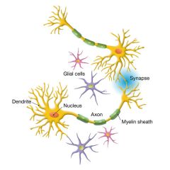
Neurons - can generate and transmit electrical signals (nerve impulses, or action potentials)
Glia - do not conduct action potentials |
|
|
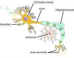
Parts of a neuron
Cell Body Dendrites Nucleus Axon Axon Soul Myelin Sheath Schwann Cells Nodes of Ranvier
|
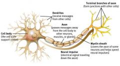
Cell Body - The largest part, contains the nucleus and much of the cytoplasm, most of the metabolic activity of the cell, including the generation of ATP and synthesis of protein for cytosol/cytoskelton in axon. Huge, active nucleus and RER. Also huge nucleolus to make ribosomes and Nissl bodies (Nissl is an RNA stain).
Dendrites - Short branch extensions spreading out from the cell body. Dendrites receive stimulus and carry impulses from the environment or from other neurons TOWARD THE CELL BODY (excitatory or inhibitory inputs). Cytoskeleton has MAP2 and tubulin, RNA for local protein synthesis. Dendrites also contain Nissl bodies, ER, ribosomes, and messenger RNA.
Axon - A long fiber that carries impulses (action potentials) away from the cell body. Action Potentials are all or none. The Axon ends in a series of small swellings called axon terminals. Neurons may have dozens or even hundreds of dendrites but usually only one axon. They have tau and tubulin. The axon does not contain ribosomes nor much ER or RNA.
Axon Soul -Reads the map and decides whether to make an AP or not. Location matters.
Myelin Sheath - lipid layer that covers axons. Insulates and speeds up transmission of action potentials through the Axon. Oligodendrocytes.
Schwann Cells -Cells that produce myelin in the Peripheral Nervous System, surround the Axon.
Nodes of Ranvier - gaps in the myelin sheath along the axon |
|
|
Resting membrane potential |
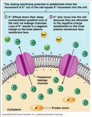
Resting Membrane Potential - electric charge difference across the plasma membrane, with the inside of the cell negative to the outside.
All cells have more potassium ions (K+) inside and more sodium ions (Na+) outside.
There are leak currents in the cell membrane due to channels that allow only certain ions—mostly K+—to “leak” passively across.
Because there are more potassium ions inside the cell than outside, K+ diffuses out of the cell down a concentration gradient. But when K+ leaks out of the cell, it leaves behind an unbalanced negative charge that tends to pull K+ back into the cell. An equilibrium is reached when the tendency for K+ to diffuse out is countered by the electrical charge pulling K+ back in.
The resting potential is slightly different than K, due to other ions (we need the Goldman Eq to calculate)
|
|
|
Generation of the resting membrane potential |
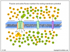
Sodium–potassium pump - shoots Na+ out, brings K+ in
Uses ATP for energy (needs energy to push these ions against their chemical gradients)
The Na+–K+ pump keeps the concentration of K+ inside the cell greater than that of the extracellular fluid.
The concentration differences established by this antiporter mean that K+ would diffuse out of the cell and Na+ would diffuse in if the ions could cross the lipid bilayer.
The direction and magnitude of the net movement of ions through a channel depend on the concentration gradient of that ion type across the plasma membrane, as well as on as the voltage difference across that membrane. These two motive forces acting on an ion are termed its electrochemical gradient.
Although the electrochemical gradient drives the movement of ions through channels across bilayers, movement through channels can be modified by gates that open and close channels.
Potassium channels are the most common open, or leak, chan- nels in the plasma membranes of resting (non-stimulated) neu- rons. As a consequence, resting neurons are more permeable to K+ than to any other ion.
As positively charged K+ potassium ions diffuse out of the cell, they leave behind unbalanced negative charges, generating an electric potential across the membrane that tends to pull K+ back into the cell. |
|
|
Action potentials |
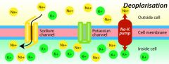
Neurons can generate rapid, large changes in membrane potential.
Action potentials = sudden large shifts in membrane potential (sometimes referred to as nerve impulses).
An action potential is generated by sudden openings and closings of ion channels (Na+ and K+ channels)
Action potentials are rapid, all-or-none changes in membrane potential that are conducted along axons from the neuron body to the axon terminals.
Travel via saltatory conduction |
|
|
Generation of an Action Potential:
Depolarization Hyperpolarization Graded membrane potential Axon Hillcock Threshold potential |
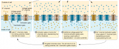
The ion channels called leak channels are always open, but other ion channels are open under some conditions and closed under other conditions.
The action potential is generated by the actions of voltage-gated Na+ and K+ channels in the plasma membrane of the axon. At the resting potential, most of these channels are closed. Depolarization of the membrane causes them to open.
Voltage-gated channels open or close in response to a change in the voltage across the plasma membrane.
Chemically gated channels open or close depending on the presence or absence of a specific molecule that binds to the channel protein, or to a separate receptor that in turn alters the channel protein.
Mechanically gated channels open or close in response to mechanical force applied to the plasma mem- brane. Gated channels play important roles in neural function.
Openings and closings of gated channels alter the resting potential.
If sodium channels in the plasma membrane open, Na+ diffuses into the neuron down its electrochemical gradient, the inside of the cell becomes less negative.
If gated K+ channels open, K+ leaves cell, the membrane potential becomes even more negative.
Depolarization - when the inside of the neuron becomes less negative in comparison to its resting condition
Hyperpolarization - when the inside of the neuron becomes less negative in comparison to its resting condition
A local change in membrane potential causes a flow of ions that spreads the change in membrane potential to adjacent regions of the membrane.
When Na+ enters a neuron through open sodium channels at one location, those positively charged ions are attracted to adjacent areas on the inside of the membrane that are more negative, and thus there is a rapid flow of electric current (movement of ions) away from the site of the open Na+ channels. However, this local flow of electric current decays as it spreads and therefore does not spread very far. Electric currents do not spread far in cells because cell membranes are not completely impermeable to ions.
Graded membrane potential - a change from the resting potential. Such changes can be due to chemical or mechanical influences on ion channels.
If a neuron is stimulated sufficiently to cause the plasma membrane of its cell body to depolarize, that depolarization can spread by local current flow to the axon hillock (the region of the cell body at the base of the axon).
Voltage-gated Na+ channels are concentrated in the axon hillock. When the plasma membrane in this area depolarizes, some of these voltage-gated channels open briefly—for less than a millisecond.
When these channels open, Na+ rushes into the axon and depolarizes the membrane even more, causing more Na+ channels to open—a positive feedback effect.
Threshold potential - When the membrane is depolarized to a certain point (about 5 to 10 mV above the resting potential), a large number of sodium channels open, and the membrane potential becomes positive—an action potential.
|
|
|
Steps of Action Potential
|
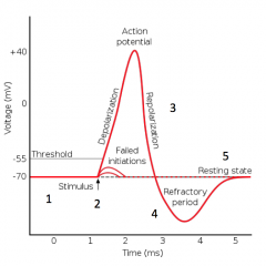
1. Negative resting potential - K+ leak channels determine potential
2. Depolarization - Na+ channels open - Na+ in
3. Repolarization - Na+ channels close, K+channels open = K+ out
4. Hyperpolarization (refractory period) - Na+ channels inactivated, K+ channels remain open
5. Return to resting potential - K+ channels close (normal leak channels remain open)
The rising phase of the action potential halts abruptly in 1 to 2 milliseconds, and the membrane potential rapidly becomes negative once again from the voltage-gated Na+ channels closing and voltage-gated K+ channels opening.
The voltage-gated K+ channels open more slowly than the Na+ channels and stay open longer, allowing K+ to carry excess positive charges out of the axon. As a result, the membrane potential returns to a negative value and usually becomes even more negative than the resting potential until the voltage-gated K+ channels close.
Another feature of the voltage-gated Na+ channels is that once they open and close, they have a refractory period of 1 to 2 mil- liseconds during which they cannot open again.
|
|
|
Saltatory conduction
Nodes of Ranvier |
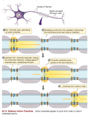
Action potentials can travel over long distances with no loss of signal.
Saltatory Conduction - rapid impulse propagation down an axon
Normally, an action potential is propagated in only one direction. It cannot reverse itself because the voltage-gated Na+ channels in the region of the membrane it came from are still in their refractory period.
They travel faster in myelinated than in nonmyelinated axons because they can “jump” from one node to another without traversing the intervening space.
The leakage of ions across the regions of the plasma membrane that are wrapped in myelin is reduced, so electric current can spread farther along the in- side of a myelinated axon than it can along a nonmyelinated axon.
Nodes of Ranvier - spaced gaps in myelin along axons
Voltage-gated ion channels are clustered at the nodes of Ranvier. Thus an axon can fire action potentials only at nodes, and those action potentials cannot be propagated through the adjacent patch of membrane covered with myelin. The positive charges that flow into the axon at the node do, however, flow down the inside of the axon in the form of electric current. When the current reaches the next node, the plasma membrane at that node is depolarized to threshold and fires another action potential. Action potentials therefore appear to jump from node to node along the axon.
The speed of conduction is increased in these myelin-wrapped axons because electric current flows much faster through the cytoplasm than ion channels can open and close. |
|
|
Oligodendrocytes & Schwann Cells |
Myelin is a plasma membrane with a few glycoproteins—some are called myelin basic protein in CNS and protein P0 in the periphery.
The so called proteolipid protein (plp) holds together the stacks of membrane wrappings (it’s a very hydrophobic protein located in the “extracellular clefts”
Within in Schwann cells there are clefts that are cytoplasmic zones.
Schmidt-Lanterman lines are regions of Schwann cell cytoplasm that are still present through the wrapping of the Schwann cell membranes.
One Schwann cell myelinates one axon at a time, but oligodendroglia myelinate more than one neuron at a time. |
|
|
Synapse
Electrical synapse Chemical synapse Neuromuscular junctions Axon terminals Neurotransmitter Acetylcholine Motor End Plate Synaptic Cleft |
Neurons communicate with each other and with other cells at synapses.
Electrical synapses - action potential spreads directly from presynaptic to postsynaptic cell via passive diffusion through gap junctions. These are rare.
Chemical synapse - neurotransmitters re- leased from a presynaptic cell induce changes in a postsynap- tic cell (most common).
Neuromuscular junctions -synapses between neurons and the skeletal muscle cells they innervate. The neuromuscular junction has a chemical synapse)
A motor neuron has only one axon, but close to its target cell that axon can branch into numerous axon terminals that form many synapses with muscle cells.
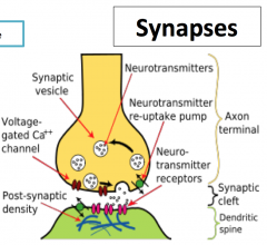
At each axon terminal an enlarged knob or buttonlike structure contains vesicles filled with neurotransmitter molecules.
Acetylcholine - The neurotransmitter used by neuromuscular synapses.
ACh is released by exocytosis when the membrane of a vesicle fuses with the presynaptic membrane of the axon terminal.
Motor End Plate - The postsynaptic membrane of the neuromuscular junction. It is a modified part of the muscle cell plasma membrane. It appears as a depression in the muscle cell membrane, and the terminals of the motor neuron sit in the depression.
Synaptic Cleft - The space between the presynaptic membrane and the postsynaptic membrane. In chemical synapses it is about 20 to 40 nanometers wide.
ACh released into the cleft by the presynaptic cell diffuses across to the postsynaptic membrane.
|
|
|
Transmission of signal across a synapse
Ca2+ AcCh Receptors |
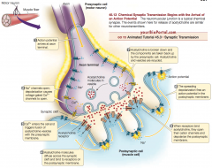
Neurotransmitter is released when an action potential arrives at the axon terminal.
This causes the opening of voltage-gated Ca2+ channels in the presynaptic membrane.
Because the Ca2+ concentration is greater outside the cell than inside, Ca2+ enters the axon terminal near the sites of vesicle exocytosis.
The increase in Ca2+ inside the axon terminal causes the vesicles containing neurotransmitter to fuse with the presynaptic membrane and empty their contents into the synaptic cleft. In neuromuscular synapses, vesicle fusion and emptying is all-or-none.
The vesicle membrane is incorporated into the presynaptic membrane, which actually gets larger as a result—at least until the extra membrane is recycled through endocytosis. The recycled membrane is processed through endosomes to become new vesicles that are then refilled with neurotransmitter.
When ACh is released at a synapse, some of it diffuses across the synaptic cleft and binds to ACh receptors on the postsynaptic membrane.
The ACh receptors are channels that allow both Na+ and K+ to flow through, but since the electrochemical gradients favor a net influx of Na+, the response of the motor end plate to ACh is to depolarize.
That graded potential reflecting the number of receptors activated spreads to the depths of the folds of the motor end plate membrane and to surrounding muscle cell membrane, which contain voltage-gated Na+ channels.
If the axon terminal of a motor neuron releases sufficient amounts of ACh to adequately depolarize a motor end plate, that spreading depolarization will activate the voltage-gated Na+ channels and cause the firing of an action potential.
This action potential is then conducted throughout the muscle cell’s system of membranes, causing the cell to contract.
Neither a single ACh molecule nor the contents of an entire vesicle (about 10,000 ACh molecules) will bring the plasma membrane of a muscle cell to threshold. However, a single action potential in an axon terminal releases the contents of about 100 vesicles, which is more than enough to fire an action potential in the muscle cell and cause it to contract.
|
|
|
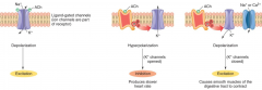
Excitatory vs Inhibitory Synapses |
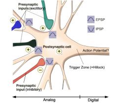
Excitatory Synapse - graded potential makes membrane potential more positive (bringing it closer to an action potential).
Synapses between motor neurons and muscle cells are always
Inhibitory Synapse - graded potential makes membrane potential more negative (bringing it further from an action potential).
(Ex. If the receptor on the postsynaptic membrane is a Cl– channel opened by a neurotransmitter, the effect of that neurotransmitter will be to cause Cl– ions to enter the postsynaptic cell and hyperpolarize it. Recall that there are more chloride ions (Cl–) outside the cell than inside it.)
Axon terminals from many neurons may form synapses with dendrites and with the cell body. The axon terminals of different presynaptic neurons may store and release different neurotransmitters, and the plasma membrane of the dendrites and cell body of a postsynaptic neuron may have receptors for a variety of neurotransmitters.
The mix of synaptic activity on a cell will cause it to have a graded membrane potential that may be either more positive or more negative than its rest-ing potential.
The sum of excitatory and inhibitory postsynaptic potentials creates a graded membrane potential in the postsynaptic cell body. This summation ability is the major mechanism by which the nervous system integrates information. At any one time, the information from all of the active inputs are translated into the rate at which that neuron generates action potentials in its axon.
For most neurons, summation takes place in the axon hillock at the base of the axon. The plasma membrane of the axon hillock is not insulated by glia and has many voltage-gated Na+ channels.
Excitatory and inhibitory postsynaptic potentials from synapses anywhere on the dendrites or the cell body may spread to the axon hillock by local current flow.
If the resulting graded potential depolarizes the axon hillock to threshold, it fires an action potential.
|
|
|
Spatial Summation
Temporal Summation |
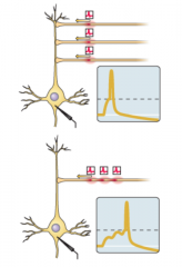
Because postsynaptic potentials decrease in strength as they spread from the site of the synapse, a synapse at the tip of a dendrite has less influence than a synapse on the cell body, near the axon hillock.
Excitatory and inhibitory postsynaptic potentials are summed over space (spatial summation and over time.
Spatial summation - adds up the simultaneous influences of synapses at different sites on the postsynaptic cell
Temporal summation - adds up postsynaptic potentials generated at the same site in a rapid sequence |
|
|
Ionotropic Receptors vs. Metabotropic Receptors |
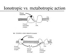
Ionotropic receptors (FAST) - ion channels themselves.
Neurotransmitter binding to an ionotropic receptor causes a direct change in ion movement across the plasma membrane of the postsynaptic cell.
These proteins enable fast, short-lived responses.
The ACh receptor of the motor end plate is an example of an ionotropic receptor.
Metabotropic receptors (SLOW) - not ion channels.
They induce signaling cascades in the postsynaptic cell that secondarily lead to changes in ion channels.
Metabotropic receptors are also transmembrane proteins, but instead of acting as ion channels, they initiate an intracellular signaling process that can result in the opening or closing of an ion channel.
|
|
|
Complexity of signal via receptors
Nicotinic Muscarinic |
Neurotransmission is complex in part because each neuro- transmitter has multiple receptor types.
The action of a neurotransmit-ter depends on the receptor to which it binds
ACh has two receptor types:
nicotinic receptors - ionotropic
muscarinic receptors - metabotropic
Ex. in PNS, ACh acting through nicotinic receptors causes the smooth muscle of the gut to increase its motility, but ACh acting through muscarinic re- ceptors causes cardiac muscle to hyperpolarize and therefore to slow down. |
|
|
3 ways to get rid of NT from the synaptic cleft
Acetylcholinesterase |
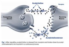
If released neurotransmitter molecules simply remained in the synaptic cleft, the postsynap- tic membrane would become saturated with neurotransmitter, and receptors would be constantly activated. As a result, the postsy- naptic cell would remain hyperpolarized or depolarized and would be unresponsive to short-term changes in the presynaptic cell. Thus, neurotransmitter must be cleared from the synaptic cleft shortly after it is released by the axon terminal.
Neurotransmitter action may be terminated in several ways:
1. Enzymes may destroy the neurotransmitter. Acetylcholinesterase - enzyme that rapidly degrades ACh. It is present in the synaptic cleft in close association with the ACh receptors on the postsynaptic membrane.
Some of the most deadly nerve gases developed for chemical warfare work by inhibiting acetylcholinesterase. As a result, ACh lingers in the synaptic cleft, causing the victim to die of spastic (contracted) muscle paralysis. Some agricultural insecticides, such as malathion, also inhibit acetylcholinesterase and can poison farm workers if used without safety precautions.
2. Neurotransmitter also may simply diffuse away from the cleft
3. NT can be taken up via active transport by nearby cell membranes.
The drug commonly prescribed under the brand name Prozac to treat depression slows the reuptake of the neurotransmitter sero- tonin, thus enhancing its activity at the synapse. |
|
|
3 types of polarity in neurons |
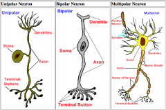
Monopolar (AKA unipolar) = sensory
1 process leaves the cell body. This single process then divides close to the cell body into a trunk to supply the branching dendrites for incoming signals and an axon for outgoing signals. Unipolar neurons are typically sensory neurons with receptors located within the skin, joints, muscles, and internal organs. Their axons are usually long, terminating in the spinal cord, while the length of their dendritic trunks vary.
Bipolar = special sensory
2 processes leave the cell body. The dendritic tree emerges from one end of the cell body, while the axon emerges from the opposite end. The dendritic branching of bipolar neurons is typically limited, and the axons of such neurons are usually short in length. Bipolar neurons are often sensory neurons associated with receptor organs of the visual and auditory systems. The narrow fields created by the short dendrites of these neurons underlie the concise encoding of visual and auditory information representing physical signals from the external world. Without this narrow encoding of sensory information, the resolution of vision and hearing would be reduced.
Multipolar = everything else
A neuron from which multiple branches leave the cell body. The many dendrites of the multipolar neuron allow for extensive integration of information coming from many other neurons. The axons of such neurons are usually long, allowing this integrated information to affect distant regions of the nervous system. (The majority of neurons.) |
|
|
Synthesis & Transport of NTs |
Some neurotransmitters, such as ACh, are synthesized in the axon terminal and packaged in vesicles.
The enzymes required for ACh biosynthesis, however, are produced in the cell body of the motor neuron and are transported along microtubules down the axon to the terminals.
In contrast, peptide neurotransmitters are produced in the cell body and packaged into membrane-bound vesicles by the Golgi apparatus. These vesicles are rapidly trans- ported down the axon to the terminals. |
|
|
3 types of axonal transport
Tau MAP2 |
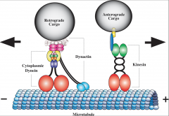
Fast Anterograde - kinesin, carries vesicles to terminals
Slow Anterograde - carries cytosolic/cytoskeletal elements for axon
Retrograde - dynein, carries viruses/toxins, rabies
The cell bodies synthesize the proteins and then transport proteins from the cell body by anterograde transport to axon terminal via microtubules/dynein. There is both Fast transport (synaptic vesicles) and Slow transport (tubulin, actin and other housekeeping proteins) both exist.
Axons also demonstrate retrograde transport of vesicles to recycle synaptic vesicles and organelles to deposit them in the lysosomes.
Retrograde transport is a method of recycling organelles—but do note that tetanus toxin uses this mechanism to reach the cell body. Virus also can reach the cell body via the synaptic tubule.
Tau (Axons) MAP2 (Dendrites)
Both of these hold microtubules bundles together. |
|
|
Small vs. Large Stimulus |
Small stimulus = release of small NT from docked vesicles that were packaged regionally (vesicle recycling maintains membranes volume)
Large stimulus = release of peptide NT (120-250 nm) are packaged in the Golgi
|
|
|
Dendrites as transmission |
Some cells have dendrites but no axon suggesting that dendrites not only receive information (toward the cell body), but also can transmit information
dendro->axonic; and dendro-> dendritic communication (information moving away from cell body)
All sorts of interactions including axo-axonic, axo-dendritic interactions etc.
All permutations are possible. |
|
|
Regulatory proteins -
Synapsin
SNARE proteins
Synaptotagmin
Botulinum
Acetylcholine
|
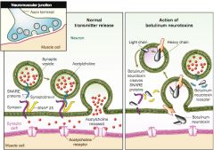
Synaptic functions involves hundreds of proteins that are responsible for various aspects of the process: vesicle formation, transport of neurotransmitter into vesicles, anchoring of vesicles to cytoskeletal elements, docking of the vesicles with the presynaptic membrane, fusion of the vesicular and cell membranes, and endocytosis of the vesicle membrane for recycling.
Some of these proteins are the targets of toxins.
A neuromuscular junction contains the active zone where AcetylCholine is released. Vesicles are lined up in a patch of membrane called the active zone.
Synapsin - tethers vesicles to actins (microfilaments) near the active zone. Ca-calmodulin activation causes docking of these reserve vesicles at the plasma membrane.
SNARE proteins – Pull vesicle and membrane together. mediate fusion of vesicles to synaptic membranes with the help of…
Hence synaptic transmission is halted and recycling is not possible.
This causes paralysis.
It can take weeks for regeneration of the cohort of vesicles.
|
|
|
Neurotransmitters
Simple Amino Acids Amino Acid Derivatives Peptides |
ACh also plays roles in certain synapses between neurons in the CNS, but it accounts for only a small percentage of the total neurotransmitter content of the CNS.
The workhorse neurotransmitters of the CNS are simple amino acids:
glutamate (excitatory) glycine (inhibitory) GABA (inhibitory)
Another important group of neuro- transmitters in the CNS is the monoamines, which are derivatives of amino acids (small amines):
Dopamine (derivative of tyrosine) Norepinephrine (derivative of tyrosine) Epinephrine (derivative of tyrosine) Serotonin (a derivative of tryptophan) Histamine Acetylcholine
Peptides also function as neurotransmitters:
Endorphins Enkephalins Substance P
Also: Nitrous Oxide
|
|
|
Central Nervous System (CNS)
Peripheral Nervous System (PNS) |
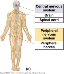
Central Nervous System (CNS) - Brain + Spinal Cord
most cells of the nervous system are found in the CNS
sites of most information processing, storage, and retrieval
Information is transmitted from sensory cells to the CNS and from the CNS to effectors via neurons that extend or reside outside the brain and the spinal cord
Peripheral Nervous System (PNS) - neurons and their supporting cells outside the spinal cord |
|
|
• Afferent neurons • Efferent neurons • Interneurons |
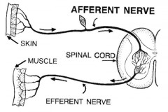
• Afferent neurons - carry sensory information into the nervous system. That information comes from specialized sensory neurons that convert various kinds of sensory stimuli (e.g., light, heat, pressure) into action potentials
• Efferent neurons - carry commands to physiological and behavioral effectors such as muscles and glands
• Interneurons integrate and store information and communicate between afferent and efferent neurons |
|
|
Glia
Astrocytes Oligodendrocytes Microglia |
Non-neuronal cells in the nervous system that serve supportive functions
There is no connective tissue in the brain, glial cells are the surrounding tissues (not fibroblasts, no connective tissues).
There are many more glia than neurons in the human brain.
They do not generate or transmit electrical signals, but they can release neurotransmitters.
Astrocytes - structural support for neurons & blood vessels
They look like stars.
Contribute to the blood–brain barrier, which protects the brain from toxic chemicals in the blood.
Blood vessels throughout the body are very permeable to many chemicals, including toxic ones, which would reach the brain if this bar- rier did not exist. Astrocytes help form the blood–brain bar- rier by surrounding the smallest, most permeable blood vessels in the brain. The barrier is not perfect, however. Since it consists of plasma membranes, it is permeable to fat-soluble substances such as anesthetics and alcohol, which explains why these substances have such rapid and marked effects on the nervous system. The blood–brain barrier usually prevents antibodies in the general circulation from entering the CNS.
Astrocytes contain IF filaments made of Glial Fibrillary Acidic Protein; These cells also have many desmosomal junctions on other cells ie their IF’s and their junctions with other cells give them tensile strength. In contrast, collagen is not present in the brain.
Oligodendrocytes - provide myelin sheath for multiple axons in the CNS.
In the CNS, glia called oligodendrocytes wrap around the axons of neurons, covering them with concentric layers of in- sulating plasma membrane. In the PNS, glia called Schwann cells perform this function. Myelin is the covering produced by oligodendrocytes and Schwann cells, and it gives many parts of the nervous system a glistening white appearance.
Microglia - non-neuronal derived immune cells involved in surveillance
To provide the CNS with immune defenses, microglia, which originate during de- velopment from stem cells in the bone marrow, come to reside in the CNS and act as macrophages and mediators of inflam- matory responses. |
|
|
Multiple Sclerosis
Guillain-Barre Syndrome |
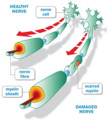
Multiple Sclerosis - “multiple scars”
The most common demyelinating disease
Occurs in about 1 in 700 people in the United States.
Individuals with this autoimmune disease produce antibodies to proteins in the myelin in the CNS. The symptoms and damage from the disease depend on where in the CNS the antibody attacks take place.
Guillain-Barre Syndrome - a demyelinating disease that affects the PNS.
Environmental factors such as pesticide exposure can also damage myelin.
There are no known cures for demyelinating diseases. |
|
|
Layers of Meninges in Brain (outside in) |
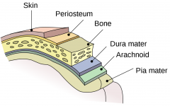
Periosteum Skull Dura Mater (dense connective tissue) Arachnoid Mater (loose connective tissue) CSF (in sub-arachnoid space) Pia Mater (adherent to brain parenchyma) Brain |
|
|
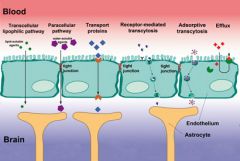
Blood-brain barrier (BBB) |
Protects the brain from "foreign substances" in the blood that may injure the brain.
Protects the brain from hormones and neurotransmitters in the rest of the body.
Maintains a constant environment for the brain.
The BBB is semi-permeable: it allows some materials to cross, but prevents others from crossing. In most parts of the body, the smallest blood vessels, called capillaries, are lined with endothelial cells. Endothelial tissue has small spaces between each individual cell so substances can move readily between the inside and the outside of the vessel. However, in the brain, the endothelial cells fit tightly together and substances cannot pass out of the bloodstream. (Some molecules, such as glucose, are transported out of the blood by special methods.)
Large molecules do not pass through the BBB easily.
Low lipid (fat) soluble molecules do not penetrate into the brain. However, lipid soluble molecules, such as barbituate drugs, rapidly cross through into the brain.
Molecules that have a high electrical charge are slowed.
-Formed by tight junctions in brain capillaries, with astrocyte foot processes attaching to capillaries providing support
-Little transcytosis from blood to brain parenchyma
-Astrocytes also provide endocytosis of unwanted foreign substances |
|
|
CSF (Cerebrospinal Fluid)
Ependymal Cells Choroid Plexus |
a clear colorless bodily fluid found in the brain and spine.
acts as a shock absorber
involved in autoregulation of cerebral blood flow
The CSF occupies the subarachnoid space (the space between the arachnoid mater and the pia mater) and the ventricular system around and inside the brain and spinal cord.
It constitutes the content of the ventricles, cisterns, and sulci of the brain, as well as the central canal of the spinal cord.
Produced by ependymal epithelial cells (no tight junctions in most areas, and their fluid is secreted into the brain extracellular spaces as well as into the ventricles of the brain —ventricles are the inner hollow cavities of the brain).
Produced by filtration (~75%) and secretion (~25%)
Filtration - in ependymal cells which line ventricles (no tight junctions, unlike rest of brain capillaries)
Secretion - in specialized cells of the choroid plexus (do have tight junctions - secrete CSF elements)
From the ventricles, the fluid is driven into the subarachnoid spaces via the Foramen of Magendie and Luschka.
The CSF is circulates and finally is reabsorbed into the venous system across arachnoid granulations, a system of valves from CSF into the blood system. |
|
|
Arachnoid Granulations |
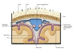
Function like pressure valves to deliver CSF from sub arachnoid space into venous sinuses as needed |