Reading...
![]()
Play button
![]()
Play button
![]()
Use LEFT and RIGHT arrow keys to navigate between flashcards;
Use UP and DOWN arrow keys to flip the card;
H to show hint;
A reads text to speech;
9 Cards in this Set
- Front
- Back
- 3rd side (hint)
Describe the iliofemoral joint:
1. Type 2. Articular surfaces 3. Capsule 4. Ligaments 5. Special features 6. Movements |
1. Type
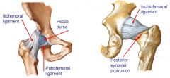
- Largest joint in the body - Synovial ball and socket joint 2. Articular surfaces: - Acetabulum = socket of hip - Femoral head - Deepened by the Acetabular Labrum = horse shoe shaped fibrocartilage 3. Capsule - Strong but loose - Proximally attached to margins of acetabulum & transverse ligament - Distally attached to the intertrochanteric line anteriorly and 0.5cm in from trochanteric crest posteriorly. - Reflected to form retinacula - Zona orbicularis = deep fibres form circular collar of fibres around the neck of femur - Thick parts of the spiralling capsule fibres form orbicular ligaments of the hip joint which help stabilise it and wind and unwind in extension and flexion - Synovial Membrane covers non articular surfaces. Anteriorly forms Psoas bursa and there is a protrusion posteriorly 4. Ligaments: - Transverse ligament between lunate surfaces with a gap for the artery to the head of the femur to pass to the non-articular acetabular surface - 3 ligaments reinforce the capsule: • Iliofemoral (Y shaped) • Ischiofemoral • Pubofemoral 5. Special features: - Proximal femoral fractures are named based on whether the intracapsular or extracapsular 6. Movements: - Flexion 120 - Extension 30 - Abduction 45 - Adduction 30 - Internal rotation 35 - External rotation 45 - Ligaments limit movement |
|
|
Label this diagram
|
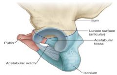
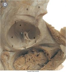
1 acetabular fossa
2 acetabular labrum 4 articular surface 5 ligamentum teres 10 transverse ligament |
|
|

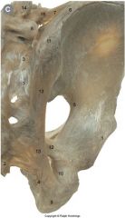
Label this diagram of the important relationships of the hip joint
|
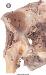
1 AIIS 2 ASIS
3 bursa for psoas 7greater trochanter 10 iliacus 11 iliofemoral ligament 16 inguinal ligament 17 intertrochanteric line & attachment of joint capsule 19 lesser trochanter 20 lumbar sacral trunk 24/25 obturator nerve 26 obturator vessel 29 pundendal nerve |
|
|
|
Describe the blood supply to the hip joint
|
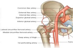
1. Branch of obturator vessel = runs underneath the transverse ligament to help supply the head of the femur. Little supply in the adult
2. Medial and circumflex femoral arteries = branches of the deep femoral artery that anastomose around the femoral neck and give off retinacular vessels to the joint capsule 3. Also has some supply from the trochanteric and the cruciate anastomosis (so receives blood from gluteal, circumflex and ascending perforating arteries) |
|
|
|
Why is the blood supply to the femoral head and neck clinically important?
|
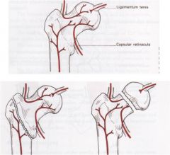
- In child the artery to the head of the femur must carry lots of blood as neck arteries don't penetrate the epipyseal plate
- In an adult neck arteries can supply the head, and so the artery to the head of the femur regresses to a ligament with only a trickle of blood - Therefore if there is a break in the neck of the femur → avascular necrosis so MUST check x-rays (will see white granular sclerosis) and replace joint - If the break is through the intertrochanteric region should be fine |
|
|
|
Describe the innervation of the hip joint
|
Hilton's Law
- 3 major and 3 minor - Major: 1. Femoral nerve 2. Obturator nerve 3. Sciatic nerve - Minor: 4. Accessory obturator nerve 5. Superior gluteal nerve 6. Nerve to quadratus femoris - Note that some of these nerves also supplt the knee joint so that pain in the knee may be referred and caused by pathology in the hip → so vital to examine the joint above and below - Femoral nerve is prone to injury anteriorly in the femoral triangle - Sciatic nerve posterior is prone to injusy from posterior dislocations and ill-judged injections |
|
|
|
Describe the features of the sacro-iliac joint:
1. Type 2. Articular surfaces 3. Capsule 4. Ligaments 5. Special features 6. Movements |
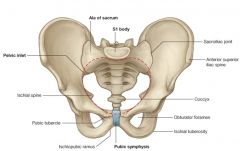
1. Type = synovial planar
2. Bones : auriculum of sacrum & ilium 3. Sacrum covered hyaline cartilage, the ilial surface covere by fibrocartilage 4. Ligaments : interosseous, anterior & posterior sacroiliac (strongest lig in body ) 5. Special features: Note the sacrotuberous & sacrospinous = these ligaments join the sacrum to the ischium and convert the sciatic notches into foramen 6. Movements : gliding |
|
|
過ち
|
/(n) fault/error/indiscretion/(P)/
|
[あやまち]
|
|
|
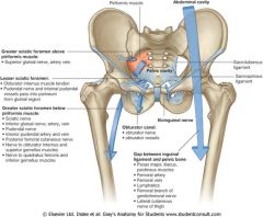
What are the structures passing through:
1. Greater sciatic foramen above piriformis (1) 2. Greater sciatic foramen below piriformis (7) 3. Lesser sciatic foramen (2) 4. Obturator canal (1) 5. Over the pubic tubercle (1) 6. Between the inguinal ligament and the pelvic bone (8) |
|
|