Reading...
![]()
Play button
![]()
Play button
![]()
Use LEFT and RIGHT arrow keys to navigate between flashcards;
Use UP and DOWN arrow keys to flip the card;
H to show hint;
A reads text to speech;
11 Cards in this Set
- Front
- Back
|
Describe the fasciae of the axilla
|
3 Major fasciae
1. Prevertebral fascia: - Fascia covering scalene mm. - Gives rise to the axillary sheath which encloses the axillary neurovascular bundle 2. Clavipectoral fascia Lies between pectoralis major and the thoracic wall - Invests pectoralis minor and subclavius 3. Axillary fascia: - From latissimus dorsi to pectoralis major - Dips inward to form the “armpit” |
|
|
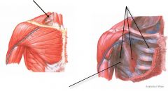
Describe the boundaries of the axilla
|
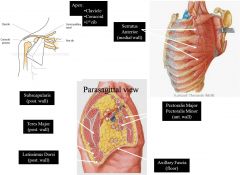
- Apex:
• Clavicle • Coracoid process • 1st rib - Borders: Anterior: pectoralis major & minor • Posterior: lats dorsi, teres major, subscapularis • Medial: serratus anterior, ribs, intercostals • Lateral: tendon of long head of biceps brachii - Base: • Axillary fascia • Runs between latissimus dorsi and pectoralis major ie. Hairy armpit |
|
|
Describe the contents of the axilla
|

- Passageway for vital structures passing to/from the neck & thoracic cavity to/from the upper limb
- Contains: • axillary a. & v. • brachial plexus = cords & branches • lymph nodes • tail of breast • fat - The axillary artery, vein & brachial plexus are enclosed in the axillary sheath - The brachial plexus gives rise to nerves supplying most of the shoulder muscles and all of the upper limb muscles |
|
|
Describe the landmarks of the brachial plexus
|
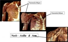
- Runs posterior to pectoralis major and minor
- Brachial artery can also be felt close to it |
|
|
Describe the lymphatics of the axilla
|
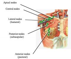
Axillary Lymph Nodes (deep nodes):
- Filter lymph from the upper limb, breast, ant. & post. trunk - Subdivided into 5 groups (named for their location) • Lateral (humeral) • Posterior (subscapular) • Anterior (pectoral) • Central • Apical - Drainage: • Lateral, posterior, anterior → Central → Apical → Subclavian lymphatic duct → Thoracic duct (left) /right lymphatic duct (right) → Subclavian v. - Axiallary nodes are the most common sites of breat cancer metastases |
|
|
Describe the couse and divisions of the axillary artery
|
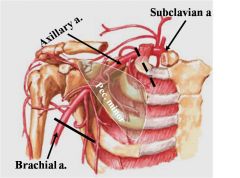
- Begins at lateral border 1st rib
- Becomes brachial a. at the inferior border of teres major - Divided into 3 parts by pectoralis minor - 1st Part = one branch proximal to pectoralis minor • superior thoracic a. - 2nd Part = two branches deep to pectoralis minor • thoracoacromial trunk • lateral thoracic a. - 3rd Part = 3 branches distal to pectoralis minor • subscapular a. • medial circumflex humeral a. • lateral circumflex humeral a. |
|
|
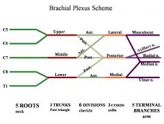
What is the brachial plexus? Describe how it is divided
|
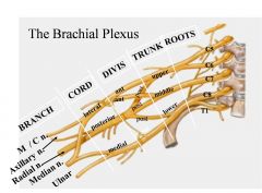
- Plexus: from the Latin root plex, meaning “interweave”
- It is an interweaving of the cervical ventral rami of C5-T1 - Gives rise to the nerves supplying the upper limb - Carries somatic motor, somatic sensory and sympathetic fibers - Composed of: • ROOTS: → C5-T1 ventral rami • TRUNKS: → upper, middle, lower named for position relative to each other • DIVISIONS: → 3 anterior, 3 posterior • CORDS: → medial, lateral, posterior named for where they sit in relationship to the axillary a. • TERMINAL BRANCHES: → terminal named nerves axillary, radial, median, musculocutaneous, ulnar n. |
|
|
What is a prefixed and postfixed brachial plexus?
|
- Prefixed brachial plexus = C4-C8
- Postfixed brachial plexus = C6-T2 → problems as likely to get compression of inferior trunk by 1st rib |
|
|
Describe how the brachial plexus is formed
|
|
|
|
Describe the muscles that the terminal branches of the brachial plexus go on to supple
|
- Musculocutaneous nerve → anterior compartment of arm = flexors of the elbow
• Biceps brachii • Brachialis • Coracobrachialis - Axillary nerve • Teres minor • Deltoid - Radial nerve → posterior compartment of arm and forearm = elbow and wrist extensors • Triceps brachii • Anconeus (• Brachioradialis = elbow flexor) • ECRL • ECRB • ECU • EDM • APL • EPB • EPL • EI - Median nerve → anterior compartment of forearm = wrist flexors • FCP • PL • FDS • FDP (1/2) • FPL - Ulnar nerve = intrinsic hand muscles (• FCU = wrist flexor) (• FDP 1/2) |
|
|
Describe the relation of nerves to the humerus
|
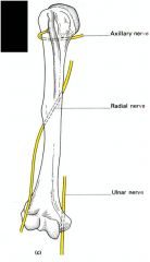
- The axillary and radial nerves especially run close to the humerus at common points of breakage → potential injury on fracture
|