![]()
![]()
![]()
Use LEFT and RIGHT arrow keys to navigate between flashcards;
Use UP and DOWN arrow keys to flip the card;
H to show hint;
A reads text to speech;
106 Cards in this Set
- Front
- Back
|
What are the three type of muscle tissue?
|
* Skeletal Muscle * Cardiac Muscle * Smooth Muscle |
|
|
What is skeletal muscles? |
Skeletal Muscles are organs composed mainly of skeletal muscle tissue, but they also contain connective tissues, nerves, and blood vessels. Each cell in the skeletal muscle tissue is a single muscle fiber. |
|
|
What are the six (6) functions of skeletal muscle? |
1) Produce Skeletal Movement 2) Maintain Posture and Body Position 3) Support Soft Tissues 4) Guard Body Entrances and Exits 5) Maintain Body Temperature 6) Store Nutrients |
|
|
What are the three layers of connective tissue in muscles? |
1) An epimysium 2) A perimysium 3) An endomysium |
|
|
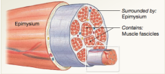
What is the epimysium? |
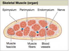
The epimysium is a dense layer of collagen fibers that surrounds the entire muscle. It separates the muscle from nearby tissues and organs. |
|
|
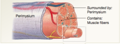
What is the perimysium? |
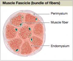
The perimysium divides the skeletal muscle into a series of compartments. Each compartment contains a bundle of muscle fibers called a fascicle. The perimysium contains blood vessels and nerves that supply the muscle fibers with the fascicles. |
|
|
What is the endomysium? |
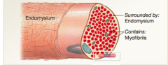
Within a fascicle, the delicate connective tissue of the endomysium surrounds the individual skeletal muscle cells, called muscle fibers, and loosely interconnects adjacent muscle fibers. |
|
|
At the end of the epimysium, the perimysium, and the endmysium come together to form either a bundle known as ______, or a broad sheet called ________. |
1) Tendon 2) Aponeurosis |
|
|
Skeletal muscle fibers are multinucleate. What does that mean? |
Multinucleate means that each contains hundreds of nuclei just internal to the plasma membrane. The more copies of these genes, the faster these proteins can be produced. |
|
|
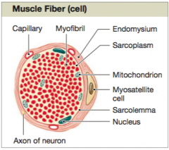
Each muscle fiber contains hundreds to thousands of cylindrical structures called ______. What are they? |
Myofibrils. The active shortening of myofibrils is responsible for skeletal muscle fiber contraction. Myofibrils consist of bundles of protein filaments called myofilaments - thin filaments and thick filaments. |
|
|
What is the name of the plasma membrane of a muscle fiber? |
The name of the plasma membrane of a muscle fiber is called sarcolemma. The sarcolemma surround the cytoplasm which is called sarcoplasm. |
|
|
What is the sarcoplasmic reticulum (SR)? |
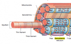
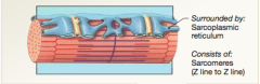
In skeletal muscle fibers, a membrane complex called sarcoplasmic reticulum (SR) forms a tubular network around each individual myofibril. The SR is similar to the smooth endoplasmic reticulum of other cells. |
|
|
Myofilaments are organized into repeating functional units called______. |
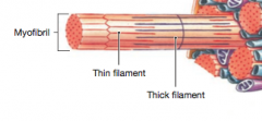
Sarcomeres. (They are the myofilaments within the myofibril. |
|
|
What are sarcomeres? |
The sarcomeres are the smallest functional units of the muscle fibers. *Interactions between the thick and thin filaments within the sarcomeres are responsible for muscle contraction. |
|
|
What does a sarcomere contain? |
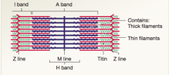
1) Thick filaments 2) Thin filaments 3) Proteins that stabilize the positions of the which and thin filaments 4) Proteins that regulate the interactions between thick and thin filaments. |
|
|
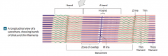
What are the dark bands called? What are the light bands called? |
The dark bands are called A band, derived from anisotropic. The light bands are called I bands, derived from isotropic. |
|
|
What are the different levels of Functional Organization in a Skeletal Muscle? |
1) Skeletal Muscle
2) Muscle Fascicle 3) Muscle Fiber 4) Myofibril 5) Sacromere |
|
What level of function is this? |
This is the Skeletal Muscle level |
|
What level of function is this? |
This is the Muscle Fascicle level
|
|
What level of function is this? |
This is the Muscle Fiber level |
|
What level of function is this? |
This is the Myofibril level |
|
What level of function is this? |
This is the Sarcomere level |
|
|
Depolarization vs Hyperpolarization vs Repolarization |
An influx of sodium ions leads to depolarization. (More positive) The movement of potassium ions out of a cell leads to hyper polarization. (Less positive) A return to the resting potential is called repolarization. |
|
|
What is a synapse? |
This is where communication between a neuron and another cell occurs at a synapse. |
|
|
What are neuromusclar junction (NMJ)? |
When the other cell is a skeletal muscle fiber, the synapse is known as neuromuscular junction. The NMJ is made up of an axon terminal of a neuron, a specialized region of the sarcolemma called the motor end plate, and, in between, a narrow space called the synaptic cleft. |
|
|
Describe isotonic contraction? |
In an isotonic contraction, tension increases and the skeletal muscle's length changes. * Lifting an object off a desk * Walking * Running involve isotonic contractions. |
|
|
What are the two isotonic contractions? |
Concentric and eccentric |
|
|
What is concentric contraction? |
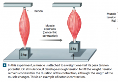
In concentric contraction, the muscle tension exceeds the load and the muscle shortens. |
|
|
What is eccentric contraction? |
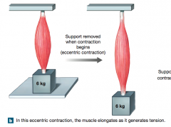
In an eccentric contraction, the peak tension developed is less than the load, and the muscle elongates due the the contraction of another muscle or the pull of gravity. Think of a tug of war team trying to stop a moving car. |
|
|
What is isometric contractions? |
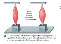
In an isometric contraction, the muscle as a whole does not change length, and the tension produced never exceeds the load. Examples of isometric contractions include carrying a bag of groceries and holding our heads up. |
|
|
What is aerobic metabolism? |
Aerobic metabolism normally provides 95 percent of the ATP demands of a resting cell. In this process, mitochondria absorb oxygen, ADP, phosphate ions, and organic substrates (such as pyruvate) from the surrounding cytoplasm. The molecules then enter the citric acid cycle. |
|
|
What is glycolysis? |
Glycolysis is the anaerobic breakdown of glucose to pyruvate in the cytoplasm of a cell. It is an anaerobic process because it does not require oxygen. Glycolysis provides a net gain of 2 ATP molecules and generates 2 pyruvate molecules from each glucose molecule. |
|
|
What is the cori cycle? |
The shuffling of lactate to the liver and glucose back to the muscle cells . |
|
|
What is excess post exercise oxygen consumption (EPOC)? |
It is the amount of oxygen required to restore normal, pre-exertion condition. |
|
|
The human body has three major types of skeletal muscle fibers, what are they? |
* fast fibers * slow fibers * intermediate fibers |
|
|
What are fast fibers? |
Most of the skeletal muscle fibers in the body are called fast fibers, because they can reach peak twitch tension in 0.01 sec or less after stimulation. |
|
|
What are slow fibers? |
One main characteristics of slow muscle fibers is that they are surrounded by a more extensive network of capillaries than is typical of fast muscle tissue. For this reason, they have a dramatically higher oxygen supply to support mitochondrial activity. Slow fibers also contain the red pigment myoglobin. Myoglobolin reversibly bind oxygen molecules. |
|
|
What are intermediate fibers? |
Intermediate fibers are intermediate between those of fast fibers and slow fibers. In appearance, intermediate fibers most closely resemble fast fibers, for they contain little myoglobin and are relatively pale. |
|
|
What is hypertrophy? |
Hypertrophy is an enlargement of the stimulated muscle. The number of muscle fibers does not change significantly, but the muscle as a whole enlarges because each muscle fiber increased in diameter. |
|
|
What is atrophy? |
The reduction in muscle size, tone, and power in muscle is called atrophy. |
|
|
What is anaerobic endurance? |
Anaerobic endurance is the length of time muscular contraction can continue to be supported by glycolysis and by the existing energy reserve of ATP and cp. |
|
|
What is aerobic endurance? |
Aerobic endurance is the length of time a muscle can continue to contract while supported by mitochondrial activities. Aerobic activities do not promote muscle hypertrophy. |
|
|
What is automaticity? |
Cardiac muscle tissue contracts without neural stimulation. This is called automaticity. |
|
|
A skeletal muscle contains ______. |
A skeletal muscle contains muscle tissue, connective tissues, blood vessels, and nerves. |
|
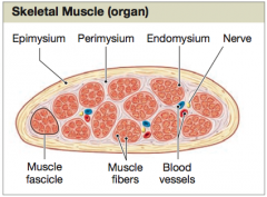
What is the epimysium, perimysium, and endomysium? |
A skeletal muscle consists of fascicles (bindles of muscle fibers) enclosed by the epimysium. The bundles themselves are separated within the epimysium by perimysium. The fibers within a bundle (fascicle) is surrounded by endomysium. Each muscle fiber has many superficial nuclei, as well as mitochondria and other organelles. |
|
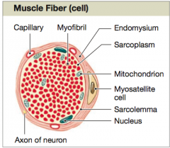
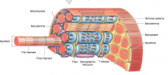
What is this an image of? Describe what you see. |
This is an image of a muscle fiber. Several of these fibers are called fascicle. What we see are the individual fibers called Myofibril bundled together and are immersed in sarcoplasm. This is all enclosed by endomysium. We see that within the fascicle there are several mitochondria, nucleus, and capillaries. ` |
|
|
What are myosatellite cells and where are they found? |
Myosatellite cells are stem cells and they are found in the endomysium. |
|
|
Are skeletal muscle considered voluntary or involuntary muscles? |
They are considered to be voluntary muscle, because we have voluntary control over their contractions. |
|
|
What are myoblasts? |

Myoblast cells are a group of embryonic cells. They fuse forming individual multinucleate skeletal muscle fibers. Each nucleus in a skeletal muscle fiber reflects the contribution of a single myoblast. |
|
|
What happens with the myoblast cells that do not fuse to develop into muscle fibers? |
These unfused cells remain in adult skeletal muscle tissue as the myosatellite cells. After an injury, myosatellite cells may enlarge, divide, and fuse with damaged muscle fibers, thereby assisting in the repair of the tissue. |
|
|
The sarcoplasm, the plasma liquid that surrounds the individual myofibrils, are enclosed by what plasma membrane? And what is this plasma membrane enclosed by? |

The sarcoplasm is enclosed by the plasma membrane sarcolemma. The sarcolemma is enclosed by the endomysium. |
|
|
There is a membrane potential in the sarcolemma to cause muscle contraction. The signal distribution must be quick. How is it done? |
This signal is conducted through the transverse tubules. The transverse tubules or the T tubules are narrow tubes whose surfaces are continuous with the sarcolemma and extend deep into the sarcoplasm. They are filled with extracellular fluid and form passageways through the muscle fiber, like a network of tunnels through a mountain. |
|
|
Where is the Sarcoplasmic Reticulum found? |
Wherever a T tubule encircles a myofibril, the tubule is tightly bound to the membranes of the SR. On either side of a T tubule the tubules of the SR enlarge and form expanded chambers called terminal cisternae. This combination of a pair of terminal cisternae plus a T tubule is known as a triad. |
|
|
When does a muscle contraction begin? |
A muscle contraction begins when stored calcium ions are released into the cytosol. These ions then diffuse into individual contractile until called SARCOMERES . |
|
|
In the sacomeres, there are difference in the size, density, and distribution of thick filaments and thin filaments. What are these known as and what are the differences? |
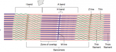
The Sarcomeres that have dark bands are called A bands and the light bands are called I bands.
The A bands are the thick filaments and the I bands are the thin filaments. |
|
|
A single Thin filament contains four proteins, what are they? |
1) F-actin 2) Nebulin 3) Tropomyosin 4) Troponin |
|
|
What is the sliding filament theory? |
When a skeletal muscle fiber contracts, a sarcomere bands narrow. These observations make sense only if the thin filaments are sliding toward the center of each sarcomere, alongside the thick filaments. |
|
|
Where would you expect to find the greatest concentration of Ca2+ in resting skeletal muscle? |
You would expect the greatest concentration of calcium ions in resting skeletal muscle to be in the terminal cisternae of the sarcoplasmic reticulum. |
|
|
What is Acteylycholine (ACh)? |
Acetylcholine (ACh) is a neurotransmitter, a chemical released by a neuron to change the permeability or other properties of another cell’s plasma membrane. |
|
|
The link between the generation of an action potential in the sarcolemma and the start of a muscle contraction is called _______. |
Excitation-Contraction Coupling. *this coupling occurs at the triads. |
|
|
What are the steps that initiate a muscle contraction? |
1) ACh is released at the neuromuscular junction and binds to ACh receptors on the sarcolemma. 2) An action potential is generated and spreads across the membrane surface of the muscle fiber and along the T tubules. 3) The sarcoplasmic reticulum releases stored calcium ions. 4) Calcium ions bind to troponin, exposing the active sites on the thin filaments. Cross-bridges from when myosin heads bind to those active sites. 5) The contraction cycle begins as repeated cycles of cross-bridge binding, pivoting, and detachment occur - all powered by ATP. |
|
|
What are the steps that end a muscle contraction? |
1) ACh is broken down by acetylcholinesterase (AChE), ending action potential generation. 2) Sarcoplasmic reticulum reabsorbs Ca2+. As the calcium ions are reabsorbed, their concentration in the cytosol decreases. 3) Active sites covered, and cross-bridge formation ends. Without calcium ions, the tropomyosin returns to its normal position and active sites are covered again. 4) Contraction ends. Without cross-bridge formation, contraction ends. 5) Muscle relaxation occurs. The muscle returns passively to its resting length. |
|
|
What is treppe? |
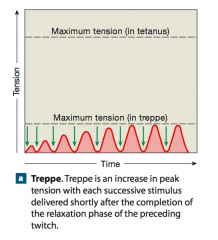
If a skeletal muscle is stimulated a second time immediately after the relaxation phase has ended, the resulting contraction will develop a slightly higher maximum tension than did the first contraction. The rise is thought to result from a gradual increase in the concentration of Ca2+ in the cytosol, in part because the calcium ion pumps in the SR have too little time to recapture the ions between stimulations. |
|
|
Do skeletal muscle go through treppe? |
Most skeletal muscles do not undergo treppe, however treppe is a phenomenon in cardiac muscle. |
|
|
What is wave summation? |
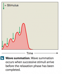
When a second stimulus arrives before the relaxation phase has ended, a second, more powerful contraction occurs. The addition of one twitch to another in this way in the summation of twitches. |
|
|
What is incomplete tetanus? |
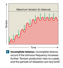
If a stimulation continues and the muscle is never allowed to relax completely, tension will increase until it reaches a peak value roughly four times the maximum produced by treppe. A muscle producing almost peak tension during rapid cycles of contraction and relaxation is in incomplete tetanus. |
|
|
What is complete tetanus? |
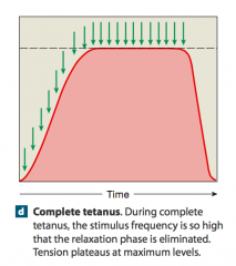
When a higher stimulation frequency eliminates the relaxation phase, complete tetanus occurs. Action potentials arrive so rapidly that the SR does not have time to reclaim the Ca2+. The high Ca2+ concentration in the cytosol prolongs the contraction, making it continuous. |
|
|
What is muscle tone? |
In any skeletal muscle, some motor units are always active, even when the entire muscle is not contracting. Their contractions do not produce enough tension to cause movement, but they do tense and firm the muscle. This resting tension in a skeletal muscle is called muscle tone. Resting muscle tone stabilizes the positions of bones and joints. For example, in muscle involved with balance and posture, enough moto units are stimulated to produce the tension needed to maintain body position. |
|
|
Why does a muscle that has been overstretched produce less tension? |
A muscle's ability to contract depends on the formation of cross-brides between the myosin and actin myofilaments in the muscle. In a muscle that is overstretched, the myofilaments would overlap very little, so very few cross-bridges between myosin and actin could form, and thus the contraction would be weak . If the myofilaments did not overlap at all, then no cross bridges would form and the muscle could not contract. |
|
|
Can a skeletal muscle contract without shortening? |
Yes, a skeletal muscle can contract without shortening. The muscle can shorten (isotonic, concentric contraction), elongate (isotonic, eccentric contraction), or remain the same length (isometric contraction), depending on the relationship between the load (resistance) and tension produced by actin-myosin interactions. |
|
|
True or False. At rest, a skeletal muscle fiber produces more ATP than it needs. |
True. Under these conditions, ATP transfers energy to creatine. |
|
|
What is creatine? |
Creatine is a small molecule that muscle cells assemble from fragments of amino acids. |
|
|
The energy transfer from ATP to creatine creates another high energy compound called what? |
Creatine Phosphate (CP) ATP + CREATINE -> ADP + CREATINE PHOSPHATE |
|
|
What does creatine kinase do? |
It is the enzyme that catalyzes the reaction to convert ADP back to ATP. ADP + CREATINE PHOSPHATE -> ATP + CREATINE |
|
|
For each molecule of pyruvate "fed" into the citric acid cycle, the cell gains ________. |
17 ATP molecules |
|
|
What are the two important drawbacks of the anaerobic production of energy from glycolysis? |
1) The underutilized pyruvate molecules produced through glycolysis are converted to lactic acid. Because it releases hydrogen ions, production of lactic acid can lower the intracellular pH. 2) Glycolysis is a relatively inefficient way to generate ATP. Under anaerobic conditions, each glucose molecule generates 2 pyruvate molecules, which are converted to lactic acid. In cell gains 2 ATP molecules through glycolysis. Had those pyruvate molecules been catabolized aerobically in a mitochondrion, the cell would have produced 34 additional ATP. |
|
|
What is muscle fatigue? |
Muscle fatigue is when a skeletal muscle can no longer perform at the required level of activity. Muscle fatigue has been correlated with 1) depletion of metabolic reserves within the muscle fibers 2) Damage to the sarcolemma and sarcoplasmic reticulum 3) A decline in pH within the muscle fibers and the muscle as a whole, which decreases calcium ion binding to troponin and alters enzyme activities 4) A sense of weariness and a reduction in the desire to continue the activity , due to the effects of low blood pH and sensations of pain. |
|
|
What is a twitch? What are the three phases in a twitch? |
A twitch is a single stimulus-contraction-relaxation sequence in a muscle fiber. The three phases are the latent period, the contraction phase, and the relaxation phase. |
|
|
What is the difference between the latent period, the contraction phase, and the relaxation phase? |
In the latent period, stimulation being and lasts about 2msec. In the contraction phase, tension increases to a peak. As the tension increases, calcium ions are binding to troponin, active sites on thin filaments are being exposed, and cross bridge interactions are occurring. In the relaxation phase, the Ca2+ levels are decreasing, active sites are being covered by tropomyosin, and the number of active cross bridges is declining as they detach. |
|
|
What is a motor unit? |
All the muscle fibers controlled by a single motor neuron constitute a motor unit. |
|
|
What is the name of the structure that contains gap junctions and desmosomes and joins the plasma membranes of two cardiac muscle cells together? |
Intercalated Discs |
|
|
What is plasticity? |
The ability to function over a wide range of lengths. |
|
|
Thin filaments are composed primarily of ______. |
Actin. |
|
|
Thick filaments are composed primarily of ______. |
Myosin. |
|
|
True or False. The arrangement of myosin and actin fibers in skeletal, cardiac, and smooth muscles is identical. |
False. While skeletal and smooth muscle have thick and thin filaments that are arranged similarly, smooth muscle filaments have a more scattered arrangement that affects the shape and the muscle cell when it contracts. |
|
|
Nerves and blood vessels are contained within the connective tissues of the _________. |
All three layers. Epimysium, perimysium, and endomysium. |
|
|
The thin filaments consist of _________. |
A pair of F-Actin molecules twisted together. |
|
|
The thick filaments consists of _______. |
About 300 myosin molecules twisted around one another. |
|
|
Why is control over leg muscles less precise than control over the muscles of the eye? |
Many muscle fibers are controlled by a SINGLE motor neuron. |
|
|
The amount of tension produced by an individual muscle fiber ultimately depends on the __________. |
Number of pivoting cross-bridges. |
|
|
In an isotonic contraction, _______. |
Cross bridges must produce ENOUGH tension to EXCEED the load to be moved. |
|
|
What is an example of isometric contraction? |
Holding a heavy stack of books above the ground. *no skeletal structures are moved. |
|
|
What type of muscle tissue do not contain sacomeres? |
Smooth |
|
|
Structurally, how do smooth muscle cells differ from skeletal muscle cells? |
Smooth muscle cells lack myofibrils and sacomeres. |
|
|
What is necessary for smooth muscle contraction? |
Calcium ions must interact with calmodulin to trigger muscle contraction. *This involves a protein not found in skeletal muscle. |
|
|
The cardiovascular system uses which types of muscle? |
Cardiac and smooth |
|
|
The area of the A band in the sarcomere consists of _________. |
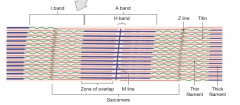
* M Line * H Line * Zone of Overlap |
|
|
Excitation-contraction coupling forms the link between __________. |
Electrical activity in the sarcolemma and the initiation of a contraction. |
|
|
What are the two mechanisms used to generate ATP from glucose? |
* Aerobic respiration * Glycolysis |
|
|
In glycolysis, glucose is broken down to pyruvic acid, which is converted to ______. |
Lactic Acid |
|
|
Which contraction does not involve bones moving? |
Isometric |
|
|
Do smooth muscle fibers have multiple nucleus? |
No. Each cell is spindle shaped and has a single, centrally located nucleus. |
|
|
Do smoother muscle fibers have T tubules? |
A smooth muscle fiber has no T tubules, and the sarcoplasmic reticulum forms a loose network throughout the sarcoplasm. |
|
|
Why are smooth muscles nonstriated? |
They are nonstriated because they lack myofibrils and sarcomeres. |
|
|
Which of the three types of muscles have the most cells? |
Smooth muscle cells have more myosin heads per thick filament. |
|
|
How is the excitation-contraction coupling in smooth muscle different than that of skeletal and cardiac muscle? |
The trigger for smoother muscle contraction is the appearance of free calcium ions in the cytoplasm. On stimulation, a surge of calcium ions enters the cell from the extracellular fluid, and the sarcoplasmic reticulum release additional calcium ions. One in the sarcoplasm, the calcium ions interact with calmodulin, a calcium-binding protein. Calmodulin then activates the enzyme myosin light chain kinase, which in turn enables myosin heads to attach to actin. ƒa band |