![]()
![]()
![]()
Use LEFT and RIGHT arrow keys to navigate between flashcards;
Use UP and DOWN arrow keys to flip the card;
H to show hint;
A reads text to speech;
207 Cards in this Set
- Front
- Back
|
VORTEX |
The large area of hair that slants toward the umbilicus
|
|
|
COMMON INTEGUMENT- EPIDERMIS, DERMIS |
Fold of skin which consists of an outer epithelium, the epidermis, and an underlying thicker layer of connective tissue, the dermis |
|
|
AREOLAR TISSUE |
Areolar tissue appears as a thin layer of loose, irregularly arranged connective tissue that often contains fat |
|
|
APONEUROSIS |
It has the same consistency as a tendon but the fibrous tissue is arranged as a thin sheet of tissue |
|
|
ORIGIN |
The more proximal attachment of the muscle that moves the least |
|
|
INSERTION |
The more distal attachment of the muscle or the part that moves the most |
|
|
TENDON |
It consists of dense, regularly arranged fibrous connective tissue organized into a small, well defined bundle |
|
|
LIGAMENT |
It is a dense fibrous connective tissue between bones, although the term may also be used to describe thin fibrous connections between organs or between an organ and the body wall |
|
|
CUTANEUS TRUNCI |
It is a thin sheet of muscle that covers most of the dorsal, lateral, and ventral walls of the the thorax and abdomen. It has no direct obny attachments Innervation: Lateral Thoracic Nerve |
|
|
FASCIA |
It is deeper than the areolar tissue and more regularly arranged thin layer of connective tissue that envelops the body beneath the skin and encloses individual muscles or groups of muscles. |
|
|
SUPERFICIAL AND DEEP FASCIA |
The superficial is deep to the areolar, forming the deep portion of the subcutneous tissue that covers the entire body. It blends with the deep fascia that is more firmly attached to the muscles. |
|
|
PECTORAL MUSCLES- SUPERFICIAL PECTORAL (2) DESCENDING PECTORAL TRANSVERSE PECTORAL |
ORIGIN: The first two sternebrae and usually a part of the third, fibrous raphe between adjacent muscles
INSERTION: The whole crest of the greater tubercle of the humerus
ACTION: To adduct the limb when it is not bearing weight or to prevent the limb from being abducted when bearing weight
INNERVATION: Cranial pectoral nerve |
|
|
DEEP PECTORAL MUSCLE
|
ORIGIN: The ventral part of the sternum and fibrous raphe between fellow muscles; the deep abdominal fascia in the region of the xiphoid cartilage
INSERTION: Majorty ison the lesser tubercule of the humerus;an aponeurosis to the greater tubercule and its crest; the caudal part ot the medial brachial fascia
ACTION: When the limb is advanced and fixed: to pull the trunk cranially and to extend the shoulder joint. When the limb is not supporting weight: to draw the limb caudally and flex the shoulder joint. To adduct the limb
INNERVATION: Caudal pectoral nerves |
|
|
BRACHIOCEPHALICUS |
ATTACHMENTS: All attachments are movable but the clavicle or clavicular intersection is considered the origin. The cleidobrachialis attaches to the distal end of the cranial border of the humerus. The cervical part of the cleidocephalicus attaches to the cranial half of the mid-dorsal fibrous raphe and sometimes to the nuchal crest of the occipital bone. Its mastoid part attaches to the mastoid part of the temporal bone with the sternomastoideus muscle
ACTION: To advance the limb; to extend the shoulder joint and draw the neck and head to the side
INNERVATION: Accessory nerve and ventral branches of cervical spinal nerves |
|
|
STERNOCEPHALICUS |
INSERTION: The mastoid part of the temporal bone and the nuchal crest of the occipital bone
ACTION: To draw the head and neck to the side
INNERVATION: Accessory nerve and ventral branches of cervical spinal nerves |
|
|
STERNOTHYROIDEUS |
ORIGIN: The first costal cartilage
INSERTION: The caudolateral surface of the thyroid cartilage
ACTION: To draw the larynx and tongue caudally
INNERVATION: Ventral branches of cervical spinal nerves |
|
|
STERNOHYOIDEUS |
ORIGIN: The first sternebra and the first costal cartilage
INSERTION: The basihyoid bone
ACTION: To pull the tongue and larynx caudally
INNERVATION: Ventral branches of cervical spinal nerves |
|
|
OMOTRANSVERSRIUS |
ATTACHMENTS: The distal end of the spine of the scapula; cranially, the transverse wing of the atlas
ACTION: To advance the limb or flex the neck laterally
INNERVATION: Accessory nerve |
|
|
TRAPEZIUS |
ORIGIN: The median raphe of the neck and the supraspinous ligament from the level of the third cervical vertebra to the level of the ninth thoracic vertebra
INSERTION: The spine of the scapula
ACTION: To elevate and abduct the forelimb
INNERVATION: Accessory nerve |
|
|
RHOMBOIDEUS- RHOMBOIDEUS CAPITIS RHOMBOIDEUS CERVICIS RHOMBOIDEUS THORACIS |
ORIGIN: The nuchal crest of the occipital bone; the median fibrous raphe of the neck; the spinous processes of the first seven thoracic vertebrae
INSERTION: The dorsal border and adjacent surfaces of the scapula
ACTION: To elevate the forelimb and draw the scapula against the trunk
INNERVATION: Ventral branches of cervical and thoracic spinal nerves |
|
|
LATISSIMUS DORSI |
ORIGIN: The thoracolumbar fascia from the spinous processes of the lumbar and the last seven or eight thoracic vertebrae; a muscular attachment to the last two or three ribs
INSERTION: The teres major tuberosity of the humerus and the teres major tendon
ACTION: To draw the free limb caudally as in digging; to flex the shoulder joint
INNERVATION: Thoracodorsal nerve |
|
|
SERRATUS VENTRALIS CERVICIS SERRATUS VENTRALIS THORACIS |
ORIGIN: The transverse processes of the last five cervical vertebrae and the first seven or eight ribs ventral to their middle
INSERTION: The dorsomedial third of the scapula (serrated face)
ACTION: To support the trunk and depress the scapula
INNERVATION: Ventral branches of cervical spinal nerves and the long thoracic nerve |
|
|
DELTOIDEUS |
ORIGIN: The spine and acromial process of the scapula
INSERTION: The deltoid tuberosity
ACTION: to flex the shoulder
INNERVATION: Axillary nerve |
|
|
INFRASPINATUS |
ORIGIN: The infraspinous fossa
INSERTION: A small, circumscribed area on the lateral side of the greater tubercule of the humerus.
ACTION: To extend or flex the joint depending on position of joint. To abduct the shoulder and to rotate the shoulder laterally. To prevent medial rotation when weight bearing and provide lateral stability to the shoulder joint
INNERVATION: Suprascapular nerve |
|
|
TERES MINOR |
ORIGIN: The infraglenoid tubercule and distal third of the caudal border of the scapula
INSERTION: The teres minor tuberosity of the humerus
ACTION: To flex the shoulder, rotate the shoulder laterally, prevent medial rotation when bearing weight
INNERVATION: |
|
|
SUPRASPINATUS |
ORIGIN: The supraspinous fossa
INSERTION: The greater tubercule of the humerus by a thick tendon
ACTION: To extend and stabalize the shoulder joint
INNERVATION: Suprascapular nerve |
|
|
SUBTENDINOUS SYNOVIAL BURSA |
A bursa is a closed connective tissue sac containing synovial fluid, which reduces friction |
|
|
SUBSCAPULARIS |
ORIGIN: The subscapular fossa
INSERTION: The lesser tubercle of the humerus
ACTION: To adduct, extend, and medially stabilize the shoulder joint. To rotate the shoulder medially and prevent lateral rotation when bearing weight
INNERVATION: Subscapular nerve |
|
|
TERES MAJOR |
ORIGIN:
INSERTION: The crest of the lesser tubercule of the humerus proximal to the teres major tuberosity
ACTION: To adduct, extend, and stabalize the shoulder joint
INNERVATION: Musculocutaneous nerve |
|
|
TENSOR FASCIAE ANTEBRACHII |
ORIGIN: The fascia covering the lateral side of the latissimus dorsi
INSERTION: The olecranon
ACTION: To extend the elbow
INNERVATION: Radial nerve |
|
|
TRICEPS BRACHII- LONG HEAD |
ORIGIN: The caudal border of the scapula
INSERTION: The olecranon tuber
ACTION: To extend the elbow and flex the shoulder
INNERVATION: Radial nerve |
|
|
TRICEPS BRACHII-LATERAL HEAD |
ORIGIN: The tricipital line of the humerus
INSERTION: The olecranon tuber
ACTION: To extend the elbow
INNERVATION: Radial nerve |
|
|
TRICEPS BRACHII- ACCESSORY HEAD |
ORIGIN: The neck of the humerus
INSERTION: The olecranon tuber
ACTION: To extend the elbow
INNERVATION: Radial nerve |
|
|
TRICEPS BRACHII- MEDIAL HEAD |
ORIGIN: The crest of the lesser tubercle near the teres major tuberosity
INSERTION: The olecranon
ACTION: To extend the elbow
INNERVATION: Radial nerve |
|
|
ACONEUS |
ORIGIN: The lateral supracondylar crest and the lateral and medial epicondyles of the humerus
INSERTION: The lateral surface of the proximal end of the ulna (the olecranon)
ACTION: To extend the elbow
INNERVATION: Radial nerve |
|
|
BICEPS BRACHII |
ORIGIN: The supraglenoid tubercule
INSERTION: The ulnar and radial tuberosities
ACTION: To flex the elbow and extend the shoulder
INNERVATION: Musculocutaneous nerve |
|
|
BRACHIALIS |
ORIGIN: The proximal third of the lateral surface of the humerus
INSERTION: The ulnar and radial tuberosities
ACTION: To flex the elbow
INNERVATION: Musculocutaneous nerve |
|
|
EXTENSOR CARPI RADIALIS |
ORIGIN: The lateral supracondylar crest
INSERTION: The small tuberosities on the dorsal surfaces of the base of metacarpals II and III
ACTION: To extend the carpus
INNERVATION: Radial nerve |
|
|
COMMON DIGITAL EXTENSOR |
ORIGIN: The lateral epicondyle of the humerus
INSERTION: The extensor processes of the distal phalanges of digits II, III, IV, and V
ACTION: To extend the joints of the four principal digits and the carpus
INNERVATION: Radial nerve |
|
|
LATERAL DIGITAL EXTENSOR |
ORIGIN: The lateral epicondyle of the humerus
INSERTION: The proximal ends of all the phalanges of digits III, IV, and V, but mainly the extensor processes of the distal phalanges of these digits
ACTION: To extend the carpus and joints of digits III, IV, V
INNERVATION: Radial nerve |
|
|
ULNARIS LATERALIS |
ORIGIN: The lateral epicondyle of the humerus
INSERTION: The lateral aspect of the proximal end of metacarpal V and the accessory carpal bone
ACTION: To abduct the carpal joint and support the carpus when extended to support weight
INNERVATION: Radial nerve |
|
|
SUPINATOR |
ORIGIN: The lateral epicondyle of the humerus
INSERTION: The cranial surface of the proximal fourth of the radius
ACTION: To rotate the forearm laterally so that the palmar side of the paw faces medially (supination); to flex the elbow
INNERVATION: Radial nerve |
|
|
ABDUCTOR DIGITI I LONGUS |
ORIGIN: The lateral border and cranial surface of the body of the ulna; the interosseous membrane
INSERTION: The proximal end of metacarpal I
ACTION: To abduct the first digit or pollex and extend the carpal joint
INNERVATION: Radial nerve |
|
|
PRONATOR TERES |
ORIGIN: The medial epicondyle of the humerus
INSERTION: The medial border of the radius between the proximal and middle thirds
ACTION: To rotate the forearm medially so that the palmar side of the paw faces the ground (pronation); to flex the elbow
INNERVATION: Median nerve |
|
|
FLEXOR CARPI RADIALIS |
ORIGIN: The medial epicondyle of the humerus and the medial border of the radius
INSERTION: The palmar side of the base of metacarpals II and III
ACTION: To flex the carpus
INNERVATION: Median nerve |
|
|
SUPERFICIAL DIGITAL FLEXOR |
ORIGIN: The medial epicondyle of the humerus
INSERTION: The palmar surface of the base (proximal end) of the middle phalanges of digits II, III, IV, and V
ACTION: To flex the carpal, metacarpophalangeal, and proximal interphalangeal joints of digits II, III, IV, and V
INNERVATION: Medial nerve |
|
|
FLEXOR CARPI ULNARIS- ULNAR HEAD, HUMERAL HEAD |
ORIGIN: Ulnar head- the caudal border and medial surface of the olecranon; humeral head- the medial epicondyle of the humerus
INSERTION: The accessory carpal bone
ACTION: To flex the carpus
INNERVATION: Ulnar nerve |
|
|
DEEP DIGITAL FLEXOR- HUMERAL HEAD, ULNAR HEAD, RADIAL HEAD |
ORIGIN: Humeral head- the medial epicondyle of the humerus, ulnar head- the proximal three fourths of the caudal border of the ulna; radial head- the middle third of the medial border of the radius
INSERTION: The flexor tubercule on the palmar surface of the distal phalanx of each digit
ACTION: To flex the carpal and metacarpophalangeal joints and the proximal and distal interphalangeal joints of the digit
INNERVATION: |
|
|
PRONATOR QUADRATUS |
ATTACHMENTS: The apposed surfaces of the radius and ulna
ACTION: To pronate the paw
INNERVATION: Median nerve |
|
|
FLEXOR MANICA |
At the metacarpophalangeal joint, it lies around the deep flexor tendon that passes through it forming a collar |
|
|
DIGITAL SYNOVIAL SHEATH |
The superficial flexor tendon are in a common synovial membrane called the digital synovial sheath |
|
|
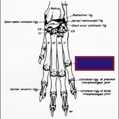
PALMAR ANNULAR LIGAMENT |
It firmly holds the superficial and deep digital flexor tendons in place at the metacarpophalangeal joint. It crosses the flexor manica |
|
|
FLEXOR RETINACULUM |
The three tendons (humeral head, ulnar head, radial head) all fuse to form a single tendon. This tendon is head in place in the carpal canal by the thick, deep part of the fibrous flexor retinaculum |
|
|
CARPAL CANAL |
It is formed by the accessory carpal bone laterally, the palmar carpal ligament and the carpal bones dorsally, and the flexor retinaculum on the palmar suface |
|
|
ANNULAR DIGITAL LIGAMENTS |
It supports the deep digital flexor tendon proximal and distal to the palmar surface of the proximal interphalangeal joint |
|
|
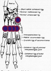
MEDIAL and LATERAL GLENOHUMERAL LIGAMENTS |
It is the poorly developed thickenings of fibrous part of the humeral joint capsule on each side |
|
|
TRANSVERSE HUMERAL RETINACULUM |
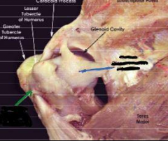
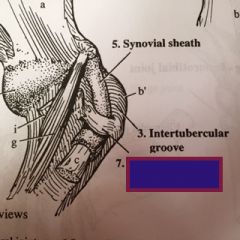
It is the collagenous thickening across the tendon of origin of the biceps at the intertubercular groove |
|
|
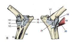
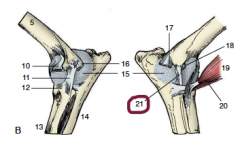
ELBOW JOINT |
It is a hinge joint formed by the condyle of the humerus, the head of the radius, and the trochlear notch of the ulna |
|
|
LATERAL and MEDIAL COLLATERAL LIGAMENTS |
They are the pronounced thickenings in the fibrous layer of the elbow joint capsule. |
|
|
INTEROSSEOUS LIGAMENT |
It is a condensation of collagenous tissue that unites the radius and ulna proximally |
|
|
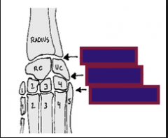
ANTEBRACHIOCARPAL JOINT |
The proximal carpal joint between the radius and ulna articulating with the intermedioradial and ulnar carpal bones. The antebrachiocarpal joint does not communicate with the other two joints of the carpus |
|
|
MIDDLE CARPAL JOINT |
The middle carpal joint between the two rows of carpal bones. The carpometacarpal and middle carpal joint compartments communicate between the distal row of carpal bones |
|
|
CARPOMETACARPAL JOINT |
The distal carpal joint between the distal row of carpal bones and the metacarpals. The carpometacarpal and middle carpal joint compartments communicate between the distal row of carpal bones |
|
|
METACARPOPHALANGEAL, PROXIMAL INTERPHALANGEAL, DISTAL INTERPHALANGEAL JOINTS |
These are the three articulations of each main digit. Medial and lateral collateral ligaments support these joints |
|
Name the arrows
|
Carpus joint, metacarpal bones, phalanges Rights side: metacarpophalangeal joints, interphalangeal joints |
|
|
Axilla |
Arm pit |
|
|
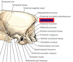
Nuchal Crest |
|

|
Wing of the atlas (C1) |
|
|
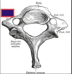
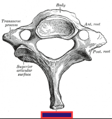
Transverse process of C3-C7 (cervical vertebrae of the neck)- (divided into ventral and dorsal tubercles) |
|
|
Transverse process (divided into ventral and dorsal tubercles) |
|
|
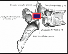
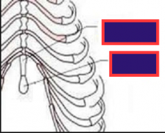
Spinous process of T1- L6 |
|
|
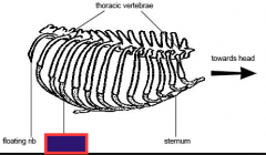
Sternum (forms floor of the thorax) |
|
|
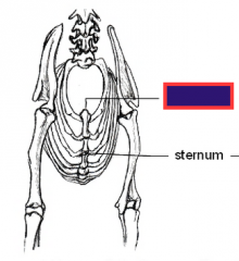
Manubrium (expanded first sternebra) |
|
|
xiphoid process (above) (the last sternebra which is capped by the xiphoid cartilage) xiphoid cartilage (below) |
|
|
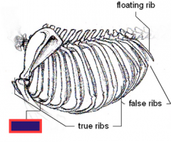
Costal Cartilage |
|
|
Clavicular intersection (on the brachiocephalicus muscle- above cleidocephalicus and below cleidobrachialis) |
|
|
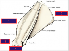
1. Spine 2. Cranial border 3. Dorsal border 4. Caudal border 5. Acromion |
|
|
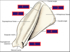
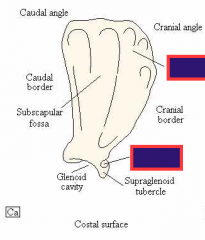
1. Supraspinous fossae 2. Infraspinous fossae 3. Superglenoid tubercle 4. Glenoid cavity |
|
|
Serrated surface (above) Coracoid process (below) |
|
|
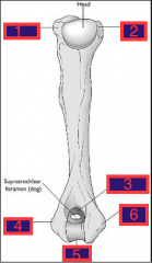
1. Greater Tubercle 2. Lesser Tubercle 3. Olecranon fossa 4. Lateral epicondyle 5. Condyle 6. Medial epicondyle |
|

|
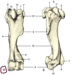
13. Intertubercular groove 4. Teres major tuberosity 5. Deltoid tuberosity b. Tricipital line a. Lateral epicondylar crest c. crest of greater tubercle (2. Head) |
|
|
formed by the ridge connecting the deltoid tuberosity to the caudal portion of the greater tubercle; lateral head of triceps arises here |
Tricipital line |
|
|
distal to brachialis groove on humerus, extends distally to the lateral epicondyle; area of insertion of the extensor carpi radialis and part of the anconeus |
lateral epicondylar crest |
|
|
Humeral shaft (cranial border) |
|
|
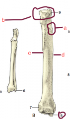
Caudal surface of the radius b. Head a. Radial tuberosity c. Lateral border d. Medial border 8. medial styloid process |
|

|
Cranial view of ulna Caudal border (can see attached to radius- smaller picture) b. Ulnar tuberosity 6. Lateral styloid process a. Olecranon (caudal border) c.Olecranon (medial border) 4'.Medial coronoid process 4. Lateral coronoid process 2. anoconeal process |
|
|
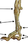
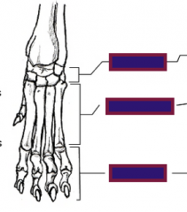
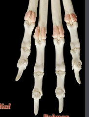
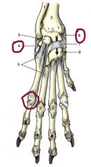
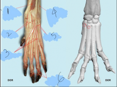
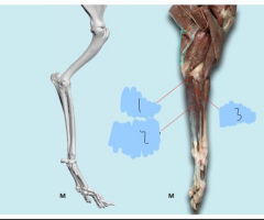
1. Carpals 2. Metacarpals 3. Phalanges |
|
|
Metacarpals I- IV (dew claw is I, so right to left I- IV) Tuberosities of metacarpals II & III |
|
|
3. Accessory Carpal 4. Ulnar Carpal 5. Radial carpal (intermedioradial) |
|
|
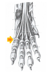
Metacarpus
Located in the interosseous tendons on the palmar surface of each metacarpophalangeal joint (digits II-V) |
|

|
Glenohumeral aka Shoulder joint (links the scapula and humerus) |
|
|
Elbow joint (links the radius and ulna) |
|
|
Carpal joints: 1. Antebrachiaocarpal 2. Middle carpal 3. Carpometacarpal |
|
|
Metacarpophalangeal joints |
|

|
Interphalangeal joints: proximal distal |
|

|
Lateral collateral ligaments of elbow |
|
|
Medial collateral ligaments of elbow |
|
|
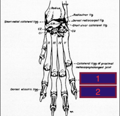
4. Lateral collateral ligament (carpal) 8. Medial collateral ligament (carpal) Collateral ligaments of metacarpophalangeal joints (specifically of the proximal joint) |
|
|
Collateral ligaments of interphalangeal joints 1. proximal 2. distal |
|
|
Palmar annular ligaments |
|
|
Annular digital ligaments (proximal and distal) -->(hold deep digital flexor and superficial digital flexor tendons in place??) |
|
|
Annular ligaments (holds tendons in place) |
|
|
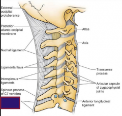
Supraspinous Ligament |
Heavy band of connective tissue running over the tops of the spinous processes from the first thoracic vertebra to the caudal vertebrae. It prevents abnormal separation of the vertebral spines during flexion of the vertebral column. It is the direct continuation of the funicular part of the nuchal ligament. |
|
|
Supraspinous Ligament |
|
|
Glenohumeral ligaments |
Thickening of the joint capsule, not true collateral ligaments |
|
medial shoulder view |
Medial glenohumeral ligament |
|
lateral shoulder view |
Lateral glenohumeral ligament |
|
|
Transverse humeral ligament |
attaches to the greater and lesser tubercles and holds the biceps tendon in the intertubercular (bicipital groove) |
|

|
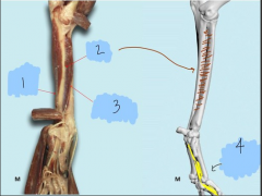
orange- antebrachial joint purple- middle carpal joint green- carpometacarpal joint |
|
|
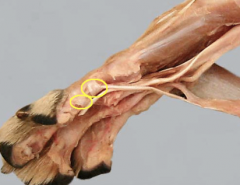
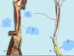
blue=medial glenohumeral ligament. green= transverse humeral retinaculum |
|
|
Transverse humeral ligament |
|
|
1. Annular ligament 2. lateral collateral ligament (caudal and cranial crura) 3. Interosseous membrane 4. Interosseous ligament |
|

|
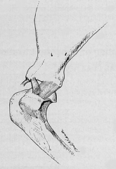
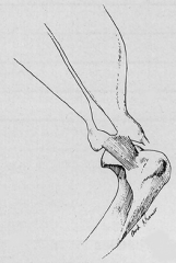
1. Dorsal elastic ligament 2. Collateral ligarment |
|

|
Dorsal elastic ligament |
|

|
Cutaneous trunci m. |
|
|
Extrinsic muscles, superficial |
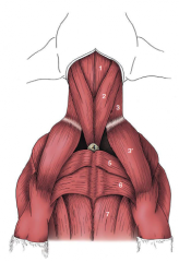
* Trapezius m. (cervicis and thoracis) * Brachiocephalicus m. (cleidobrachialis, cleidocervicalis, cleidomastoideus) * Latissimus dorsi m. * Pectoral mm. (superficial and deep) * Omotransversarius m.
|
|
|
Extrinsic muscles, deep |
Rhomboideus m. (capitis, cervicis, and thoracis) Serratus ventralis m. |
|
|
Intrinsic muscles, shoulder |
Deltoideus m. Teres major m. Infraspinatus m. Supraspinatus m. Subscapularis m. Triceps brachii m. (long, lateral, medial, and accessory heads) Biceps brachii m. Coracobrachialis m. Teres minor m. |
|
|
Intrinsic muscles, elbow |
Biceps and triceps (cross two joints) Brachialis m. Anconeus m. Supinator m. Pronator teres m. |
|
|
Extensors of carpus and digits |
Extensor carpi radialis m. Common digital extensor m. Lateral digital extensor m.
|
|
|
Flexors of carpus and digits |
Flexor carpi ulnaris m. Superficial digital flexor m. Flexor carpi ulnaris m. (ulnar and humeral heads) Deep digital flexor m. (humeral, radial, and ulnar heads) Ulnaris lateralis m. |
|
1? |
1. Abductor pollicis longus m. |
|
2? |
Pronator quadratus m. |
|
4? |
Interossei mm. |
|
|
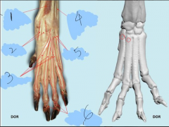
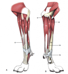
1. abductor pollicus longus m. 2. extensor carpi radialis m. 3. common digital extensor m. 4. ulnaris lateralis m. (flexor) 5. lateral digital extensor 6. dorsal ligament |
|

|
1. deep digital flexor (ulnar head) 2. pronator quadratus 3. deep digital flexor (radial head) 4. interossei |
|
|
1. extensor carpi radialis m. 2. common digital extensor m. 3. lateral digital extensor m. 4. ulnaris lateralis m. (flexor) |
|
|
1. flexor carpi ulnaris m. (flex carpus) 2. superficial digital flexor m. 3. flexor carpi radialis m. |
|
|
trapezius m. (two parts: cervicis and thoracis separated by an aponeurosis) |
|
|
Omotransversarius |
|

|
Brachiocephalicus (cleidobrachialis, cleidocervicalis, cleidomastoideus) |
|
|
Superficial pectoral mm. |
|
|
Deep pectoral mm. |
|
|
Latissimus dorsi m. |
|
|
Rhomboideus capitis m. |
|
|
Rhomboideus cervicis m. |
|

|
Rhomboideus thoracis m. |
|
|
Serratus ventralis m. |
|
|
Serratus ventralis m. |
|
1, 2, 3, 5, 8, 9, 12, 13 |
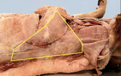
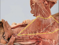
1. Rhomboideus 2. Trapezius 3. Nuchal ligament 5. Omotransversarius 8. brachiocephalicus 9. external jugular vein in jugular groove 12. trachea 13. esophagus |
|
|
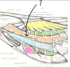
1. Combined sternohyoideus and sternothyroideus 2. sternocephalicus 3, 3'. Brachiocephalicus (cleidocervicalis, cleidobrachialis) 4. manubrium of sternum 5. pectoralis descendens 6. pectoralis transversus 7. pectoralis profundus |
|
|
1. External jugular vein 2. Groove formed by the brachiocephalic muscle 3. sternocephalic muscle |
|
|
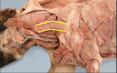
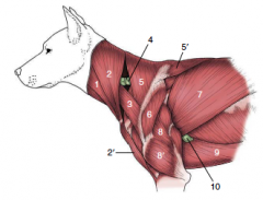
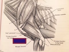
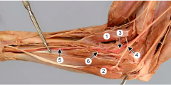
1. Sternocephalicus 2, 2'. Brachiocephalicus: cleidocervicalis and cleidobrachialis 3. Omotransversarius 4. Superficial cervical lymph node 5, 5'. Cervical and thoracic parts of the trapezius 6. Deltoideus 7. Latissimus dorsi 8, 8'. Long and lateral heads of triceps 9. pectoralis profundus (ascendens) 10. accessory axillary lymph node |
|
|
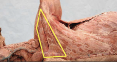
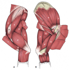
1. Rhomboideus 2. Teres major 3. Supraspinatus 4, 4'. Scapular and acromial parts of the deltoideus 5. Latissimus dorsi 6, 6', 6''. Long, lateral, and medial heads of triceps 7. Brachiocephalicus 8. Brachialis 9. Subscapularis 10. Corabrachialis 11. Tensor fasciae antebrachii 12. Biceps brachii |
|

|
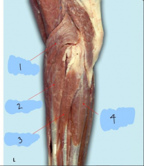
Anconeus |
|

|
Brachialis m. |
|
|
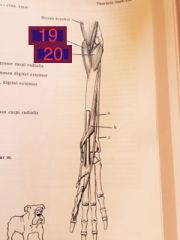
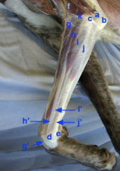
19. Pronator teres m. 20. Supinator |
|
|
Brachialis |
|
7? 15? |
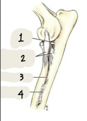
7. Extensor retinaculum 15. Flexor retinaculum |
|
|
Superficial antebrachial fascia. The antebrachial muscles are enclosed within the antebrachial fascia, cut at k where brachial muscles insert onto it. |
|
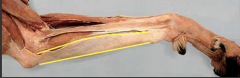
what is the proximal attachment of this muscle? Name the muscle. |
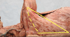
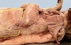
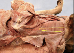
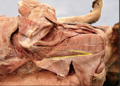
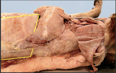
Thoracolumbar fascia. Latissimus dorsi. |
|
|
Thoracolumbar fascia |
the deep fascia of the trunk. Arises from the supraspinous ligament and spines of the thoracic and lumbar vertebrae. Fuses with the opposite fascia on the mid-ventral raphe (linea alba) |
|
|
Linea Alba |
Mid-ventral raphe. The fibrous cord formed by the joining of the aponeuroses of the abdominal muscles from both sides. It is on the ventral midline, extending from the xiphoid cartilage to the pelvic symphysis. |
|
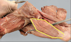
Name and what is it? |
Deep antebrachial fascia. A dense sleeve for the muscles of the forearm on the caudal surface. Unites with the periosteum of the radius. |
|
|
This type of fascia is deep to the areolar tissue, forming the deep portion of the subcutaneous tissue that covers the entire body. |
Superficial fascia |
|
|
This type of fascia is more firmly attached to the muscle that it encloses. |
Deep fascia |
|
|
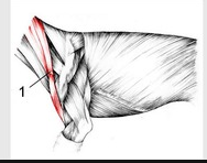
Tensor Fasciae Antebrachii. Long thin strap |
|
|
Ventral median raphe |
Runs from between the bones of the mandible |
|
|
Dorsal median raphe |

Insertion of the brachiocephalicus muscle.
|
|
|
Carpal canal |
Formed by accessory carpal bone laterally, the other carpal bones dorsally and the flexor retinaculum on the palmar side. Structures passing through the carpal canal: Tendons and synovial sheaths of the superficial and deep digital flexors, ulnar and median nerves, arteries and veins. |
|
|
1. Subscapular artery |
|
|
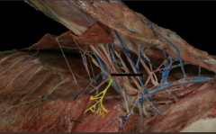
1. Axillary nerve 2. Radial nerve |
|

|
1. Radial n. |
|
|
Nerves of the brachial plexus |
1. Long thoracic - to serratus ventralis |
|

|
Brachial plexus (C5- T2 spinal nerves) |
|
|
Suprascapular nerve |
|

|
Subscapular nerve - Subscapularis m. |
|
|
Suprascapular nerve
|
|

|
Suprascapular nerve |
|
|
Courses between the Supraspinatus and Subscapularis m. near the neck of the scapula
- Supraspinatus |
Suprascapular nerve |
|

|
Subscapular nerve - Subscapularis m. |
|

|
Subscapular nerve - Subscapularis m. |
|
|
Lies between the Biceps brachii cranially and the Brachial vessels caudally. Courses deep to the insertion of the Biceps m.
- Coracobrachialis |
Musculocutaneous nerve |
|
|
Musculocutaneous nerve |
|
|
Musculocutaneous nerve |
|
|
Enters the space between the Subscapularis and Teres major m |
Axillary Nerve |
|
|
Axillary nerve |
|

|
Axillary nerve |
|
|
Arises from the same trunk as the Ulnar n. Runs to the ante brachium in contact with the caudal surface of the Brachial a. |
Median nerve |
|
|
Median nerve |
|

|
Median nerve |
|
|
Separates from the Median n. in the distal arm and crosses the elbow caudal to the medial epicondyle of the humerus. |
Ulnar nerve |
|
|
Ulnar nerve |
|

|
Ulnar nerve |
|
|
Leaves the Ulnar n. near the middle of the arm and runs caudaodistally across the medial surface of the Triceps and olecranon |
Caudal cutaneous antebrachial nerve |
|
|
Caudal cutaneous antebrachial nerve |
|
|
Caudal pectoral nerve |
|
|
Leaves the caudal portion of the brachial plexus and courses caudally between the Latissimus dorsi and Deep pectoris |
Lateral thoracic nerve |
|

|
Lateral thoracic nerve |
|
|
Lateral thoracic nerve |
|
|
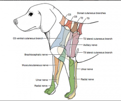
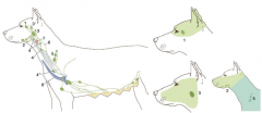
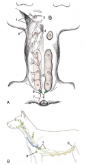
Autonomous zones |
|
|
|
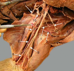
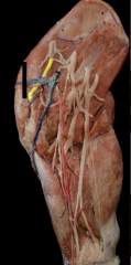
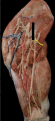
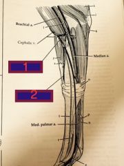
1. Cephalic vein 2. superficial radial nerve 3. axillobrachial vein 4. cephalic vein 5. external jugular vein * cleidobrachialis muscle |
|
probe as well? |
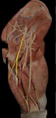
1. pronator teres muscle 2. carpi radialis m 3. brachial artery 4. median nerve 5. median artery 6. antebrachial artery probe- median artery and median nerve |
|

|
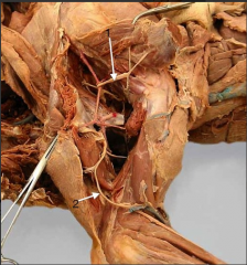
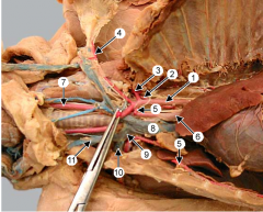
1. axillobrachial vein (joins the brachial vein to form the) 2. axillary vein 3. omobrachial vein 4. cephalic vein 5. external jugular vein 6. axillary lymph node (landmark for the lateral thoracic vessels and nerve which run past it. 7. Pectoral nn. and external thoracic vessels 8. Pectoral muscles 9. carotid artery 10. vagosympathetic trunk 11. Recurrent laryngeal nerve 12. trachea
|
|
|
During clinical examination any difficulties in turning the neck or muscle atrophy around the dorsal and ventral neck may indicate a problem with the accessory nerve. The dorsal branch innervates the brachiocephalicus, trapezius and omotransversarius muscles of the dorsal neck. The ventral branch innervates the sternocephalicus muscle. |
Accessory nerve (CN XI (11)) |
|

|
1. median artery |
|
4? |
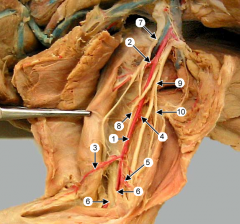
1. Left Subclavian artery 2. Vertebral artery 3. Costocervical trunk 4. Superficial cervical artery 5. internal thoracic artery 6. Brachiocephalic trunk 7. common carotid arteries |
|
5? 3? 4? |
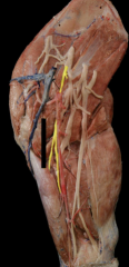
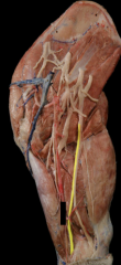
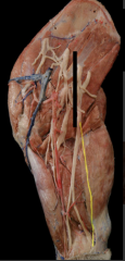
1. Brachial artery 2. Bicipital artery 3. Superficial brachial artery 4. Deep brachial artery 5. Collateral ulnar artery 6. Supracondylar foramen of the humerus 7. musculocutaneous nerve 8. median nerve 9. ulnar nerve |
|
5? |
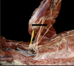
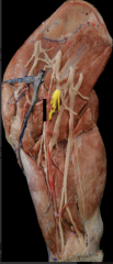
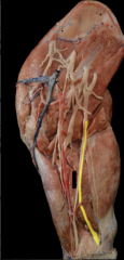
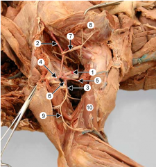
1. Axillary artery 2. Subscapular artery 3. Brachial artery 4. Thoracodorsal artery 5. Caudal circumflex humeral artery 6. Cranial circumflex humeral artery 7. Axillary nerve 8. Deltoideus muscle 9. Radial nerve 10. Brachialis muscle |
|
|
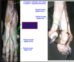
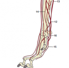
Dorsal common digital artery, vein, and nerve |
|

|
F. Suprascapular artery O. Bicipital artery P. Collateral ulnar artery |
|

|
o. Radial artery 4. Common interosseus artery 5. Caudal interosseus artery |
|
|
1.Median cubital 2. Accessory cephalic |
|
15? |
15. Superficial palmar arch |
|
15? |
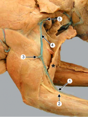
15. Superficial cervical lymph nodes |
|
5? |
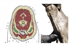
5. Superficial cervical lymph nodes |
|
1? |
1. Axillary lymph nodes |