![]()
![]()
![]()
Use LEFT and RIGHT arrow keys to navigate between flashcards;
Use UP and DOWN arrow keys to flip the card;
H to show hint;
A reads text to speech;
45 Cards in this Set
- Front
- Back
|
Central Nervous System (CNS) |
brain and spinal cord |
|
|
Peripheral Nervous System (CNS) |
cranial and spinal nerves and their branches |
|
|
I. Neurons |
nerve cell (communicating to each other/ sending messages)
|
|
|
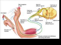
sensory neurons |
*afferent
*conduct impulses toward the CNS. Sensory Receptors respond to various stimuli
*send information from PNS to CNS |
|
|
interneurons |
*association neurons
*entirely within the CNS. Conducts impulses from sensory neurons to motor neurons. Interneurons have an integrative function
*located entirely within CNS sends information to motor neurons |
|
|
motor neurons |
*efferent
*conducts impulses away from CNS and to effectors (muscles or glands)
*send impulses toward an effector (muscle or gland) in PNS |
|
|
reflex arc |
an example of the arrangement of sensory neurons, interneurons, and motor neurons |
|
|
functions of neurons |
*excitability (irritability) *conductivity - traveling from finger to synapse *secretion of neurotransmitters |
|
|
fact |
reflexes are in the spinal cord not the brain
|
|
|
review |
|
|
|
II. Structure of Neurons - pg 396-402 |
. |
|
|
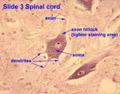
soma |
cell body, perikaryon |
|
|
axon |
conducts nerve impulses away from the soma: there is never more than one axon per neuron. (some have none) |
|
|
dendrites |
receive impulses from other neurons; receiving fibers |
|
|
myelin sheath |
insulating layer (mostly lipid) around nerve fibers, formed by neuroglia cells. Not all nerve fibers are myelinated. Those that are myelinated comprise the white matter, unmyelinated fibers comprise the gray matter |
|
|
III. Classes of Neurons - pg 398 |
. |
|
|
multipolar neurons |
one axon, multiple dendrites; most common type |
|
|
bipolar neurons |
one axon, one dendrite |
|
|
unipolar neurons |
single axon branches a short distance from soma, one branch (peripheral fiber) carries impulses from source of sensation, the other (central fiber) carries impulse into the spinal cord |
|
|
anaxonic neurons |
these have no axon, multiple dendrites. Examples in retina and brain |
|
|
IV. Neuroglia- pgs 400-402 |
These cells are not neurons, but perform various supportive roles in the nervous system. |
|
|
oligodendrocytes |
from myelin sheath in CNS |
|
|
protoplasmic astrocytes |
cover brain surfaces, formation of blood-brain barrier, remove neurotransmitters and potassium ions from intercellular fluid, regulate composition of cerebrospinal fluid |
|
|
fibrous astrocytes |
form supportive network in CNS, replace damaged nerve tissue (scar tissue) |
|
|
ependymal cells |
line cavity of brain and spinal cord. Produce and circulate CSF. (circulate by cilia) |
|
|
microgilia |
phagocytosis |
|
|
schwann cells |
form myelin sheath in peripheral nervous system |
|
|
satellite cells |
surround somas of neurons in galia function |
|
|
astrocytes |
provide structural support within neural tissue |
|
|
V. Initiation and Conduction of Nerve Impulses |
. |
|
|
resting membrane potential |
interior of nerve cell is negatively charged, relative to extracellular fluid. Cell is said to be polarized. |
|
|
action potential |
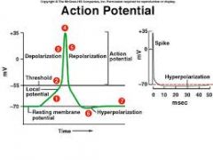
1. Na+ gates open, allowing influx of sodium ions. K+ gates begin to open. Depolarization begins 2. Na+ gate closes. K+ gate opens fully. K+ ions leave cell, bringing about beginning of repolarization 3. Both Na+ gate and K+ gate closed, repolarization complete
|
|
|
refractory period |
nerve impulse travels along a nerve fiber as a wave of depolarization. For a short time after an action potential, it is impossible to stimulate that region of a neuron to fire again |
|
|
Nodes of Ranvier |
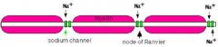
In myelinated nerve fibers ions can be exchanged with extracellular fluid only at the Nodes of Ranvier; impulse appears to jump (saltate) from node to node |
|
|
action potential |
1. all or nothing
2. irreversible
3. non-decremental (doesn't lose strength over distance) |
|
|
VI. Synaptic Transmission |
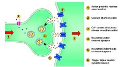
although never impulses are conducted electrically along a nerve fiber, a 20-40 nm gap (the synaptic cleft exits between neurons). this necessitates (in most cases) chemical communication between neurons
|
|
|
synaptic structures include |
*synaptic knob of pre-synaptic neuron
*synaptic vesicles (filled with neurotransmitters) in the pre-synaptic neuron
*neurotransmitter receptors in the post-synaptic membrane |
|
|
Chemical Synapses are of two types |
Ionotropic Synapses and Metabotropic Synapses |
|
|
ionotropic synapses |
nerve impulses reaches synaptic knob, causes Ca2+ gates to open. This triggers exocytosis of synaptic vesicles, which allow diffusion of neurotransmitter into the synaptic cleft. These bind receptors in the post-synaptic membrane, which open and allow passage of Na+ and K+ ions, producing post-synaptic potential |
|
|
metabotropic synapses |
bind of neurotransmitter by receptors in post-synaptic cell activates cyclic AMP production. This can turn certain metabolic pathways on or off, activate genetic transcription, and open ion gates in the membrane |
|
|
there are several classes of neurotransmitters |
*some are excitatory *others are inhibitory *others act at the neuromuscular junction |
|
|
VII. Cessation of the Signal at the Synapse |
. |
|
|
the "turning off" of the signal at the synapse is just as important as the stimulation of the post-synaptic cell. this is accomplished by |
1. re-uptake of the neurotransmitter by the pre-synaptic neuron
2. diffusion of the neurotransmitter into the extracellular fluid, where astrocytes absorb it.
3. enzymatic degradation of the neurotransmitter in the synaptic cleft. |
|
|
acetylcholinesterase |
enzyme that degrates ACH |
|
|
ACH-ase inhibitors |
*some used in treating Alzheimers patients
*some are used as pesticides
*some are used as never agents; Sarin and VX |